

Effectiveness of diffusion tensor imaging in differentiating early-stage subcortical ischemic vascular disease, Alzheimer's disease and normal ageing
- PMID: 28388630
- PMCID: PMC5384760
- DOI: 10.1371/journal.pone.0175143
Effectiveness of diffusion tensor imaging in differentiating early-stage subcortical ischemic vascular disease, Alzheimer's disease and normal ageing
Abstract
Objective: To describe and compare diffusion tensor imaging (DTI) parameters between patients with subcortical ischemic vascular disease (SIVD) and Alzheimer's disease (AD) diagnosed using structuralized neuropsychiatric assessments, and investigate potential neuronal substrates related to cognitive performance.
Methods: Thirty-five patients with SIVD, 40 patients with AD, and 33 cognitively normal control (NC) subjects matched by age and education level were consecutively recruited and underwent cognitive function assessments and DTI examinations. Comparisons among these three subgroups with regards to cognitive performance and DTI parameters including fractional anisotropy (FA) and mean diffusivity (MD) values were performed. Partial correlation analysis after controlling for age and education was used to evaluate associations between cognitive performance and DTI parameters.
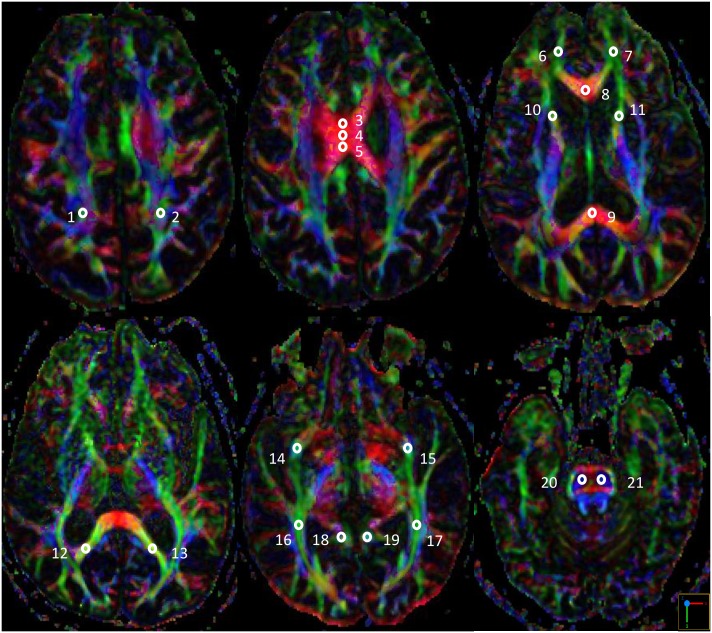
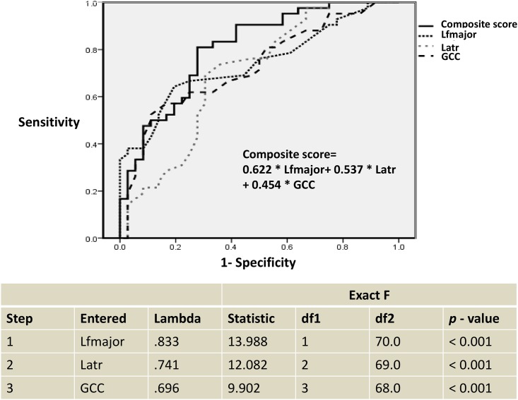
Results: With regards to cognitive performance, the patients with SIVD had lower total scores in frontal assessment battery (FAB) compared to those with AD (p < 0.05) in the context of comparable Mini-Mental Status Examination and Cognitive Abilities Screening Instrument scores. With regards to DTI parameters, there were more regions of significant differences in FA among these three subgroups compared with MD. Compared with NC group, the patients with SIVD had significant global reductions in FA (p < 0.001 ~ 0.05), while significant reductions in FA among the patients with AD were regionally confined within the left superior longitudinal fasciculus, genu and splenium of the corpus callosum, and bilateral forceps major, and the anterior thalamic radiation, uncinate fasciculus, and cingulum of the left side (p < 0.01 ~ 0.05). Analysis of FA values within the left forceps major, left anterior thalamic radiation, and genu of the corpus callosum revealed a 71.8% overall correct classification (p < 0.001) with sensitivity of 69.4%, specificity of 73.8%, positive predictive value of 69.4%, and negative predictive value of 73.8% in discriminating patients with SIVD from those with AD. In combined analysis of the patients with SIVD and AD (n = 75), the total FAB score was positively correlated with FA within the bilateral forceps minor, genu of the corpus callosum, left forceps major, left uncinate fasciculus, and right inferior longitudinal fasciculus (p = 0.001 ~ 0.038), and inversely correlated with MD within the right superior longitudinal fasciculus, genu and body of the corpus callosum, bilateral forceps minor, right uncinate fasciculus, and right inferior longitudinal fasciculus (p = 0.003 ~ 0.040).
Conclusions: Our findings suggest the effectiveness of DTI measurements in distinguishing patients with early-stage AD from those with SIVD, with discernible changes in spatial distribution and magnitude of significance of the DTI parameters. Strategic FA assessments provided the most robust discriminative power to differentiate SIVD from AD, and FAB may serve as an additional cognitive marker. We also identified the neuronal substrates responsible for FAB performance.
Conflict of interest statement
Figures
Similar articles
-
Comparison of neuropsychiatric symptoms and diffusion tensor imaging correlates among patients with subcortical ischemic vascular disease and Alzheimer's disease.BMC Neurol. 2017 Jul 28;17(1):144. doi: 10.1186/s12883-017-0911-5. BMC Neurol. 2017. PMID: 28754095 Free PMC article.
-
Discriminating subcortical ischemic vascular disease and Alzheimer's disease by diffusion kurtosis imaging in segregated thalamic regions.Hum Brain Mapp. 2021 May;42(7):2018-2031. doi: 10.1002/hbm.25342. Epub 2021 Jan 8. Hum Brain Mapp. 2021. PMID: 33416206 Free PMC article.
-
The value of diffusion tensor imaging in the differential diagnosis of subcortical ischemic vascular dementia and Alzheimer's disease in patients with only mild white matter alterations on T2-weighted images.Acta Radiol. 2012 Apr 1;53(3):312-7. doi: 10.1258/ar.2011.110272. Epub 2012 Mar 13. Acta Radiol. 2012. PMID: 22416261
-
The role of diffusion tensor imaging and fractional anisotropy in the evaluation of patients with idiopathic normal pressure hydrocephalus: a literature review.Neurosurg Focus. 2016 Sep;41(3):E12. doi: 10.3171/2016.6.FOCUS16192. Neurosurg Focus. 2016. PMID: 27581308 Review.
-
Diffusion tensor imaging of white matter degeneration in early stage of Alzheimer's disease: a review.Int J Neurosci. 2020 Mar;130(3):243-250. doi: 10.1080/00207454.2019.1667798. Epub 2019 Sep 24. Int J Neurosci. 2020. PMID: 31549530
Cited by
-
Joint diffusional kurtosis magnetic resonance imaging analysis of white matter and the thalamus to identify subcortical ischemic vascular disease.Sci Rep. 2024 Jan 31;14(1):2570. doi: 10.1038/s41598-024-52910-x. Sci Rep. 2024. PMID: 38297073 Free PMC article.
-
Stage-Dependent Cerebral Blood Flow and Leukoaraiosis Couplings in Subcortical Ischemic Vascular Disease and Alzheimer's Disease.J Alzheimers Dis. 2022;86(2):729-739. doi: 10.3233/JAD-215405. J Alzheimers Dis. 2022. PMID: 35124651 Free PMC article.
-
The Associations Between White Matter Disruptions and Cognitive Decline at the Early Stage of Subcortical Vascular Cognitive Impairment: A Case-Control Study.Front Aging Neurosci. 2021 Aug 2;13:681208. doi: 10.3389/fnagi.2021.681208. eCollection 2021. Front Aging Neurosci. 2021. PMID: 34408641 Free PMC article.
-
Greater Social Engagement and Greater Gray Matter Microstructural Integrity in Brain Regions Relevant to Dementia.J Gerontol B Psychol Sci Soc Sci. 2021 Jun 14;76(6):1027-1035. doi: 10.1093/geronb/gbaa173. J Gerontol B Psychol Sci Soc Sci. 2021. PMID: 33219690 Free PMC article.
-
Frequency-dependent white-matter functional network changes associated with cognitive deficits in subcortical vascular cognitive impairment.Neuroimage Clin. 2022;36:103245. doi: 10.1016/j.nicl.2022.103245. Epub 2022 Oct 25. Neuroimage Clin. 2022. PMID: 36451351 Free PMC article.
References
-
- World Health Organization. Dementia Fact sheet. 2016. http://www.who.int/mediacentre/factsheets/fs362/en/
-
- Herrera E Jr, Caramelli P, Silveira AS, Nitrini R. Epidemiologic survey of dementia in a community-dwelling Brazilian population. Alzheimer Dis Assoc Disord. 2002;16(2):103–8. - PubMed
-
- Englund E, Brun A. Histopathology. White matter changes in dementia of Alzheimer's type: the difference in vulnerability between cell compartments. 1990; 16(5):433–9. - PubMed
-
- O’Brien JT, Ames D, Schwietzer I. White matter in depression and Alzheimer’s disease: a review of magnetic resonance imaging studies. International Journal of Geriatric Psychiatry. 1996;11:681–94.
-
- Kalaria RN, Ballard C. Overlap between pathology of Alzheimer disease and vascular dementia. Alzheimer Dis Assoc Disord. 1999;13 Suppl 3:S115–23. - PubMed
MeSH terms
LinkOut - more resources
Full Text Sources
Other Literature Sources
Medical
