. 2017 Apr 7;13(4):e1006726.
doi: 10.1371/journal.pgen.1006726. eCollection 2017 Apr.

The bHLH transcription factor SPATULA enables cytokinin signaling, and both activate auxin biosynthesis and transport genes at the medial domain of the gynoecium

Affiliations

The bHLH transcription factor SPATULA enables cytokinin signaling, and both activate auxin biosynthesis and transport genes at the medial domain of the gynoecium

J Irepan Reyes-Olalde et al. PLoS Genet. .

Abstract

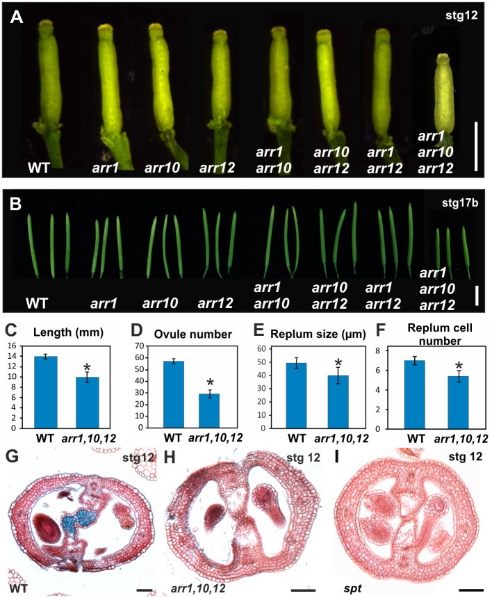
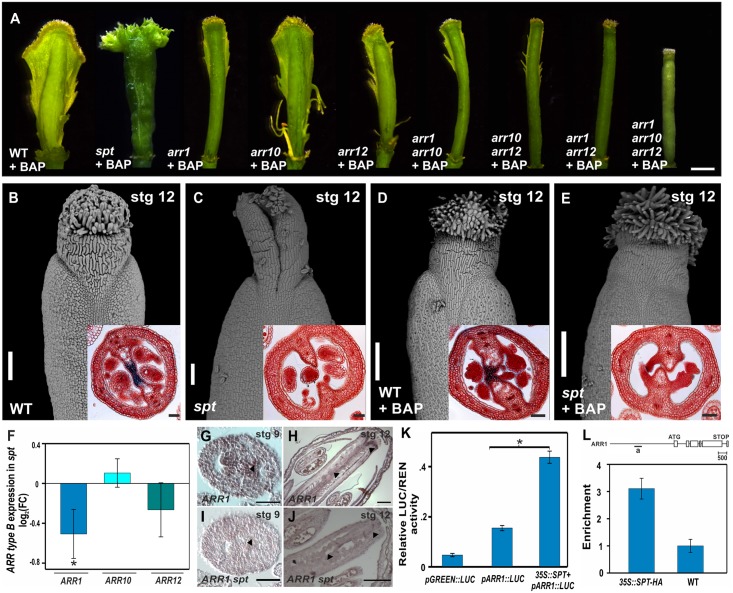
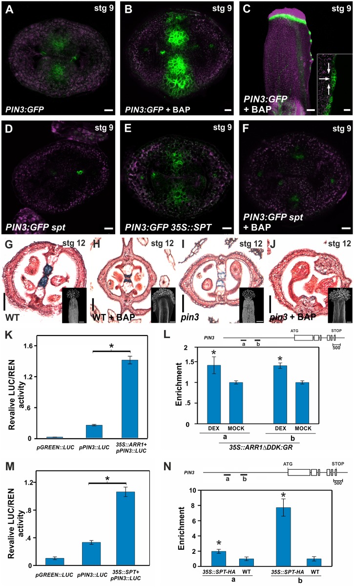
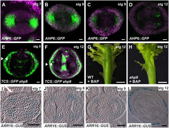
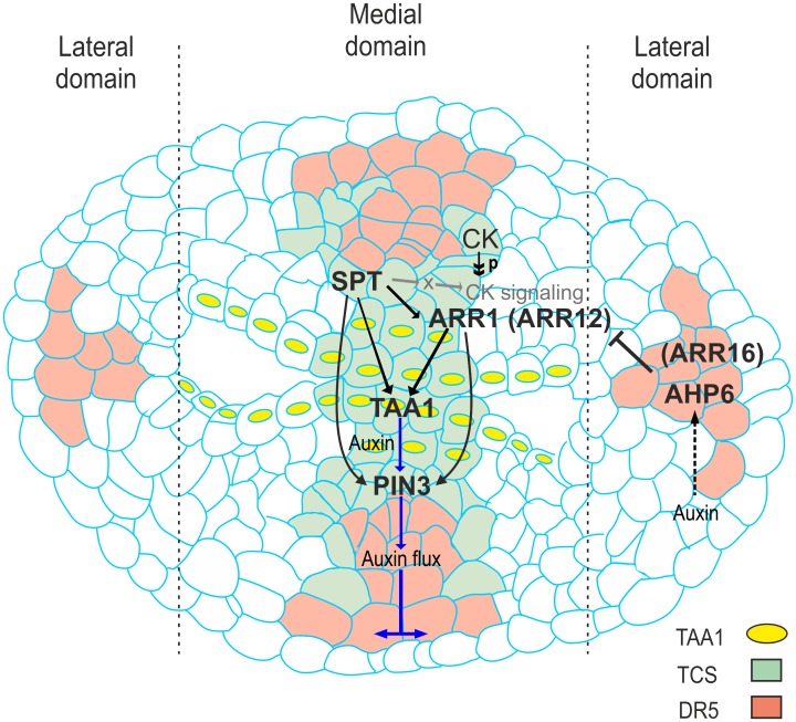
Fruits and seeds are the major food source on earth. Both derive from the gynoecium and, therefore, it is crucial to understand the mechanisms that guide the development of this organ of angiosperm species. In Arabidopsis, the gynoecium is composed of two congenitally fused carpels, where two domains: medial and lateral, can be distinguished. The medial domain includes the carpel margin meristem (CMM) that is key for the production of the internal tissues involved in fertilization, such as septum, ovules, and transmitting tract. Interestingly, the medial domain shows a high cytokinin signaling output, in contrast to the lateral domain, where it is hardly detected. While it is known that cytokinin provides meristematic properties, understanding on the mechanisms that underlie the cytokinin signaling pattern in the young gynoecium is lacking. Moreover, in other tissues, the cytokinin pathway is often connected to the auxin pathway, but we also lack knowledge about these connections in the young gynoecium. Our results reveal that cytokinin signaling, that can provide meristematic properties required for CMM activity and growth, is enabled by the transcription factor SPATULA (SPT) in the medial domain. Meanwhile, cytokinin signaling is confined to the medial domain by the cytokinin response repressor ARABIDOPSIS HISTIDINE PHOSPHOTRANSFERASE 6 (AHP6), and perhaps by ARR16 (a type-A ARR) as well, both present in the lateral domains (presumptive valves) of the developing gynoecia. Moreover, SPT and cytokinin, probably together, promote the expression of the auxin biosynthetic gene TRYPTOPHAN AMINOTRANSFERASE OF ARABIDOPSIS 1 (TAA1) and the gene encoding the auxin efflux transporter PIN-FORMED 3 (PIN3), likely creating auxin drainage important for gynoecium growth. This study provides novel insights in the spatiotemporal determination of the cytokinin signaling pattern and its connection to the auxin pathway in the young gynoecium.


Conflict of interest statement

The authors have declared that no competing interests exist.

Figures

Fig 1
Fig 1. Overview of the gynoecium and SPT is necessary for cytokinin signaling in the young gynoecium.
(A) Schematic overview and false-coloured transverse section of a stage 8 and of a stage 12 Arabidopsis thaliana gynoecium (pistil). The medial (M) and lateral (L) domains of the gynoecium are indicated. The CMM in the medial domain (stage 8 gynoecium; left side) is indicated and its derived structures can be seen in a stage 12 gynoecium (right side). L, lateral domain; M, medial domain. Orange, abaxial valve (abv); blue, adaxial valve (adv); white, abaxial replum (abr); pink, adaxial replum (adr); green, ovule primordium (op); red, septum primordium (sp); CMM, carpel margin meristem; septum (S); replum (R); transmitting tract (TT); ovule (O); funiculus (F). (B-M) Expression of the cytokinin response reporter TCS::GFP in transverse sections of gynoecia at stage 7, 8, 9, and 12 of wild-type (B-E), spt-2 (F-I), and 35S::SPT (J-M).(N-U) Expression of the reporter TCS::GFP in transverse sections of gynoecia at stage 7, 8, 9, and 12, after 48 hours of 6-benzylaminopurine (BAP; a synthetic cytokinin) treatment in wild-type (N-Q) and spt-2 (R-U). Scale bars: 20 μm (E, I, M, Q, U), 10 μm (B-D, F-H, J-L, N-P, R-T).
Fig 2
Fig 2. Phenotypes of the type-B arr mutants and of the spt mutant.
(A) Mature gynoecium size of wild-type, arr1, arr10, arr12, arr1 arr10, arr10 arr12, arr1 arr12, and arr1 arr10 arr12. (B) Mature fruit size of wild-type, arr1, arr10, arr12, arr1 arr10, arr10 arr12, arr1 arr12, and arr1 arr10 arr12. (C-F) Phenotypes of the type-B arr1 arr10 arr12 triple mutant compared to wild-type (WT): fruit length (C), ovule number (D), replum width (E), and replum cell number (F). (G-I) Transverse sections of stage 12 gynoecia of wild-type (G), arr1 arr10 arr12 (with transmitting tract and septum fusion defects) (H), and spt-2 (I). Scale bars: 1 mm (A), 5 mm (B), 50 μm (G-I). Error bars represent SD. *P < 0.05 (Student-t test). Sample numbers: (C, D) WT, n = 14 and arr1 arr10 arr12, n = 19; (E, F) WT, n = 20 and arr1 arr10 arr12, n = 19.
Fig 3
Fig 3. SPT enables cytokinin responses during early gynoecium development and regulates type-B ARR gene expression.
(A) Phenotypes of wild-type, arr1, arr10, arr12, arr1 arr10, arr10 arr12, arr1 arr12, arr1 arr10 arr12, and spt-2 gynoecia three to four weeks after receiving BAP treatment for five to ten days. (B-E) Scanning electron microscopy image of wild-type and spt-2 stage 12 gynoecia one day after either receiving mock (B, C) or BAP treatment for only 48 hours (D, E). Insets show a transverse section of the ovary. (F) Expression analysis by qRT-PCR of ARR1, ARR10, and ARR12 in wild-type and spt-12 dissected gynoecia. (G-J) In situ hybridization of type-B ARR1 mRNA in wild-type (G, H) and spt-2 (I, J) floral buds at stages 9 and 12. Arrowheads indicate the detected expression in wild-type and the absence in spt-2. (K) Luciferase reporter assay in N. benthamiana leaves co-transformed with 35S::SPT and pARR1::LUC. Ratio of firefly luciferase (LUC) to Renilla luciferase (REN) activity. (L) ChIP experiments against the ARR1 promoter region (indicated by “a” in the scheme above) using a 35S::SPT-HA line and wild-type. ACT2/7 served as a negative control. For the LUC assays and qRT-PCR experiments error bars represent the SD based on three biological replicates. ChIP results of one representative experiment are shown; error bars represent the SD of the technical replicates. *P < 0.05 (LUC: Student-t test; qRT-PCR and qPCR: ANOVA). Scale bars: 500 μm (A), 100 μm (B-E, H, J), 50 μm (insets in B-E, G, I).
Fig 4
Fig 4. Cytokinin signaling activates the auxin biosynthetic gene TAA1 in a SPT-dependent manner.
(A, B) Expression of the translational fusion TAA1::GFP-TAA1 in a transverse section of a stage 9 wild-type gynoecium that either received mock (A) or BAP treatment for 48 hours (B). (C, D) Expression of the translational fusion TAA1::GFP-TAA1 in a transverse section of a stage 9 spt-12 gynoecium that received mock (C) or BAP treatment for 48 hours (D). (E) Luciferase reporter assay in N. benthamiana leaves co-transformed with 35S::ARR1 and pTAA1::LUC. Ratio of LUC/REN activity. (F) ChIP experiments against the TAA1 promoter region (indicated by “a” in the scheme above) using an inducible 35S::ARR1ΔDDK:GR line treated with dexamethasone or mock. ACT2/7 served as a negative control. (G) Luciferase reporter assay in N. benthamiana leaves co-transformed with 35S::SPT and pTAA1::LUC. Ratio of LUC/REN activity. (H) ChIP experiments against the TAA1 promoter region (indicated by “a” in the scheme above) using a 35S::SPT-HA line and wild-type. ACT2/7 served as a negative control. Error bars represent the SD for the LUC assays based on three biological replicates. ChIP results of one representative experiment are shown; error bars represent the SD of the technical replicates. *P < 0.05 (LUC: Student-t test; qPCR: ANOVA). Scale bars: 10 μm (A-D).
Fig 5
Fig 5. The auxin transporter PIN3 is coordinately activated by cytokinin and SPT.
(A-C) PIN3 expression in stage 9 PIN3::PIN3-GFP gynoecia that either received mock (A, transverse section) or BAP treatment for 48 hours (B, transverse section and C, longitudinal view). The inset in (C) shows a magnified view of the proliferating tissue. Arrows indicate the possible auxin flow. (D-F) PIN3 expression in transverse sections of stage 9 PIN3::PIN3-GFP gynoecia in spt-2 (D), 35S::SPT (E), and in spt-2 treated for 48 hours with BAP (F). (G-J) Transverse sections of stage 12 gynoecia of wild-type (G, H) and pin3-4 (I, J). Gynoecia phenotypes after three to four weeks of mock (G, I) or BAP treatment for five days (H, J). Insets show a scanning electron microscopy image of the gynoecium. (K) Luciferase reporter assay in N. benthamiana leaves co-transformed with 35S::ARR1 and pPIN3::LUC. Ratio of LUC/REN activity. (L) ChIP experiments against the PIN3 promoter regions (indicated by “a” and “b” in the scheme above) using an inducible 35S::ARR1ΔDDK:GR line treated with dexamethasone or mock. ACT2/7 served as a negative control. (M) Luciferase reporter assay in N. benthamiana leaves co-transformed with 35S::SPT and pPIN3::LUC. Ratio of LUC/REN activity. (N) ChIP experiments against the PIN3 promoter regions (indicated by “a” and “b” in the scheme above) using a 35S::SPT-HA line and wild-type. ACT2/7 served as a negative control. Error bars represent the SD for the LUC assays based on three biological replicates. ChIP results of one representative experiment is shown and the error bars represent the SD of the technical replicates. *P < 0.05 (LUC: Student-t test; qPCR: ANOVA). Scale bars: 10 μm (A-F), 100 μm (G-J, G-J insets). Ovule primordium (op).
Fig 6
Fig 6. The cytokinin signaling repressors AHP6 and ARR16 likely block cytokinin responses in lateral tissues.
(A-D) Expression of the transcriptional reporter AHP6::GFP in transverse sections of stage 7, 8, 9, and 12 gynoecia. (E, F) Expression of the cytokinin response reporter TCS::GFP in transverse sections of stage 9 and 12 gynoecia in an ahp6-1 mutant background. Arrowheads indicate the absence of GFP signal in the epidermis of the valves. (G, H) Phenotypes of wild-type (G) and ahp6-1 (H) gynoecia one week after receiving BAP treatment for two weeks. (I-L) Expression of the transcriptional reporter ARR16::GUS (type-A ARR) in transverse sections of stage 7, 8, 9, and 12 gynoecia. Scale bars: 10 μm (A-C, E), 20 μm (D, F), 1 mm (G, H), 100 μm (I-L).
Fig 7
Fig 7. Model of the regulatory network in early gynoecium development integrating SPT, cytokinin signaling, auxin biosynthesis, and auxin transport.
Model of the regulatory network in early gynoecium development. This regulatory network integrates the results that SPT, an important player of gynoecium development, enables cytokinin signaling in the medial domain of the young gynoecium by activating the transcription of type-B ARR genes (at least ARR1 and ARR12; likely ARR1 directly and ARR12 indirectly), which proteins become active upon phosphorylation because of a phosphorelay cascade initiated when cytokinin is present, and then together activate auxin biosynthesis (TAA1) and transport important (PIN) for growth. It is likely that SPT also affects other components of the cytokinin signaling pathway (indicated by gray arrows). Solid black arrows indicate a positive regulation and a T-bar indicates a repression function, a broken black arrow indicates possible positive regulation by auxin, a double arrowhead indicates phosphorylation, purple arrows indicate possible auxin flow; CK, cytokinin; P, phosphate group.
Fig 8
Fig 8. Dynamic GRN Boolean model active during early gynoecium development.
(A) The topology of the Gene Regulatory Network (GRN) model visualized using the computational and graphical platform BioTapestry [71]. Regulatory relations among genes are based on the experimental evidence (this work). The two coherent feed-forward subcircuits are formed by starting with SPT regulating ARR1 and both together regulate TAA1, and also both together regulate PIN3. TAA1 also has other positive regulators, but for simplicity only SPT and ARR1 are depicted. ARR1 is depicted, but ARR12 is likely part of the network too. CK: cytokinin; Aux: auxin; P: phosphorylation. (B) All regulatory interactions were fed to the computational tool GeNeTool [72] to create the Boolean vector equations and for modeling of the GRN. The coherent feed-forward subcircuits are both configured as an AND-gate, i.e., cooperate regulation. Boolean output for gene active = 1 and for gene inactive = 0. (C-F) Alterations of the topology of the GRN model after perturbations. Boolean output was calculated by GeNeTool for each perturbation and visualized with BioTapestry. Gray color of lines and genes means it is inactive. The GRN is affected after the following perturbation experiments: SPT off (C), CK signaling off (D), PIN3 off (E), and TAA1 off (F). In all cases the GRN is altered, which predicts that gynoecium development will also be altered, and this happens in the mutants, validating our GRN model.

Similar articles

Cited by

References

    1. Reyes-Olalde JI, Zuñiga-Mayo VM, Chavez Montes RA, Marsch-Martinez N, de Folter S (2013) Inside the gynoecium: at the carpel margin. Trends Plant Sci 18: 644–655. 10.1016/j.tplants.2013.08.002 - DOI - PubMed
    1. Alvarez-Buylla ER, Benitez M, Corvera-Poire A, Chaos Cador A, de Folter S, et al. (2010) Flower development. Arabidopsis Book 8: e0127 10.1199/tab.0127 - DOI - PMC - PubMed
    1. Bowman JL, Baum SF, Eshed Y, Putterill J, Alvarez J (1999) Molecular genetics of gynoecium development in Arabidopsis. Curr Top Dev Biol 45: 155–205. - PubMed
    1. Chávez Montes RA, Herrera-Ubaldo H, Serwatowska J, de Folter S (2015) Towards a comprehensive and dynamic gynoecium gene regulatory network. Current Plant Biology 3–4: 3–12.
    1. Sehra B, Franks RG (2015) Auxin and cytokinin act during gynoecial patterning and the development of ovules from the meristematic medial domain. Wiley Interdiscip Rev Dev Biol. - PubMed

Publication types

MeSH terms