Review
. 2017 Jul:65:1-7.
doi: 10.1016/j.ceca.2017.03.006. Epub 2017 Mar 29.

Enamel: Molecular identity of its transepithelial ion transport system

Affiliations
Review

Enamel: Molecular identity of its transepithelial ion transport system

Rodrigo S Lacruz. Cell Calcium. 2017 Jul.

Abstract

Enamel is the most calcified tissue in vertebrates. It differs from bone in a number of characteristics including its origin from ectodermal epithelium, lack of remodeling capacity by the enamel forming cells, and absence of collagen. The enamel-forming cells known as ameloblasts, choreograph first the synthesis of a unique protein-rich matrix, followed by the mineralization of this matrix into a tissue that is ∼95% mineral. To do this, ameloblasts arrange the coordinated movement of ions across a cell barrier while removing matrix proteins and monitoring extracellular pH using a variety of buffering systems to enable the growth of carbonated apatite crystals. Although our knowledge of these processes and the molecular identity of the proteins involved in transepithelial ion transport has increased in the last decade, it remains limited compared to other cells. Here we present an overview of the evolution and development of enamel, its differences with bone, and describe the ion transport systems associated with ameloblasts.

Keywords: CRAC channel; Enamel; Ion transport; pH.


Figures

Figure 1
Figure 1. Schematic of the histology of the enamel organ
Ameloblasts undergo a number of important morphological changes. Ameloblasts originate from undifferentiated epithelium at the cervical loop (CL) before elongating and developing a secretory Tomes’ process (TP) distally. These tall polarized cells are the secretory ameloblasts. The adjacent stratum intermedium (SI) and stellate reticulum (SR) cells also form part of the enamel organ during the secretory stage. At this stage the matrix is poorly mineralized (light blue enamel). Secretory ameloblasts will transition to maturation stage ameloblasts increasing their capacity for ion transport to fully mineralize the enamel (dark blue enamel). The SR and SI cells also give rise to the papillary layer (PL) of cells. Organics are removed in maturation stage while Ca2+ and PO43− transport increases. The maturation stage contains two distinct cell morphologies. The dominant ruffle-ended ameloblasts (RA) cycle into smooth-ended cells (SA). The former has been associated with active transport. Ameloblasts regress and undergo apoptosis at the end of the life cycle prior to eruption of the tooth into the oral cavity.
Figure 2
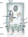
Figure 2. Model of ion channels and transport mechanisms in ameloblasts
The schematic represents a maturation-stage ruffled ended ameloblast. This model is based on reports of mRNA and protein expression as well as cellular localization. Thus in some cases the functional roles of these proteins have not been established. At the basal pole, HCO3 is transported into the cytosol by the Na+/ HCO3 exchanger NBCe1. Intracellular HCO3 can also be produced by carbonic anhydrases (CA2). The removal of H+ that are generated during CA2 activity and other sources of cytosolic H+ might be mediated by the Na+/H+ exchanger NHe1. Extrusion of HCO3 is mediated by the anion exchanger AE2 at the lateral membrane pole and by members of the SLC26a family (at least 3 members) at the apical pole. Chloride (Cl) is transported in and out of the cytoplasm by a number of proteins. AE2 and SLC26a exchange Cl for HCO3 whereas the cystic fibrosis transmembrane conductance regulator (CFTR) removes cytosolic Cl at the distal pole. Ca2+ enters the cell largely via the activity of the Ca2+ release activated Ca2+ channel mediated by ORAI1 and STIM1 proteins. Removal of cytosolic Ca2+ into ER is modulated by the sarco-endoplasmic reticulum SERCA2a pump, and it is released from this organelle by inositol triphosphate receptors (IP3R). The expression of L-type voltage-gated Ca2+ channels has not been reported in enamel cells but patients with dysfunctional channel show abnormal enamel. It might be the case that it plays a role in the functioning of the cell’s membrane potential. The bulk of Ca2+ extrusion is likely mediated by the Na+/Ca2+/K+ exchanger NCKX4, but also to a lesser extent by the plasma membrane Ca2+-ATPases (PMCA) and the Na+/Ca2+ exchanger NCX. Phosphate transport is poorly known but the Na+/ PO43− proteins NaPi have been localized at the distal cell pole. The cation Mg2+ can be moved into the cytosol by TRMP7 (transient receptor potential cation channel subfamily M member 7) whereas CNN4 (cyclin and CBS domain divalent metal cation transport mediator 4) can remove Mg2+ out of the cell in exchange for Na+. N= nucleus. ER= endoplasmic reticulum. GJ=gap junctions.

References

    1. Smith CE. Cellular and chemical events during enamel maturation. Crit Rev Oral Biol Med. 1998;9(2):128–61. - PubMed
    1. Boyde A. Microstructure of enamel. Ciba Found Symp. 1997;205:18–27. discussion 27–31. - PubMed
    1. Hubbard MJ. Abundant calcium homeostasis machinery in rat dental enamel cells. Up-regulation of calcium store proteins during enamel mineralization implicates the endoplasmic reticulum in calcium transcytosis. Eur J Biochem. 1996;239(3):611–23. - PubMed
    1. Berridge MJ, Lipp P, Bootman MD. The versatility and universality of calcium signalling. Nat Rev Mol Cell Biol. 2000;1(1):11–21. - PubMed
    1. Hubbard MJ. Calcium transport across the dental enamel epithelium. Crit Rev Oral Biol Med. 2000;11(4):437–66. - PubMed

Publication types

LinkOut - more resources