. 1988 Jul 15;241(4863):353-7.
doi: 10.1126/science.2838909.

Abnormalities in structure and expression of the human retinoblastoma gene in SCLC

Affiliations

Abnormalities in structure and expression of the human retinoblastoma gene in SCLC

J W Harbour et al. Science. .

Abstract

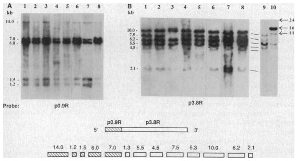
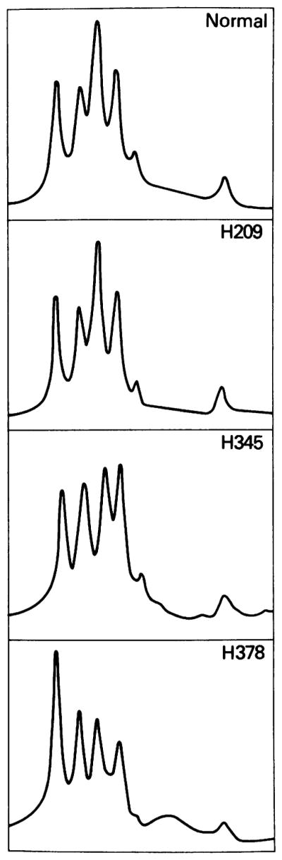
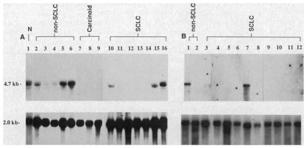
Small cell lung cancer (SCLC) has been associated with loss of heterozygosity at several distinct genetic loci including chromosomes 3p, 13q, and 17p. To determine whether the retinoblastoma gene (Rb) localized at 13q14, might be the target of recessive mutations in lung cancer, eight primary SCLC tumors and 50 cell lines representing all major histologic types of lung cancer were examined with the Rb complementary DNA probe. Structural abnormalities within the Rb gene were observed in 1/8 (13%) primary SCLC tumors, 4/22 (18%) SCLC lines, and 1/4 (25%) pulmonary carcinoid lines (comparable to the 20 to 40% observed in retinoblastoma), but were not detected in other major types of lung cancer. Rb messenger RNA expression was absent in 60% of the SCLC lines and 75% of pulmonary carcinoid lines, including all samples with DNA abnormalities. In contrast, Rb transcripts were found in 90% of non-SCLC lung cancer lines and in normal human lung. The finding of abnormalities of the Rb gene in SCLC and pulmonary carcinoids (both neuroendocrine tumors) suggests that this gene may be involved in the pathogenesis of a common adult malignancy.


Figures

Fig. 1
Fig. 1
Genomic restriction endonuclease analysis of the Rb in a representative panel of lung cancer lines. DNA (10 μg) extracted from seven lung cancer cell lines (H345, H510, H889, H187, H378, H209, and H679; lanes 1 to 7, respectively), normal human thymus (lane 8), and two primary SCLC tumor specimens (lanes 9 to 10, panel B) were digested to completion with Hind III and transferred to nitrocellulose after 0.8% agarose gel electrophoresis. Filters were then hybridized with either the p0.9R (A) or the p3.8R (B) 32P random-primed cDNA probes for 18 hours under standard hybridization conditions (26). For p0.9R, a 675-bp Eco RI–Hpa I fragment was used as a probe since this removed a GC-rich region that conferred a high background on hybridization. Filters were washed twice at room temperature in 2× SSC/0.1% SDS for 30 minutes and then twice in 0.1× SSC/0.1% SDS at 55°C for 1 hour. A diagram at the bottom of the figure depicts the previously described Rb cDNA clone and the Hind III genomic fragments (5, 6) detected by the p0.9R probe (hatched boxes) and the p3.8R probe (open boxes).
Fig. 2
Fig. 2
Densitometric analysis. The top panel shows the densitometric tracing observed with normal human DNA (from fresh thymus tissue) digested to completion with Hind III and hybridized with the p3.8R probe. The five peaks from left to right represent, respectively, the Hind III fragments at 10, 7.5, 6.2, 5.5, 4.5, and 2.1 kb (see Fig. 1B). The tracings were obtained on a Hoefer scanning densitometer (model GS300). Thirteen tumor lines, including H209 shown above, demonstrated densitometric patterns similar to that obtained for normal human DNA. In contrast, cell lines H345 and H378, shown in the lower two panels, had anomalous patterns.
Fig. 3
Fig. 3
Rb gene expression in a representative panel of lung cancer lines. (A) Total RNA (15 μg) was extracted (27) from normal lung tissue obtained from a surgical specimen (lane 1), five non-SCLC lines (H820, H1355, H1373, H1404, and H1437; lanes 2 to 6), three carcinoid lines (H720, H835, and H679; lanes 7 to 9), and seven SCLC lines (H1092, H1622, H748, H1304, H774, H1436, and H1105; lanes 10 to 16). (B) Two micrograms of poly(A)-selected RNA (28) was extracted from two additional non-SCLC lines (H125 and H23; lanes 1 and 2) and ten additional SCLC lines (N592, H345, H378, H510, H209, H187, H526, H524, H82, and N417; lanes 3 to 12). The above RNA samples were transferred to nitrocellulose after formaldehyde-denaturing gel electrophoresis (29) and filters were hybridized sequentially with the 32P random-primed p3.8R probe which detects the 4.7-kb Rb transcript (–7) or with a β-actin cDNA probe which detects a 2-kb transcript (30). Autoradiographs were developed on XAR-5 film after 5 days at −70°C for p3.8R and after overnight exposure for β-actin. N, normal lung.

Similar articles

Cited by

References

    1. Knudson AG., Jr Proc Natl Acad Sci USA. 1971;68:820. - PMC - PubMed
    1. Benedict WF, et al. Science. 1983;219:973. - PubMed
    2. Cavenee WK, et al. Nature. 1983;305:779. - PubMed
    3. Hansen MF, et al. Proc Natl Acad Sci USA. 1985;82:6216. - PMC - PubMed
    1. Friend SH, Dryja TP, Weinberg RA. N Engl J Med. 1988;318:618. - PubMed
    1. Knudson AG, Jr, Meadows AT, Nichols WW, Hill R. 1976;295:1120. ibid. - PubMed
    2. Yunis JJ, Ramsay N. Am J Dis Child. 1978;132:161. - PubMed
    3. Sparkes RS, et al. Science. 1983;219:971. - PubMed
    1. Friend SH, et al. Nature. 1986;323:643. - PubMed

Publication types