. 2017 Jun 7;25(6):1306-1315.
doi: 10.1016/j.ymthe.2017.03.016. Epub 2017 Apr 4.

Optimizing Cardiac Delivery of Modified mRNA

Affiliations

Optimizing Cardiac Delivery of Modified mRNA

Nishat Sultana et al. Mol Ther. .

Abstract

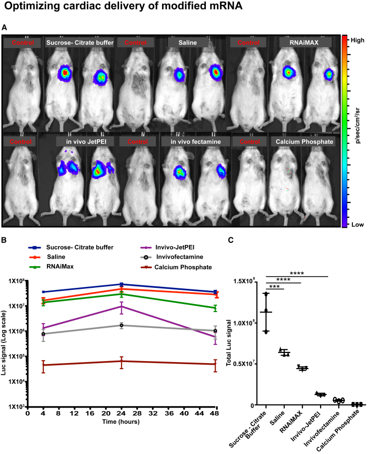
Modified mRNA (modRNA) is a new technology in the field of somatic gene transfer that has been used for the delivery of genes into different tissues, including the heart. Our group and others have shown that modRNAs injected into the heart are robustly translated into the encoded protein and can potentially improve outcome in heart injury models. However, the optimal compositions of the modRNA and the reagents necessary to achieve optimal expression in the heart have not been characterized yet. In this study, our aim was to elucidate those parameters by testing different nucleotide modifications, modRNA doses, and transfection reagents both in vitro and in vivo in cardiac cells and tissue. Our results indicate that optimal cardiac delivery of modRNA is with N1-Methylpseudouridine-5'-Triphosphate nucleotide modification and achieved using 0.013 μg modRNA/mm2/500 cardiomyocytes (CMs) transfected with positively charged transfection reagent in vitro and 100 μg/mouse heart (1.6 μg modRNA/μL in 60 μL total) sucrose-citrate buffer in vivo. We have optimized the conditions for cardiac delivery of modRNA in vitro and in vivo. Using the described methods and conditions may allow for successful gene delivery using modRNA in various models of cardiovascular disease.

Keywords: delivery; heart; modified mRNA.


Figures

Figure 1
Figure 1
Optimizing ModRNA Modification to Increase Translation and Expression Kinetics in Vitro and In Vivo Luc mRNA expression was compared between mRNA with or without nucleotide modifications (unmodified), using the IVIS imaging system, in rat neonatal CMs in vitro or CFW mouse heart in vivo. The nucleotide modifications tested were as follows: 100% replacement of uridine by 2-Thiouridine-5′-Triphosphate (2-thio ψU) or by 1-Methylpseudouridine-5′-Triphosphate (1-mψU) or 100% replacement of uridine by Pseudouridine-5′-Triphosphate and cytidine by 5-Methylcytidine-5′-Triphosphate (ψU + 5mC). (A and B) Representative images of transfected (with RNAiMAX) neonatal CMs or mice. Images were taken 24 hr post-transfection with 2.5 μg/well in a 24-well plate or intramyocardial injection with 100 μg modified or non-modified Luc mRNA, respectively. (C and D) Kinetics or pharmacokinetics of modified or non-modified Luc mRNA in vitro or in vivo, respectively. (E and F) Efficient translation of Luc modRNA with different nucleotide modifications. Total Luc signal was measured over 5 or 10 days post-transfection in vitro or in vivo, respectively. Results represent two independent experiments with n = 2 or n = 3 (total n = 5 wells or mice; ****p < 0.0001, one-way ANOVA with Bonferroni post hoc test).
Figure 2
Figure 2
Optimizing Vehicle and ModRNA Dose for CM Transfection In Vitro Rat neonatal or hPSC-derived CMs were isolated and plated onto 24-well tissue culture plates. Different transfection regents were used to transfect the CMs with 2.5 μg nGFP modRNA (1-mψU)/per well. Then 18 hr post-transfection, cells were fixed and stained for actinin (red) and GFP (green). ImageJ was used to calculate the number of nGFP+ and Actinin+ cells in each well. (A and B) Representative images of transfected rat neonatal CMs (A) or hPSC-derived CMs (B) with RNAiMAX-nGFP modRNA complex 18 hr post-transfection. (C and D) Quantification of transfection efficiency of rat neonatal (C) or hPSC-derived CMs (D) using different transfection reagents. (E and F) Optimal doses per well were tested using optimal transfection reagent (RNAiMAX) using different doses of nGFP in rat neonatal CMs (E) or in hPSC-derived CMs (F). (G–J) FACS analysis was used 18 hr post-transfection with RNAiMAX and different nGFP modRNA doses to measure median GFP intensity and cell death (percentage Annexin V of viable cells) for rat neonatal CMs (G and I) or hPSC-derived CMs (H and J), respectively. Results represent two independent experiments with n = 3 wells (*p < 0.05 and ***p < 0.001, one-way ANOVA with Bonferroni post hoc test; scale bar, 10 μm).
Figure 3
Figure 3
Vehicle Optimization for ModRNA Cardiac Tissue Transfection In Vivo Luc modRNA at a dose of 100 μg (1-mψU)/heart, complexed with different commercially available RNA transfection reagents, was injected directly into myocardium of CFW mice in an open-chest surgery. The IVIS imaging system was used to calculate gene expression at different time points (4, 24, and 48 hr). (A) Representative images of transfected mice 24 hr post-injection directly into myocardium with different commercially available RNA transfection reagents. (B and C) Pharmokinetics (B) or efficient translation (C) of Luc modRNA transfected with different RNA transfection reagents and measured at different time points (4, 24, and 48 hr) using IVIS. Results represent three independent experiments with n = 2 or 3 mice (****p < 0.0001 and ***p < 0.001, one-way ANOVA with Bonferroni post hoc test).
Figure 4
Figure 4
Optimizing ModRNA Dose for Cardiac Tissue Transfection In Vivo Different doses (50, 100, and 200 μg) of Luc or Cre modRNA (1-mψU)/heart, delivered in sucrose-citrate buffer, were injected directly into the myocardium of CFW or Rosa26mTmG mice, respectively, in an open-chest surgery. (A) Representative image of transfected CFW mice 24 hr post-intramyocardial injection with different Luc modRNA doses. (B and C) Pharmokinetics (B) or efficient translation (C) of Luc modRNA transfected with different doses and measured at different time points (4, 24, and 48 hr) using IVIS. (D) Representative images of a transfected heart (transfected cells are green and non-transfected cells are red) or heart cross-sections (short axis view) of Rosa26mTmG mouse 24 hr post-injection of 100 μg Cre modRNA directly into myocardium. (E) Quantification of the biodistribution of different doses of Cre modRNA post-transfection in vivo. Results represent two independent experiments with n = 2 or 3 mice (total n = 3–5 mice; **p < 0.01 and *p < 0.05; N.S., not significant; one-way ANOVA with Bonferroni post hoc test).
Figure 5
Figure 5
Optimized Cardiac Delivery of ModRNA Cardiac delivery of modRNA with 100% replacement of uridine with 1-mψU yields the highest gene expression and longest pharmacokinetics in comparison to other nucleotide modifications, both in vitro and in vivo. CM transfection in vitro (rat neonatal or hPSC) is optimal with a positively charged transfection reagent, such as RNAiMAX, TransIT, jetMESSENGER, or MessengerMAX, in a concentration of 0.013 μg modRNA/1 mm2 well diameter. For in vivo cardiac delivery in mice, our data show that 100 μg/heart (1.6 μg modRNA/μL in 60 μL total) in sucrose-citrate buffer directly injected into myocardium yields the highest gene expression and covers close to a quarter of the left ventricle of mouse heart.

Comment in

References

    1. del Monte F., Hajjar R.J. Efficient viral gene transfer to rodent hearts in vivo. Methods Mol. Biol. 2003;219:179–193. - PubMed
    1. Del Monte F., Ishikawa K., Hajjar R.J. Gene Transfer to Rodent Hearts In Vivo. Methods Mol. Biol. 2017;1521:195–204. - PubMed
    1. Ylä-Herttuala S., Bridges C., Katz M.G., Korpisalo P. Angiogenic gene therapy in cardiovascular diseases: dream or vision? Eur. Heart J. 2017:ehw547. - PMC - PubMed
    1. Andrieu-Soler C., Bejjani R.A., de Bizemont T., Normand N., BenEzra D., Behar-Cohen F. Ocular gene therapy: a review of nonviral strategies. Mol. Vis. 2006;12:1334–1347. - PubMed
    1. Chaanine A.H., Kalman J., Hajjar R.J. Cardiac gene therapy. Semin. Thorac. Cardiovasc. Surg. 2010;22:127–139. - PMC - PubMed

Publication types

LinkOut - more resources