Review
. 2017 Apr 7;474(8):1417-1438.
doi: 10.1042/BCJ20160499.

RNA-binding proteins with prion-like domains in health and disease

Affiliations
Review

RNA-binding proteins with prion-like domains in health and disease

Alice Ford Harrison et al. Biochem J. .

Abstract

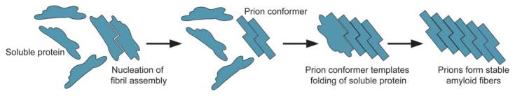
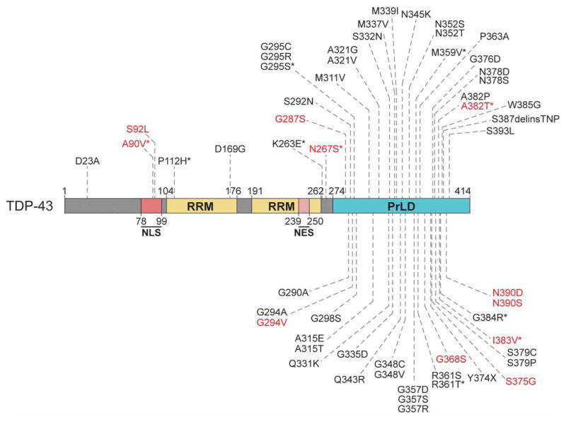
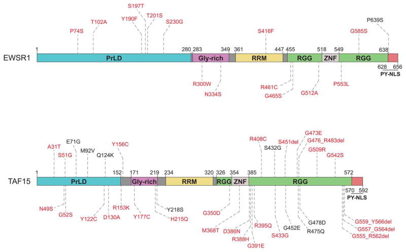
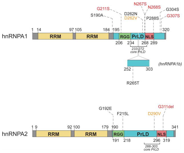
Approximately 70 human RNA-binding proteins (RBPs) contain a prion-like domain (PrLD). PrLDs are low-complexity domains that possess a similar amino acid composition to prion domains in yeast, which enable several proteins, including Sup35 and Rnq1, to form infectious conformers, termed prions. In humans, PrLDs contribute to RBP function and enable RBPs to undergo liquid-liquid phase transitions that underlie the biogenesis of various membraneless organelles. However, this activity appears to render RBPs prone to misfolding and aggregation connected to neurodegenerative disease. Indeed, numerous RBPs with PrLDs, including TDP-43 (transactivation response element DNA-binding protein 43), FUS (fused in sarcoma), TAF15 (TATA-binding protein-associated factor 15), EWSR1 (Ewing sarcoma breakpoint region 1), and heterogeneous nuclear ribonucleoproteins A1 and A2 (hnRNPA1 and hnRNPA2), have now been connected via pathology and genetics to the etiology of several neurodegenerative diseases, including amyotrophic lateral sclerosis, frontotemporal dementia, and multisystem proteinopathy. Here, we review the physiological and pathological roles of the most prominent RBPs with PrLDs. We also highlight the potential of protein disaggregases, including Hsp104, as a therapeutic strategy to combat the aberrant phase transitions of RBPs with PrLDs that likely underpin neurodegeneration.

Keywords: RNA-binding proteins; disaggregase; neurodegeneration; phase separation; prion-like domain.


Conflict of interest statement

Competing Interests

The Authors declare that there are no competing interests associated with the manuscript.

Figures

Figure 1
Figure 1. Prions self-replicate conformation by templating the folding of soluble protein to the prion conformation
Prions are protein conformers that self-replicate by templating the folding of natively folded proteins of the same amino acid sequence to the prion conformation [18,35]. Prions typically form stable amyloid fibers with a hallmark ‘cross-β’ structure in which β-strands run perpendicular to the axis of the fiber [18,35]. These amyloid assemblies are typically resistant to denaturation by heat, proteases, and detergents [18,37].
Figure 2
Figure 2. Mutations that cause ALS and FTD cluster in the PrLD of TDP-43
TDP-43 is an RBP with two canonical RRMs and a C-terminal PrLD [48,77,78,196]. Mutations that have been identified in patients with ALS and FTD are shown, and cluster in the PrLD [71,196]. Mutations identified in patients reported to have features of FTD, with or without a clinical ALS phenotype, are denoted by asterisks [,–227]. Mutations in red have also been observed in healthy control individuals [,,–230]. Disease-associated mutations were identified from Buratti [231], Cady et al. [228], Floris et al. 2015 [69], Lagier-Tourenne et al. [71], Peters et al. [196], the ALS data browser (http://alsdb.org) [134], and the ALS Online Genetics Database (http://alsod.iop.kcl.ac.uk/) [232].
Figure 3
Figure 3. ALS- and FTD-causing mutations in FUS cluster in LC domains and the PrLD
FUS has an N-terminal PrLD (residues 1–239) that is rich in glutamine, serine, tyrosine, and glycine. Bioinformatic analysis predicts that the PrLD also includes a portion of an adjacent glycine-rich region [93]. FUS has a single RRM, two RGG-rich regions, and a zinc-finger domain [93,196]. Mutations in FUS that have been associated with ALS and FTD cluster in the PrLD, RGG-rich region, and PY-NLS [196,233]. Mutations identified in patients reported to have symptoms of FTD, with or without a clinical ALS phenotype, are denoted by asterisks [–239]. Mutations in red have also been observed in healthy control individuals [,,,,,,–244]. Disease-associated mutations were identified from Belzil et al. [240], Corrado et al. [85], Huey et al. [236], Kwiatkowski et al. [86], Lagier-Tourenne et al. [71], Lattante et al. [233], Mackenzie et al. [30], Peters et al. [196], Rademakers et al. [87], Yan et al. [239], the ALS Online Genetics Database (http://alsod.iop.kcl.ac.uk/) [232], and the ALS Data Browser (http://alsdb.org) [135].
Figure 4
Figure 4. FET proteins EWSR1 and TAF15 have domain architectures similar to the domain architecture of FUS
FUS, TAF15, and EWSR1 are members of the FET protein family and are similar in domain structure and function [83,98,99]. Like FUS (Figure 3), EWSR1 and TAF15 each have an N-terminal PrLD, a glycine-rich region, and a single RRM, with C-terminal RGG-rich regions, a zinc-finger domain, and a PY-NLS [,,,,,–247]. Mutations shown were identified in ALS patients and compiled from Cady et al. [228], Couthouis et al. [48], Couthouis et al. [98], Couthouis et al. [136], Ticozzi et al. [100], and the ALS Data Browser (http://alsdb.org) [135]. Those in red have also been observed in healthy control individuals [98,135,136,228,229,241].
Figure 5
Figure 5. MSP-causing mutations affect a conserved aspartate residue in the hnRNPA1 and hnRNPA2 PrLDs
hnRNPA1 and hnRNPA2 contain two N-terminal RRMs and a C-terminal PrLD [101]. The PrLDs contain an RGG motif and a PY-NLS that mediates nuclear import [101,110,248]. A 52 amino acid stretch that occurs in the longer isoform of hnRNPA1 (hnRNPA1b) is depicted [249]. Missense mutations in hnRNPA1 and hnRNPA2 that cause MSP are noted in orange [101]. All other mutations were identified in patients with sporadic or familial ALS and compiled from Couthouis et al. [136], Kim et al. [101], Liu et al. [137], and the ALS Data Browser (http://alsdb.org) [135]. Those in red have been observed in healthy control individuals [229].
Figure 6
Figure 6. MSP- and ALS-associated mutations are predicted to increase the fibrillization propensity of hnRNPA1 and hnRNPA2
ZipperDB, a structure-based algorithm, calculates the propensity of hexapeptide fragments to form steric zippers [131]. Steric zippers, which are self-complementary β-sheets that form the backbone of an amyloid fibril, are predicted to form when the Rosetta energy of a hexapeptide is below the empirically determined ‘high fibrillization propensity’ threshold of −23 kcal/mol [131]. Several of the described mutations in hnRNPA1 and hnRNPA2 introduce a predicted steric zipper motif or strengthen an existing zipper [101,131]. For example, the D262V substitution in hnRNPA1 creates a potent SYNVFG zipper, whereas the D262N substitution also strengthens the GSYNDF zipper [101,131].
Figure 7
Figure 7. Cytoplasmic RNP granules include stress granules and P bodies
Stress granules are cytoplasmic assemblies that form in response to environmental stress and are sites of stalled translation initiation [49,140,144]. They contain polyadenylated mRNA transcripts, RBPs, translation initiation factors, and small ribosomal subunits [141]. P bodies are constitutively present but also form in response to stressful conditions [140]. They serve as sites of mRNA degradation and are characterized by the elements of the mRNA decapping and decay machinery [49,140]. Shown are many protein components of mammalian stress granules and P bodies [49,140,144].

References

    1. Khurana V, Lindquist S. Modelling neurodegeneration in Saccharomyces cerevisiae: why cook with baker’s yeast? Nat Rev Neurosci. 2010;11:436–449. doi: 10.1038/nrn2809. - DOI - PubMed
    1. Lepesant JA. The promises of neurodegenerative disease modeling. C R Biol. 2015;338:584–592. doi: 10.1016/j.crvi.2015.06.018. - DOI - PubMed
    1. Swinnen B, Robberecht W. The phenotypic variability of amyotrophic lateral sclerosis. Nat Rev Neurol. 2014;10:661–670. doi: 10.1038/nrneurol.2014.184. - DOI - PubMed
    1. Li YQ, Tan MS, Yu JT, Tan L. Frontotemporal lobar degeneration: mechanisms and therapeutic strategies. Mol Neurobiol. 2016;53:6091–6105. doi: 10.1007/s12035-015-9507-5. - DOI - PubMed
    1. Ferrari R, Kapogiannis D, Huey ED, Momeni P. FTD and ALS: a tale of two diseases. Curr Alzheimer Res. 2011;8:273–294. doi: 10.2174/156720511795563700. - DOI - PMC - PubMed

MeSH terms