. 2017 May 31;83(12):e00505-17.
doi: 10.1128/AEM.00505-17. Print 2017 Jun 15.

New Insights into PhaM-PhaC-Mediated Localization of Polyhydroxybutyrate Granules in Ralstonia eutropha H16

Affiliations

New Insights into PhaM-PhaC-Mediated Localization of Polyhydroxybutyrate Granules in Ralstonia eutropha H16

Stephanie Bresan et al. Appl Environ Microbiol. .

Abstract

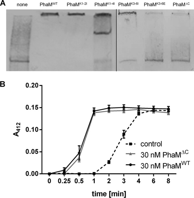
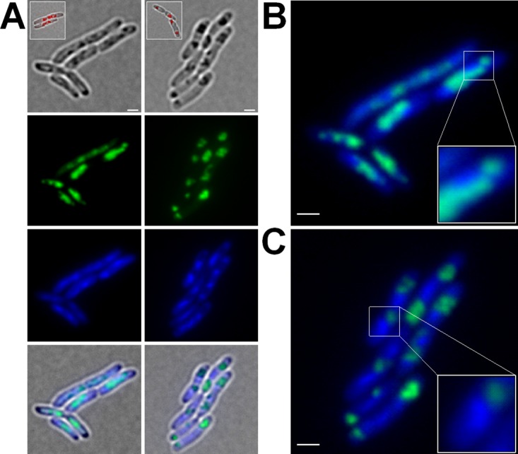
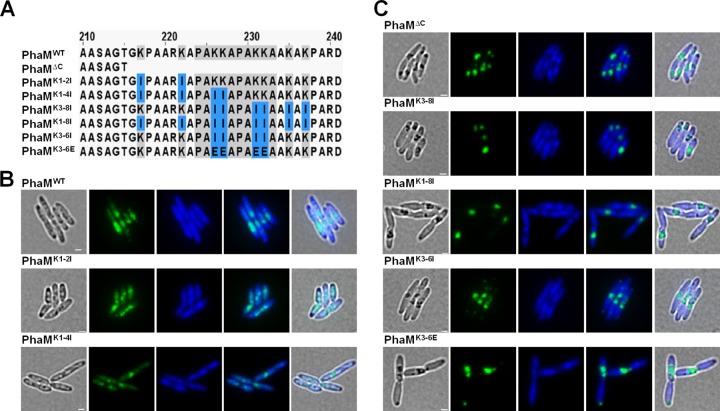
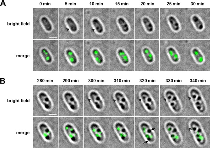
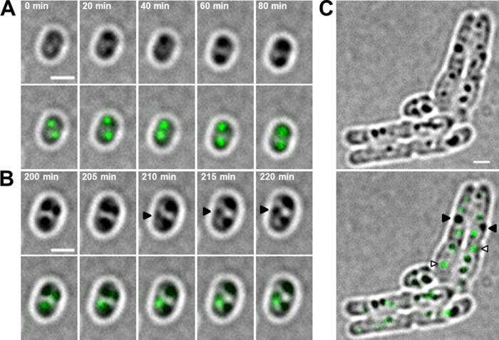
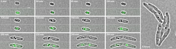
The formation and localization of polyhydroxybutyrate (PHB) granules in Ralstonia eutropha are controlled by PhaM, which interacts both with the PHB synthase (PhaC) and with the bacterial nucleoid. Here, we studied the importance of proline and lysine residues of two C-terminal PAKKA motifs in PhaM for their importance in attaching PHB granules to DNA by in vitro and in vivo methods. Substitution of the lysine residues but not of the proline residues resulted in detachment of formed PHB granules from the nucleoid. Instead, formation of PHB granule clusters at polar regions of the rod-shaped cells and an unequal distribution of PHB granules to daughter cells were observed. The formation of PHB granules was studied by the expression of chromosomally anchored gene fusions of fluorescent proteins with PhaM and PhaC in different backgrounds. PhaM and PhaC fusions showed a distinct colocalization at formed PHB granules in the nucleoid region of the wild type. In a ΔphaC background, PhaM and the catalytically inactive PhaCC319A protein were not able to form fluorescent foci, indicating that correct positioning requires the formation of PHB. Furthermore, time-lapse experiments revealed that PhaC and PhaM proteins detach from formed PHB granules at later stages, resulting in a nonhomogeneous population of PHB granules. This could explain why growth of individual PHB granules stops under PHB-permissive conditions at a certain size.IMPORTANCE PHB granules are storage compounds for carbon and energy in many prokaryotes. Equal distribution of accumulated PHB granules during cell division is therefore important for optimal fitness of the daughter cells. In R. eutropha, PhaM is responsible for maximal activity of PHB synthase, for initiation of PHB granule formation at discrete regions in the cells, and for association of formed PHB granules with the nucleoid. Here we found that four lysine residues of C-terminal PhaM sequence motifs are essential for association of PHB granules with the nucleoid. Furthermore, we followed PHB granule formation by time-lapse microscopy and provide evidence for aging of PHB granules that is manifested by detachment of previously PHB granule-associated PhaM and PHB synthase.

Keywords: PHB accumulation; Ralstonia; Ralstonia eutropha; biopolymer.


Figures

FIG 1
FIG 1
Electrophoretic mobility shift assay (EMSA) of constructed PhaM variants. (A) A PCR-generated DNA fragment labeled with biotin-11-dUTP (eYFP gene DNA, 824 bp, 0.62 nM in each experiment) was incubated with purified His6-tagged PhaM variants for 20 min at room temperature. After electrophoresis (6% native polyacrylamide gel) and blotting, biotinylated DNA-protein complexes were detected as described in Materials and Methods. Additions: none (control without PhaM) (lane 1), 5.4 μM PhaMWT (lane 2), 5.4 μM PhaM muteins as specified (lanes 3 to 7). (B) Effect of truncation of the C terminus of PhaM on PHB synthase activation. A continuous PHB synthase assay was performed using purified PHB synthase and PhaMWT or PhaMΔC. The dashed line corresponds to a control experiment without added PhaM.
FIG 2
FIG 2
Expression of PhaMWT-eYFP and PhaMΔC-eYFP in R. eutropha H16 cells. Cells expressing PhaMWT-eYFP and PhaMΔC-eYFP were grown on NB medium supplemented with 0.2% Na-gluconate at 30°C. (A) Diffuse fluorescent signals of cells expressing PhaMWT-eYFP colocalized with the DAPI-stained DNA (left panels; enlarged in panel B). Expression of truncated PhaM (PhaMΔC-eYFP) resulted in more focused fluorescence and in detachment from the nucleoid region (right panels; enlarged in panel C). Scale bars correspond to 1 μm. From top to bottom: bright field, eYFP channel, DAPI channel, and merge. Inlays show PHB granules of Nile red-stained cells.
FIG 3
FIG 3
Phenotypic characterization of truncated PhaM and PhaM lysine mutants in R. eutropha H16 cells. Cells were grown on NB medium supplemented with 0.2% Na-gluconate and 150 μg/ml kanamycin at 30°C. Images were taken after 4 h of PHB formation. (A) Sequence alignment of wild-type PhaM and PhaM mutants, highlighting substituted residues. (B) Replacement of lysine residues upstream of the PAKKA motifs (PhaMK1-2I) and upstream of as well as within the first PAKKA motif (PhaMK1-4I) showed phenotypes similar to that of wild-type PhaM. (C) Truncation of the C terminus of PhaM (PhaMΔC) and replacement of positively charged lysine residues by isoleucine or aspartate residues as indicated induced the formation of large PHB granule clusters. This phenotype showed strong similarity to that of PhaMΔC-expressing cells. From left to right: bright field, eYFP channel, DAPI channel, merge of fluorescent channels, and merge of all channels. Scale bars correspond to 1 μm.
FIG 4
FIG 4
Expression pattern of chromosomally integrated PhaM-eYFP in R. eutropha H16 cells in WT and ΔphaC backgrounds. (A) R. eutropha H16 WT cells harboring pBBR1MCS2-PphaC-eyfp (control) showed a uniformly distributed fluorescence within the cytoplasm (top panels). Formation of fluorescent foci was observed in R. eutropha H16 cells with a chromosomal integration of the phaM-eYFP gene (middle panels). In contrast, expression of the phaM-eYFP gene in ΔphaC cells resulted in a heterogeneously distributed fluorescence within the cytoplasm except in the areas of the cell poles (bottom panels, arrowheads). From left to right: bright field, eYFP channel, merge. (B) DsRed2EC-PhaC-driven formation of PHB granules induced the formation of fluorescent foci of PhaM-eYFP in a background of ΔphaC cells that colocalized with PHB granules (top panels). (C) Coexpression of PhaM-eYFP and DsRed2EC-PhaCC319A in the ΔphaC background showed a cytoplasmically distributed fluorescence (bottom panels). From left to right in panels B and C: bright field, eYFP channel, DsRed2EC channel, and merge of all channels. Scale bars correspond to 1 μm in all images.
FIG 5
FIG 5
SDS-PAGE of PhaM, PhaC/PhaCC319A, and mixtures of PhaM with PhaC/PhaCC319A after chemical cross-linking. Oligomerization of His6-PhaC (A) or His6-PhaCC319A (B) and/or PhaM-His6 was analyzed using 2.5% glutardialdehyde as a cross-linking agent as described in Materials and Methods. Lane 1, marker proteins as indicated. Lanes 2 to 10: 8 μM PhaM without glutardialdehyde (lane 2), 14 μM PhaC without glutardialdehyde (lane 3), mixture of 8 μM PhaM and 14 μM PhaC without glutardialdehyde (lane 4), 8 μM PhaM with glutardialdehyde (lane 5), 14 μM PhaC with glutardialdehyde (lane 6), and mixtures of 14 μM PhaC with different amounts of PhaM and glutardialdehyde (1.3, 2.6, 5.2, and 7.8 μM PhaM in lanes 7 to 10, respectively).
FIG 6
FIG 6
Time-lapse experiment with R. eutropha H16 cells expressing a chromosomally integrated phaM-eYFP gene. (A) PHB-free cells were transferred to fresh NB-gluconate agarose agar, and early PHB granule formation was immediately imaged every 5 min. At time point t = 0 min, fluorescent foci, but no PHB granules, are visible, and after 5 min a PHB granule that colocalizes with a fluorescent focus of PhaM-eYFP is formed (arrowheads at t = 5 and 10 min). (B) PHB granule formation was also imaged at later time points. Arrowheads show the formation of a PHB granule at the position of a preexisting fluorescent focus (top panels, t = 300 to 340 min). At later time points, a heterogeneity of PHB granules can be observed: while new PHB granules appear at the position of preexisting PhaM-eYFP foci, older PHB granules have lost PhaM-eYFP fluorescence (arrows in the cell at 320 min). White arrowheads in the cell at 340 min indicate a PHB granule that had a PhaM-eYFP focus at 310 to 330 min but lost this focus at 340 min. All pictures were taken with an exposure time of 500 ms. Scale bars correspond to 1 μm.
FIG 7
FIG 7
Time-lapse experiment with R. eutropha H16 cells expressing a chromosomally integrated phaC-eYFP gene. (A) PHB-free cells were transferred to fresh NB agar and dried for 10 min. Early PHB granule formation was then imaged every 20 min. (B) PHB granule formation was additionally analyzed at later time points at 5-min intervals. Arrowheads show the formation of a PHB granule at the position of a preexisting fluorescent focus. (C) After 9 h of incubation at 30°C, heterogeneity of PHB granules that do or do not colocalize with PhaC-eYFP can be observed. Black arrowheads indicate PHB granules without fluorescence, while PHB granules with a fluorescent focus are highlighted by white arrowheads. Pictures were taken with an exposure time of 500 ms. Scale bars correspond to 1 μm. Bright-field images are shown in the top panels, and overlays of bright field and the eYFP channel are shown in the bottom panels.
FIG 8
FIG 8
Time-lapse experiment with R. eutropha H16 cells expressing a chromosomally encoded phaC-eYFP gene in a ΔphaM background. PHB-free cells were transferred to fresh NB agar and dried for 10 min. PHB granule formation was imaged microscopically. At time point 0 min, the fluorescence of PhaC-eYFP was soluble, and the first fluorescent foci were observed during PHB granule formation (60 min). At later time points (260 min), the PhaC-eYFP fluorescence showed no colocalization with all formed PHB granules. Pictures were taken with an exposure time of 500 ms. Scale bars correspond to 1 μm.

References

    1. Cao C, Yudin Y, Bikard Y, Chen W, Liu T, Li H, Jendrossek D, Cohen A, Pavlov E, Rohacs T, Zakharian E. 2013. Polyester modification of the mammalian TRPM8 channel protein: implications for structure and function. Cell Rep 4:302–315. doi:10.1016/j.celrep.2013.06.022. - DOI - PMC - PubMed
    1. Tsuda H, Shiraki M, Inoue E, Saito T. 2016. Generation of poly-β-hydroxybutyrate from acetate in higher plants: detection of acetoacetyl CoA reductase and PHB synthase activities in rice. J Plant Physiol 201:9–16. doi:10.1016/j.jplph.2016.06.007. - DOI - PubMed
    1. Anderson AJ, Dawes EA. 1990. Occurrence, metabolism, metabolic role, and industrial uses of bacterial polyhydroxyalkanoates. Microbiol Rev 54:450–472. - PMC - PubMed
    1. Madison LL, Huisman GW. 1999. Metabolic engineering of poly(3-hydroxyalkanoates): from DNA to plastic. Microbiol Mol Biol Rev 63:21–53. - PMC - PubMed
    1. Pötter M, Steinbüchel A. 2006. Biogenesis and structure of polyhydroxyalkanoate granules. Microbiol Monogr 1:1–28.

Publication types

MeSH terms