. 2017 May 1;149(5):561-576.
doi: 10.1085/jgp.201611719. Epub 2017 Apr 7.

Control of Kir channel gating by cytoplasmic domain interface interactions

Affiliations

Control of Kir channel gating by cytoplasmic domain interface interactions

William F Borschel et al. J Gen Physiol. .

Abstract

Inward rectifier potassium (Kir) channels are expressed in almost all mammalian tissues and play critical roles in the control of excitability. Pancreatic ATP-sensitive K (KATP) channels are key regulators of insulin secretion and comprise Kir6.2 subunits coupled to sulfonylurea receptors. Because these channels are reversibly inhibited by cytoplasmic ATP, they link cellular metabolism with membrane excitability. Loss-of-function mutations in the pore-forming Kir6.2 subunit cause congenital hyperinsulinism as a result of diminished channel activity. Here, we show that several disease mutations, which disrupt intersubunit salt bridges at the interface of the cytoplasmic domains (CD-I) of adjacent subunits, induce loss of channel activity via a novel channel behavior: after ATP removal, channels open but then rapidly inactivate. Re-exposure to inhibitory ATP causes recovery from this inactivation. Inactivation can be abolished by application of phosphatidylinositol-4,5-bisphosphate (PIP2) to the cytoplasmic face of the membrane, an effect that can be explained by a simple kinetic model in which PIP2 binding competes with the inactivation process. Kir2.1 channels contain homologous salt bridges, and we find that mutations that disrupt CD-I interactions in Kir2.1 also reduce channel activity and PIP2 sensitivity. Kir2.1 channels also contain an additional CD-I salt bridge that is not present in Kir6.2 channels. Introduction of this salt bridge into Kir6.2 partially rescues inactivating mutants from the phenotype. These results indicate that the stability of the intersubunit CD-I is a major determinant of the inactivation process in Kir6.2 and may control gating in other Kir channels.


Figures

Figure 1.
Figure 1.
KATP channel gating and inactivation. (A) After patch excision, WT Kir6.2/SUR1 (KATP) channel currents activate to a stable level and are reversibly inhibited by application of 1 mM cytoplasmic ATP (black line). In this panel and in C, the arrow indicates patch excision. (B) Tetrameric Kir6.2 homology model depicting R192 (red) located at the CD-I between adjacent subunits (blue and green). (C) Kir6.2[R192A] channels are inhibited by ATP and then exhibit activation followed by fast current decay after removal of ATP. (D) Simplified models of KATP channel gating consisting of a closed apo (C0) state, ATP-bound stabilized closed (CA) state, inactivated (CI) state, PIP2-bound stabilized open (O) state, and short-lived closed flicker (Cf) state.
Figure 2.
Figure 2.
Conserved and unique salt bridge interactions at the CD-I in Kir channels. (A) Sequence alignment of eukaryotic Kir channel residues in the cytoplasmic domain. Charged residues that form intersubunit CD-I interactions are highlighted and colored with respect to their subunit orientation. (B and C) Homology models of mouse Kir6.2 (mKir6.2; B) and human Kir2.1 (hKir2.1; C) with adjacent subunits individually colored (blue and green) and cartoon representation of intersubunit interactions (R192–E229 in mKir6.2 and R204–E241 and D205–R235 in hKir2.1).
Figure 3.
Figure 3.
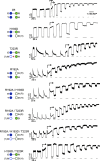
Engineering unique Kir2.1 contacts into inactivating mutant Kir6.2 channels. Macroscopic KATP channel current responses to ATP (1 mM, gray lines) in excised patches. Arrows indicate the first application of PIP2 (5 µg/ml), which was then present for the remaining duration of the recording. * indicates potential repulsion between sidechains.
Figure 4.
Figure 4.
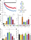
The inactivating phenotype of Kir6.2[R192A] is rescued by introduced Kir2.1 CD-I contacts. (A) Current response after initial patch excision of WT (black), R192A (blue), and R192A/H193D/T223R (red) channels, normalized to the peak current amplitude (Ipk). Steady-state current (Iss) shows the extent of channel inactivation. (B) Extent (1 − Iss/Ipk; left) and time course (τinact; right) of inactivation for CD-I mutations, from experiments as in Fig. 3. (C and D) Fold increase of steady-state (Iss; C) and initial (Ipk; D) current amplitude after PIP2 potentiation; means ± SEM; *, P < 0.05 relative to WT means; †, P < 0.05 relative to R192A means (Student’s t test).
Figure 5.
Figure 5.
CD-I strength does not contribute to the open state stability. (A and B) Normalized single-channel amplitude histogram of the corresponding traces (right) for WT (gray), R192A (blue), and R192A/H193D/T223R (red) at −50 mV (A) and average single-channel amplitudes (B). (C) Open interval distributions overlaid with the probability density function (thick line) from the entire corresponding recordings depicted in A. (D) The open state time constant (τo, ms, inset) and average MOT (ms) were calculated from fits to unbiased models consisting of three to four closed states and one open state (see Materials and methods). (B and D) Means ± SEM. (E) Microscopic recordings of R192A (left) and R192A/H193D/T223R (right) before and after onset of PIP2 application (arrows).
Figure 6.
Figure 6.
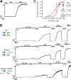
Disruption of CD-I salt bridges reduces Kir2.1 channel activity. (A) Current response to a 1-s voltage ramp from −100 to 100 mV. Currents were recorded in the cell-attached configuration (black), after patch excision (red), and after application of PIP2 (5 µg/ml; blue). (B) PIP2 potentiation of Kir2.1 channel excised currents; means ± SEM; *, P < 0.05 relative to WT means; †, P < 0.05 relative to R204A means (Student’s t test).
Figure 7.
Figure 7.
Extent of Kir2.1 PIP2 activation is controlled by CD-I stability. (A) Depletion of endogenous PIP2 with CaCl2 (10 mM) eliminates channel activity, which is reactivated by application of exogenous PIP2. (B) After CaCl2 exposure, channel activity was measured at varying concentrations of short-chain PIP2 (DiC8-PIP2), and the maximum response was determined at the end of each recording with PIP2. (C) DiC8-PIP2 dose–response relationship fit with the Hill equation (Eq. 2). Data are presented as means ± SEM; *, P < 0.05 relative to WT means (Student’s t test).
Figure 8.
Figure 8.
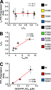
Strength of the CD-I correlates with inactivation parameters. (A and B) Ipk PIP2 potentiation versus Iss/Ipk (A) and Iss/Ipk (B) for inactivating mutants versus τinact, for Kir6.2 CD-I mutations (data from Fig. 4). (C) Correlation between Kir2.1 PIP2 potentiation and DiC8-PIP2 EC50. The corresponding correlation coefficient (r) and coefficient of determination (r2) from each linear regression fit (red) are provided (inset). Data are presented as means ± SEM.
Figure 9.
Figure 9.
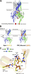
PIP2 binding to Kir2 is associated with CD-I rearrangements. (A) Kir6.2 homology model with the location of CD-I residues that cause inactivation when mutated, whether identified in HI patients (red) or experimentally introduced (orange), indicated by spheres centered on the Cα position. (B) In the presence of PIP2 (3SPI), the CD is ∼6 Å closer to the membrane than in the apo cKir2.2 (3JYC) crystal structure. (C) Translocation of CD-I salt bridge residue Cα (ΔCα) in 3JYC relative to 3SPI (structures aligned at R204). Only the R204–E241 salt bridge (3 Å) is preserved in 3JYC. The E241/R325 and D205/R235 intersubunit sidechain distances in 3JYC are beyond those necessary to form salt bridge contacts (residue numbering according to Kir2.1 sequence).
Figure 10.
Figure 10.
Proposed model of KATP channel gating. Cartoon model of proposed KATP channel gating states and inactivation caused by rupture of CD-I contacts, based on cKir2.2 structures. Stabilized CD-I interactions (red spheres) in the CO channel (modeled on the K62W mutant 5KUK) permits binding of ATP or PIP2 to transition to either ATP-bound (CA) or PIP2 bound (3SPI, 5KUM; O) states. Rupture of the CD-I contacts (orange spheres) disengages the CD from the membrane and results in transition to the apo (CI; 3JYC) state, to which ligands cannot bind.

References

    1. Alekseev A.E., Kennedy M.E., Navarro B., and Terzic A.. 1997. Burst kinetics of co-expressed Kir6.2/SUR1 clones: comparison of recombinant with native ATP-sensitive K+ channel behavior. J. Membr. Biol. 159:161–168. 10.1007/s002329900279 - DOI - PubMed
    1. Alekseev A.E., Brady P.A., and Terzic A.. 1998. Ligand-insensitive state of cardiac ATP-sensitive K+ channels. Basis for channel opening. J. Gen. Physiol. 111:381–394. 10.1085/jgp.111.2.381 - DOI - PMC - PubMed
    1. Amorós I., Dolz-Gaitón P., Gómez R., Matamoros M., Barana A., de la Fuente M.G., Núñez M., Pérez-Hernández M., Moraleda I., Gálvez E., et al. . 2013. Propafenone blocks human cardiac Kir2.x channels by decreasing the negative electrostatic charge in the cytoplasmic pore. Biochem. Pharmacol. 86:267–278. 10.1016/j.bcp.2013.04.023 - DOI - PubMed
    1. Antcliff J.F., Haider S., Proks P., Sansom M.S., and Ashcroft F.M.. 2005. Functional analysis of a structural model of the ATP-binding site of the KATP channel Kir6.2 subunit. EMBO J. 24:229–239. 10.1038/sj.emboj.7600487 - DOI - PMC - PubMed
    1. Bichet D., Haass F.A., and Jan L.Y.. 2003. Merging functional studies with structures of inward-rectifier K+ channels. Nat. Rev. Neurosci. 4:957–967. 10.1038/nrn1244 - DOI - PubMed

Substances

LinkOut - more resources