. 2018 Apr;73(4):350-360.
doi: 10.1136/thoraxjnl-2016-208977. Epub 2017 Apr 7.

Intravascular donor monocytes play a central role in lung transplant ischaemia-reperfusion injury

Collaborators, Affiliations

Intravascular donor monocytes play a central role in lung transplant ischaemia-reperfusion injury

Kate Colette Tatham et al. Thorax. 2018 Apr.

Abstract

Rationale: Primary graft dysfunction in lung transplant recipients derives from the initial, largely leukocyte-dependent, ischaemia-reperfusion injury. Intravascular lung-marginated monocytes have been shown to play key roles in experimental acute lung injury, but their contribution to lung ischaemia-reperfusion injury post transplantation is unknown.

Objective: To define the role of donor intravascular monocytes in lung transplant-related acute lung injury and primary graft dysfunction.

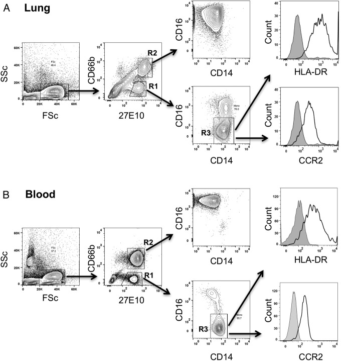
Methods: Isolated perfused C57BL/6 murine lungs were subjected to warm ischaemia (2 hours) and reperfusion (2 hours) under normoxic conditions. Monocyte retention, activation phenotype and the effects of their depletion by intravenous clodronate-liposome treatment on lung inflammation and injury were determined. In human donor lung transplant samples, the presence and activation phenotype of monocytic cells (low side scatter, 27E10+, CD14+, HLA-DR+, CCR2+) were evaluated by flow cytometry and compared with post-implantation lung function.

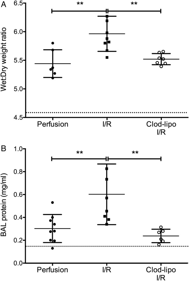
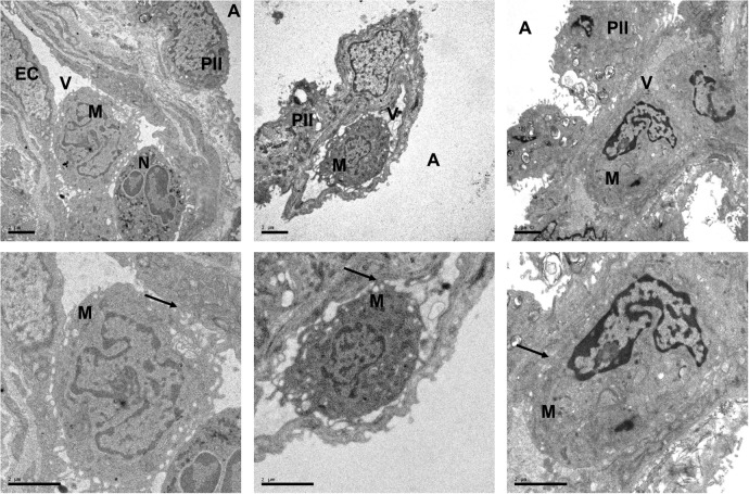
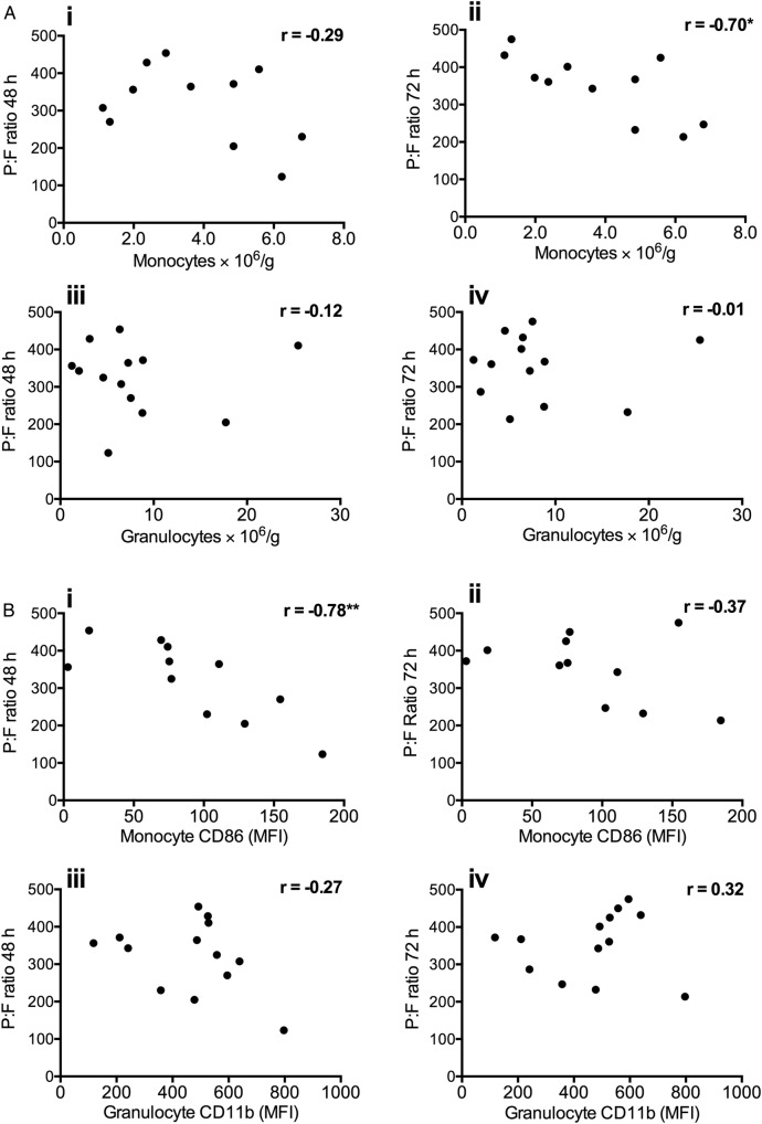
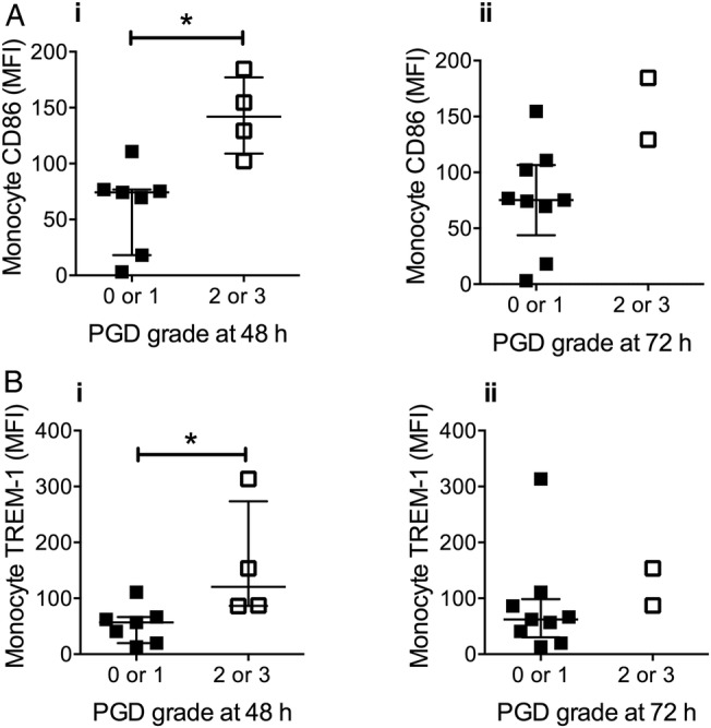
Results: In mouse lungs following ischaemia-reperfusion, substantial numbers of lung-marginated monocytes remained within the pulmonary microvasculature, with reduced L-selectin and increased CD86 expression indicating their activation. Monocyte depletion resulted in reductions in lung wet:dry ratios, bronchoalveolar lavage fluid protein, and perfusate levels of RAGE, MIP-2 and KC, while monocyte repletion resulted in a partial restoration of the injury. In human lungs, correlations were observed between pre-implantation donor monocyte numbers/their CD86 and TREM-1 expression and post-implantation lung dysfunction at 48 and 72 hours.

Conclusions: These results indicate that lung-marginated intravascular monocytes are retained as a 'passenger' leukocyte population during lung transplantation, and play a key role in the development of transplant-associated ischaemia-reperfusion injury.

Keywords: Innate Immunity; Lung Transplantation; Macrophage Biology.


Conflict of interest statement

Competing interests: None declared.

Figures

Figure 1
Figure 1
Pulmonary intravascular lung monocytes are retained during lung perfusion. The location and phenotype of monocyte subsets in the lungs were determined by dual compartment, intra-vascular and intra-alveolar leukocyte staining. Mice were injected intravenously (i.v.) with anti-CD45 (PE-CF594) before anaesthetic overdose, followed by intra-tracheal (i.t.) instillation of anti-CD45.2 (APC). To analyse mononuclear leukocytes within the lung cell suspensions, extra-alveolar leukocytes were first identified as CD45.2− and CD11b+ cells (A). In these cell populations, monocytes/macrophages were further identified as F4/80+ and subdivided into Ly6CHigh and Low subsets (B). Intravascular monocytes were identified as anti-CD45 PE-CF594+ (C and D). MHCII was used as a marker for interstitial CD11b+, F4/80+ macrophages. To evaluate intravascular monocyte removal during perfusion, lungs were flushed with open circuit perfusion for 15 min after compartmental antibody staining. Only partial reduction in intravascular monocyte numbers was observed following washout (black diamonds) compared with non-surgical controls (white boxes) with both intravascular Ly6CHigh (E) and Ly6CLow (F) populations. Numbers of interstitial Ly6CLow, MHCII+ macrophages (G) were not significantly changed, confirming their extravascular location. These numbers of intravascular monocyte subsets far exceeded those expected in an unlikely situation when residual blood within the pulmonary vasculature (estimated ∼50 µL as maximum) was not washed out, and totally preserved within the lung despite this extended perfusion, and hence can be ascribed to a marginated pool. Data are displayed as mean±SD, and analysed by t test. n=4–6, *p<0.05, ***p<0.001.
Figure 2
Figure 2
Activation of retained lung monocytes and neutrophils during ischaemia-reperfusion (I/R). Monocyte and neutrophil activation during I/R was indicated by changes in expression of their respective surface activation markers relative to 2 hours of perfusion only, with values for those from untreated (non-surgical) control mice indicated by a dotted line. Ly6CHigh monocyte activation was indicated by L-selectin shedding and CD86 upregulation (A); Ly6CLow monocytes and interstitial macrophages by increased CD86 expression (B and C); and neutrophils by L-selectin shedding and increased surface CD11b expression (D). Data are displayed as mean±SD and analysed by t tests. n=4–8, *p<0.05, **p<0.01, ***p<0.001.
Figure 3
Figure 3
Depletion of intravascular monocytes attenuates ischaemia-reperfusion (I/R)-induced lung injury. I/R-induced lung injury was indicated by increases in lung wet:dry weight ratios (A) and bronchoalveolar lavage (BAL) fluid protein production (B). BAL fluid protein and wet:dry ratios were increased with 2 hours of perfusion of the lungs alone, but they were not significantly different from untreated non-surgical controls (dotted line). Clodronate-liposome (clod-lipo) depletion of intravascular monocytes restored these indices to baseline. Data are displayed as mean±SD and analysed by one-way ANOVA with Bonferroni correction tests. n=6–9, **p<0.01.
Figure 4
Figure 4
Intravascular monocyte depletion modifies soluble mediator release during lung ischaemia-reperfusion (I/R). Lungs from normal (black squares) or clodronate-liposome pretreated (white circles) mice were subjected to I/R, with perfusate samples obtained pre ischaemia, post ischaemia and post reperfusion. Levels of soluble KC (A), MIP-2 (B) RAGE (C), MCP-1 (D) and IL-6 (E) were determined by ELISA. Increases in KC, MIP-2 and RAGE post ischaemia and post reperfusion were reduced by clodronate-induced monocyte depletion, whereas this treatment resulted in higher MCP-1 levels at all sampling points. Data are displayed as mean±SD, and analysed by two-way ANOVA with t tests with Bonferroni correction. n=4–6, **p<0.01, ***p<0.001, ****p<0.0001.
Figure 5
Figure 5
Adoptive transfer of monocytes restores ischaemia-reperfusion (I/R)-induced lung injury in monocyte-depleted lungs. Isolated lungs from monocyte-depleted mice were infused slowly (1 min) with normal blood-derived monocytes and recirculated for 10 min prior to initiation of the ischaemic period. At the end of I/R, Ly6CHigh monocyte numbers were found to be elevated in infused lungs (A). Lung wet:dry weight ratios (B) and bronchoalveolar lavage (BAL) protein levels (C) were increased in monocyte-infused lungs, indicating monocyte-dependent IR injury. Post-I/R treatment values obtained in previous experiments in normal (non-monocyte-depleted) I/R mice are indicated by a dotted line. Data are analysed by t tests (A and B; mean±SD) or by Mann–Whitney U tests (C; median±IQR). n=4, *p<0.05. clod-lipo, clodronate-liposome.
Figure 6
Figure 6
Identification of monocyte subsets in human donor lung grafts. Single cell suspensions prepared from donor lung tissue were antibody stained for surface markers, and then fixed and permeabilised for intracellular detection of the monocyte and neutrophil S100A8/A9 antigen using the 27E10 monoclonal antibody (A). Classical subset monocytes were identified in lungs as 27E10High, CD66b– (R1) and CD14+, CD16– (R3) events. Granulocytes were identified as 27E10High, CD66b+ events (R2). Using the same staining protocol, comparable staining characteristics were observed for monocytes in healthy volunteer whole blood (B), including similar expression levels of HLA-DR and CCR2.
Figure 7
Figure 7
Electron micrographs showing intravascular monocyte in pre-implantation lung biopsies. Electron micrographs from three different lung allografts are shown. Samples were obtained at the end of the cold ischaemia, prior to implantation. Scale=2 μm. The endothelial cells (ECs) covering the vascular (V) side of the alveolar–capillary barrier appear unmodified, with preserved mitochondria. Moreover, no significant changes are exhibited by the monolayer of type I pneumocytes, lining the alveolar space (A), or the type II pneumocytes (PII). Despite the flushing at the time of the retrieval, monocytes (M), with typical horseshoe-like nuclei, and neutrophils (N), with bi-lobed nuclei, are seen within the pulmonary vasculature. These micrographs also demonstrate evidence for various grades of monocyte–endothelial interactions (arrows), including multiple connections with the ECs consistent with tethering and manifest adhesion.
Figure 8
Figure 8
Correlation of donor lung monocyte numbers and activation state with P:F ratios following transplantation. Numbers of monocytes retained in donor lungs post harvest were found to negatively correlate with P:F ratios at 72 hours post implantation (p<0.5) (Aii). Donor lung granulocyte numbers did not correlate with P:F ratios at either 48 or 72 hours (A; iii–iv). Donor lung monocyte activation (CD86 expression) correlated negatively with P:F ratio at 48 hours (Bi). No correlation was seen with granulocyte activation (CD11b), at either time point (B; iii–iv). Data are analysed by Spearman's rank test (showing r values *p<0.05, **p<0.01), n=11–13.
Figure 9
Figure 9
Donor monocyte activation is associated with primary graft dysfunction (PGD) severity. Expression of CD86 and TREM-1 on donor lung monocytes pre implantation was higher in lungs that developed grade 2 or 3 PGD at 48 hours post transplantation (Ai and Bi). Data are displayed as median±IQR and analysed by Mann–Whitney U tests. n=11, *p<0.05. (NB 2 samples were lost from the donor monocyte CD86/TREM-1 analysis due to technical difficulty).

Comment in

Similar articles

Cited by

References

    1. Lee JC, Christie JD, Keshavjee S. Primary graft dysfunction: definition, risk factors, short- and long-term outcomes. Semin Respir Crit Care Med 2010;31:161–71. 10.1055/s-0030-1249111 - DOI - PubMed
    1. de Perrot M, Liu M, Waddell TK, et al. . Ischemia-reperfusion-induced lung injury. Am J Respir Crit Care Med 2003;167:490–511. 10.1164/rccm.200207-670SO - DOI - PubMed
    1. den Hengst WA, Gielis JF, Lin JY, et al. . Lung ischemia-reperfusion injury: a molecular and clinical view on a complex pathophysiological process. Am J Physiol Heart Circ Physiol 2010;299:H1283–99. 10.1152/ajpheart.00251.2010 - DOI - PubMed
    1. Zhao M, Fernandez LG, Doctor A, et al. . Alveolar macrophage activation is a key initiation signal for acute lung ischemia-reperfusion injury. Am J Physiol Lung Cell Mol Physiol 2006;291:L1018–26. 10.1152/ajplung.00086.2006 - DOI - PubMed
    1. Eppinger MJ, Jones ML, Deeb GM, et al. . Pattern of injury and the role of neutrophils in reperfusion injury of rat lung. J Surg Res 1995;58:713–18. 10.1006/jsre.1995.1112 - DOI - PubMed

Publication types