. 2017 Apr 7;7(1):719.
doi: 10.1038/s41598-017-00350-1.

The dual developmental origin of spinal cerebrospinal fluid-contacting neurons gives rise to distinct functional subtypes

Affiliations

The dual developmental origin of spinal cerebrospinal fluid-contacting neurons gives rise to distinct functional subtypes

Lydia Djenoune et al. Sci Rep. .

Abstract

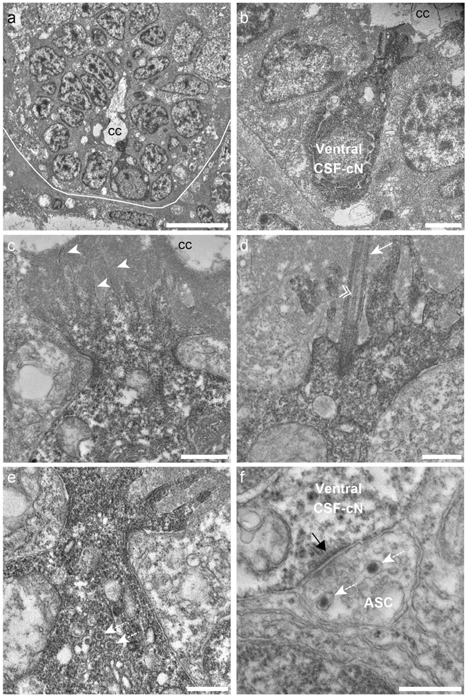
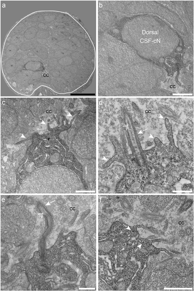
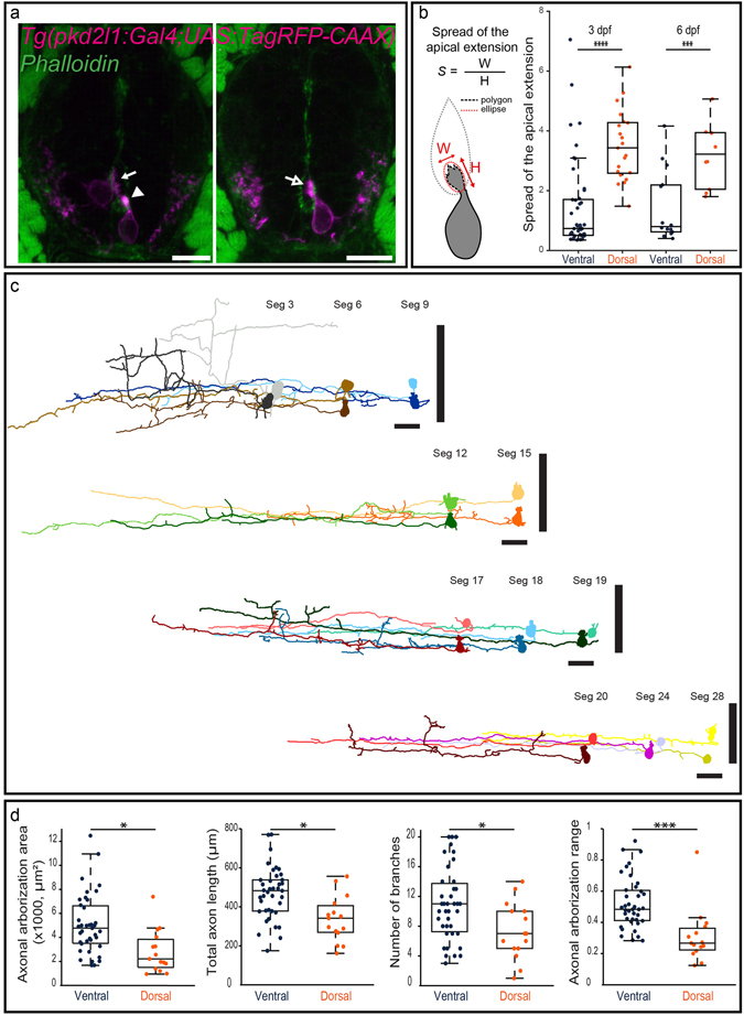
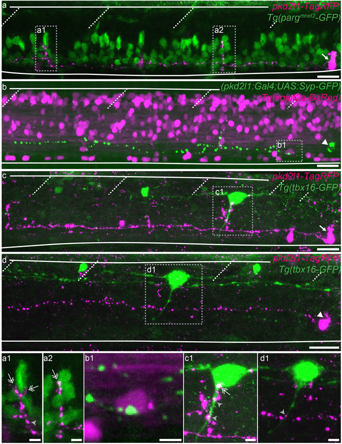
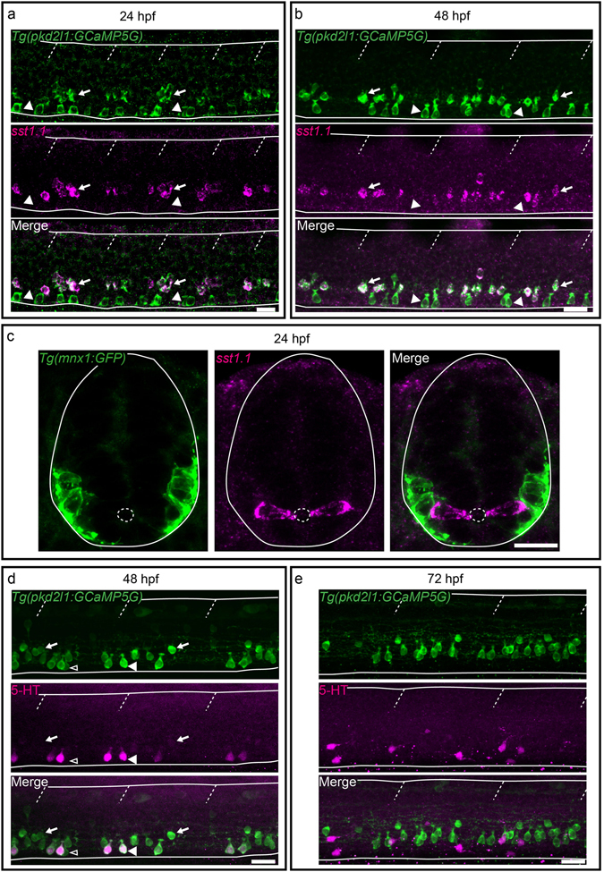
Chemical and mechanical cues from the cerebrospinal fluid (CSF) can affect the development and function of the central nervous system (CNS). How such cues are detected and relayed to the CNS remains elusive. Cerebrospinal fluid-contacting neurons (CSF-cNs) situated at the interface between the CSF and the CNS are ideally located to convey such information to local networks. In the spinal cord, these GABAergic neurons expressing the PKD2L1 channel extend an apical extension into the CSF and an ascending axon in the spinal cord. In zebrafish and mouse spinal CSF-cNs originate from two distinct progenitor domains characterized by distinct cascades of transcription factors. Here we ask whether these neurons with different developmental origins differentiate into cells types with different functional properties. We show in zebrafish larva that the expression of specific markers, the morphology of the apical extension and axonal projections, as well as the neuronal targets contacted by CSF-cN axons, distinguish the two CSF-cN subtypes. Altogether our study demonstrates that the developmental origins of spinal CSF-cNs give rise to two distinct functional populations of sensory neurons. This work opens novel avenues to understand how these subtypes may carry distinct functions related to development of the spinal cord, locomotion and posture.


Conflict of interest statement

The authors declare that they have no competing interests.

Figures

Figure 1
Figure 1
Ventral CSF-cNs exhibit an apical extension composed of microvilli and a kinocilium in the spinal cord. (a) Transverse section of the spinal cord showing restricted deposition of DAB in a ventral CSF-cN. (b) Overall view of a DAB+ ventral CSF-cN contacting the central canal (cc) and surrounded by ependymal cells. (c) Ventral CSF-cNs project at the apical pole an extension toward the central canal bearing several microvilli (arrowheads). (d) Within this extension is located a cilium (arrows) with two central microtubule along the axoneme (double arrowhead), suggesting a motile cilium. Large granular vesicles (LGV) are observed in the cytoplasm (e, dotted arrows) and axo-somatic synaptic contacts in the basal pole (f, ASC). Note the symmetry of the synaptic contact (black arrow) is reminiscent of an inhibitory synapse. Note the presence of LGV in the axon (f, dotted arrows). Scale bar = 10 μm (a), 2 µm (b), 1 μm (c), 500 nm (d,e) and 400 nm (f).
Figure 2
Figure 2
Dorsal spinal CSF-cNs also exhibit ultrastructural properties of sensory neurons. (a) Transverse section of the spinal cord showing restricted deposition of DAB in a dorsal CSF-cN. (b) Overall view of a DAB+ dorsal CSF-cN contacting the central canal (cc). (c,d) Dorsal CSF-cNs bear at the apical pole multiple microvilli (arrowheads). (d,e) In the apical pole is located a cilium (arrow) with two central microtubule singlets along the axoneme (double arrowhead), reminiscent of a motile cilium. (f) Dorsal CSF-cN also exhibit LGV distributed in the cytoplasm (dotted arrows). Scale bar = 10 μm (a), 2 μm (b), 1 µm (c,f) and 500 nm (d,e).
Figure 3
Figure 3
Morphological analysis of CSF-cNs reveals heterogeneous shapes of apical extension and axonal projections. (a) Transverse sections showing ventral and dorsal TagRFP-CAAX+ CSF-cNs (magenta) at 3 dpf reflecting the diversity of morphologies of the apical extension. The apical extension of all dorsal CSF-cNs spreads along the central canal border (arrow) while most ventral CSF-cNs (86.7%) form compact extensions (arrowhead). The small remaining subpopulation of ventral CSF-cNs exhibits the typical spread of dorsal apical extensions (arrow with empty head; Phalloidin staining, green). (b) Schematics of the analysis of the apical extension performed on each cell and statistical analysis comparing the size of the apical extension between ventral and dorsal CSF-cNs at 3 dpf (n = 45 versus 21) and 6 dpf (n = 14 versus 10). The apical extension of dorsal CSF-cNs extends more than for ventral CSF-cNs (two-sample t-tests, p < 5 · 10−7) and this difference persists at later stages (6 dpf, p < 0.002). (c) The reconstruction from dorsal (light shade) and ventral (dark shade) CSF-cNs from different segments (Seg) illustrates the diversity of axonal morphologies CSF-cNs between the two types along the spinal cord (n = 11 for each type). Vertical black bars represent the dorso-ventral limits of the spinal cord. Cells are positioned according to their dorso-ventral (D-V) position with dorsal edge set to 1 and ventral to 0. (d) Comparison of ventral and dorsal CSF-cNs for axonal arborization area, total axon length, number of branches and axonal arborization dorso-ventral range (n = 39 and 15 cells respectively). Ventral CSF-cNs have a wider axonal arborization (p < 0.003), a longer axon (p = 0.0014), reach more ventral domains of the spinal cord (p < 9 · 10−4), and cover a larger dorso-ventral (D-V) range (p < 2 · 10−4) with more axonal branches (p < 0.02). Two-sample t-tests were performed to compare the two populations. Scale bar = 10 μm (a) and 20 µm (c).
Figure 4
Figure 4
Ventral and dorsal CSF-cNs project onto distinct neuronal populations. (ad) Z projection stacks showing contact from ventral and dorsal CSF-cNs onto different spinal targets. (a) Lateral view of a ventral CSF-cN (magenta, arrow) contacting 2 CaP motor neurons (identified based on their location within the segment) labelled in green in the Tg(parg mnet2 -GFP) transgenic line (a1,a2, double headed arrows). (b) Dorsal CSF-cN (green, arrowhead) contacting a putative V0-v interneuron (magenta, based on its dorso-ventral and lateral location) in the Tg(vglut2a:DsRed) transgenic line. (c,d) Ventral (c, arrow) and dorsal (d, arrowhead) CSF-cNs (magenta) contact CoPA sensory interneurons (green) labelled in the Tg(tbx16-GFP) transgenic line. Boxes with close-ups highlight contacts between the CSF-cN and its target. Scale bar = 20 µm (ad) and 5 µm (a1d1).
Figure 5
Figure 5
Secreted factors distinguish dorsal and ventral CSF-cNs: while dorsal express the somatostatin paralog sst1.1, ventral CSF-cNs express 5-HT. (ac) Lateral views of the spinal cord show that sst1.1 expression is restricted to dorsal CSF-cNs (arrows, FISH for sst1.1 (magenta) coupled to GFP IHC (green) on Tg(pkd2l1:GCaMP5G)) (a,b) and Tg(mnx1:GFP) (c) embryos and larvae at 24 hpf (a,c) and 48 hpf (b). (c) Transverse sections show that sst1.1 (magenta) is not expressed in motor neurons (green) as previously suggested (Devos et al.). (d,e) IHC for 5-HT (magenta) and GFP (green) on Tg(pkd2l1:GCaMP5G) transgenic larvae at 48 hpf (d) and 72 hpf (e). (d) At 48 hpf, most ventral CSF-cNs express 5-HT (arrowhead, compared to negative cells shown with empty arrowhead). Note that dorsal CSF-cNs (arrows) are not labelled by 5-HT. (e) At 72 hpf, ventral CSF-cNs are not serotoninergic anymore in the rostral part of the spinal cord. Horizontal lines represent the limits of the spinal cord and slash dashed lines represent somite boundaries. Small dotted ellipses represent the limit of the central canal. Scale bars = 20 μm.

References

    1. Iliff JJ, et al. A paravascular pathway facilitates CSF flow through the brain parenchyma and the clearance of interstitial solutes, including amyloid beta. Sci Transl Med. 2012;4:147ra111. doi: 10.1126/scitranslmed.3003748. - DOI - PMC - PubMed
    1. Pappenheimer JR, Miller TB, Goodrich CA. Sleep-promoting effects of cerebrospinal fluid from sleep-deprived goats. Proc Natl Acad Sci USA. 1967;58:513–517. doi: 10.1073/pnas.58.2.513. - DOI - PMC - PubMed
    1. Martin FH, Seoane JR, Baile CA. Feeding in satiated sheep elicited by intraventricular injections of CSF from fasted sheep. Life Sci. 1973;13:177–184. doi: 10.1016/0024-3205(73)90193-8. - DOI - PubMed
    1. Lerner RA, et al. Cerebrodiene: a brain lipid isolated from sleep-deprived cats. Proc Natl Acad Sci USA. 1994;91:9505–9508. doi: 10.1073/pnas.91.20.9505. - DOI - PMC - PubMed
    1. Nishino S, et al. Low cerebrospinal fluid hypocretin (Orexin) and altered energy homeostasis in human narcolepsy. Ann Neurol. 2001;50:381–388. doi: 10.1002/ana.1130. - DOI - PubMed

Publication types

MeSH terms

LinkOut - more resources