. 2017 Apr 7;7(1):737.
doi: 10.1038/s41598-017-00462-8.

CRISPR/Cas9-mediated PD-1 disruption enhances anti-tumor efficacy of human chimeric antigen receptor T cells

Affiliations

CRISPR/Cas9-mediated PD-1 disruption enhances anti-tumor efficacy of human chimeric antigen receptor T cells

Levi J Rupp et al. Sci Rep. .

Abstract

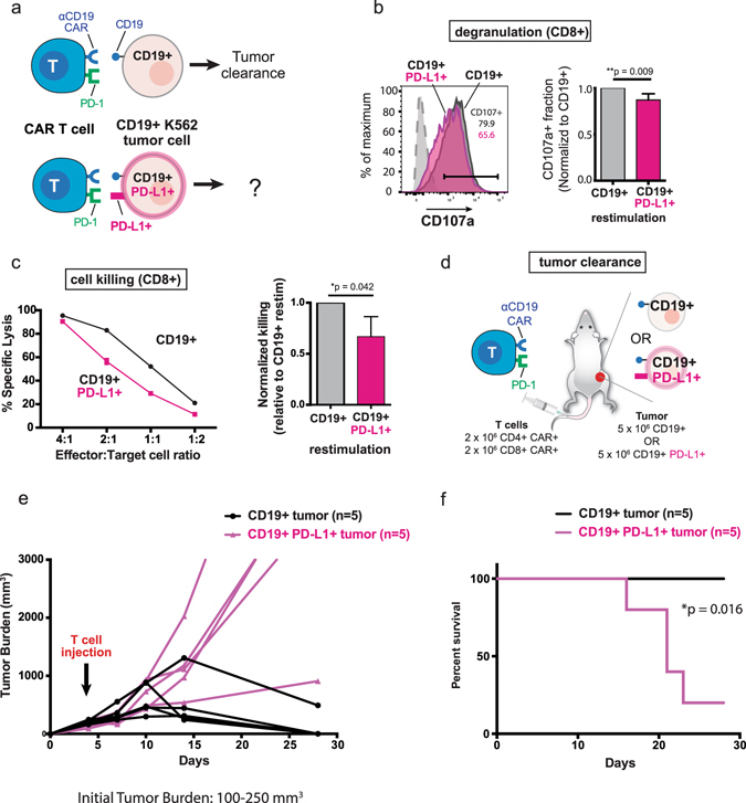
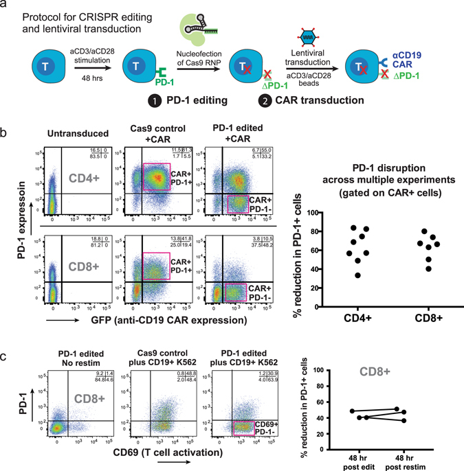
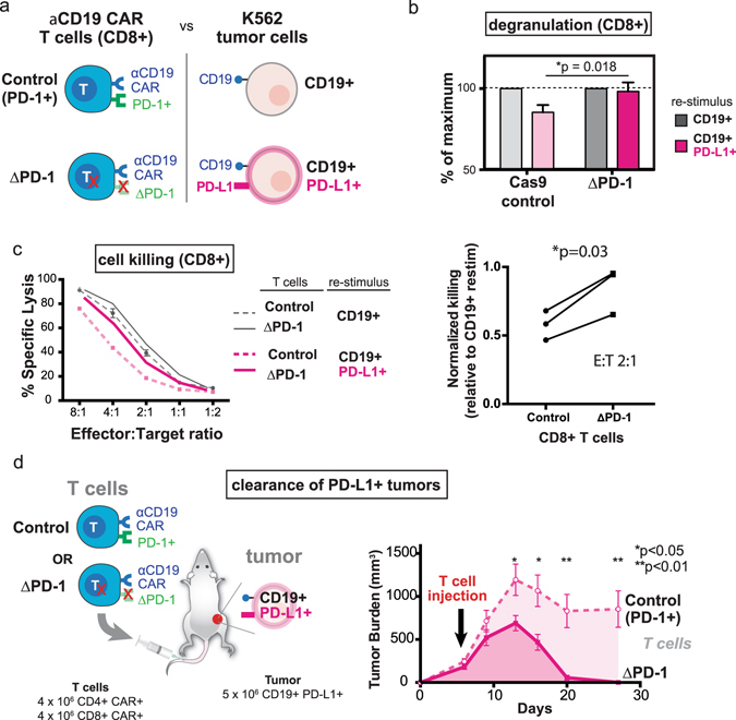
Immunotherapies with chimeric antigen receptor (CAR) T cells and checkpoint inhibitors (including antibodies that antagonize programmed cell death protein 1 [PD-1]) have both opened new avenues for cancer treatment, but the clinical potential of combined disruption of inhibitory checkpoints and CAR T cell therapy remains incompletely explored. Here we show that programmed death ligand 1 (PD-L1) expression on tumor cells can render human CAR T cells (anti-CD19 4-1BBζ) hypo-functional, resulting in impaired tumor clearance in a sub-cutaneous xenograft model. To overcome this suppressed anti-tumor response, we developed a protocol for combined Cas9 ribonucleoprotein (Cas9 RNP)-mediated gene editing and lentiviral transduction to generate PD-1 deficient anti-CD19 CAR T cells. Pdcd1 (PD-1) disruption augmented CAR T cell mediated killing of tumor cells in vitro and enhanced clearance of PD-L1+ tumor xenografts in vivo. This study demonstrates improved therapeutic efficacy of Cas9-edited CAR T cells and highlights the potential of precision genome engineering to enhance next-generation cell therapies.


Conflict of interest statement

A patent has been filed on the use of Cas9 RNPs to edit the genome of human primary T cells (A.M., K.S.). A.M. serves as an advisor to Juno Therapeutics and PACT Therapeutics and the Marson lab has received sponsored research support from Juno Therapeutics and Epinomics. L.J.R. is an employee of Cell Design Labs, K.T.R. is an advisor to Cell Design Labs, and W.A.L. is a founder of Cell Design Labs and a member of its scientific advisory board. A.M. is an advisor to Juno Therapeutics.

Figures

Figure 1
Figure 1
PD-L1 expression in human K562 myelogenous leukemia cells inhibits anti-CD19 CAR T cell function in vitro and tumor clearance in vivo. (a) Schematic representation of CAR T cell interaction with either CD19+ or CD19+ PD-L1 K562 tumor cells. (b) CD8+ anti-CD19 CAR T cells exhibit reduced degranulation (CD107a staining) upon re-stimulation with CD19+ PD-L1+ K562 cells. Representative CD107a staining is shown, gated on CD8+ CAR+ T cells. Normalized CD107a staining shows ~15% reduction in degranulation upon co-culture with CD19+ PD-L1+ targets over three independent experiments (**p = 0.009, Student’s t-test). (c) CD19+ PD-L1+ K562 cells are resistant to anti-CD19 CAR-mediated lysis in an in vitro killing assay. Left panel: a complete effector:target ratio titration is shown for a representative experiment. Mean ± S.D. for triplicate wells in a single experiment are plotted. Right panel (bar chart): CD19+ PD-L1+ K562 cells induce ~40% reduction in specific lysis relative to CD19+ K562 cells at effector:target ratio of 2:1 (*p = 0.042, Student’s t-test). The experiment was performed three times; error bars are S.D. (d) Experimental design for subcutaneous xenograft model. (e) CD19+ PD-L1+ subcutaneous xenografts impair anti-CD19 CAR mediated tumor clearance. NOD-scid-IL-2Rγ−/− (NSG) mice were injected with 5 × 106 CD19+ or CD19+ PD-L1+ K562 cells subcutaneously. Mice with established tumors (100–250 mm3) were injected intravenously with 2 × 106 CD4+ and 2 × 106 CD8+ anti-CD19 CAR T cells and tumor burden measured longitudinally by caliper. Shown are tumor burdens for individual mice (n = 5 per tumor type). (f) Kaplan-Meier curve for experiment described in Fig. 1E. A statistically significant difference in survival was observed between animals bearing CD19+ vs. CD19+ PD-L1+ tumors (*p = 0.016, Gehan-Breslow-Wilcoxon test).
Figure 2
Figure 2
Pdcd1 can be efficiently disrupted in CAR T cells using Cas9 ribonucleoproteins (Cas9 RNPs). (a) Schematic of protocol for combined CRISPR gene editing and lentiviral transduction of human primary T cells. (b) Efficient PD-1 deletion and CAR transduction in primary human T cells. PD-1 surface staining and CAR transduction were assessed 48 hours post editing. A >50% reduction in PD-1+ cells was routinely observed, with CAR transduction >70%. Right panel, individual dots represent independent editing experiments. (c) PD-1 edited CAR T cells are stable in culture. Resting PD-1 edited CD8+ anti-CD19 CAR T cells were re-stimulated with CD19+ K562 cells. Activation was measured by CD69 induction and percent reduction in PD-1+ cells measured by flow cytometry based on surface PD-1 expression; percent reduction of PD-1+ cells was similar to that observed 48 hours after editing (rightmost panel, pairwise plot).
Figure 3
Figure 3
CRISPR-mediated PD-1 editing rescues anti-CD19 CAR T cell function in vitro and enhances tumor clearance in vivo. (a) Diagram of PD-1 edited CAR T cell: K562 interactions (b) PD-1 edited CD8+ anti-CD19 CAR T cells (ΔPD-1) exhibit greater degranulation (CD107a staining) upon co-culture with CD19+ PD-L1+ K562 cells as compared to control CD8+ CAR T cells (*p = 0.018, Student’s t-test). (c) PD-1 edited CAR T cells are partially resistant to CD19+ PD-L1+ mediated inhibition of cytolysis. Left panel: percent lysis for control and PD-1 edited CD8+ anti-CD19 CAR T cells is shown across a range of effector:target ratios. Error bars are S.D. of triplicate wells in a single experiment. Right panel: normalized killing shows reduced PD-L1 dependent inhibition of killing in PD-1 edited CD8+ CAR T cells at effector:target ratio of 2:1 (*p = 0.03, paired t-test). The experiment was performed three independent times. (d) PD-1 deficient anti-CD19 CAR T cells exhibit enhanced anti-tumor efficacy and clear subcutaneous CD19+ PD-L1+ tumor xenografts. NSG mice were injected with 5 × 106 CD19+ PD-L1+ K562 cells subcutaneously. Mice with established tumors (100–250 mm3) were injected intravenously with 4 × 106 CD4+ CAR+ and 4 × 106 CD8+ CAR+ control T cells or PD-1 edited cells, and tumor burden measured longitudinally by caliper. Tumor burdens are mean ± SEM for each group (n = 6 mice per group). A statistically significant decrease in tumor burden of mice receiving PD-1 edited CAR T cells was observed at multiple points (*p < 0.05, **p < 0.01, Student’s t-test).

References

    1. Maus MV, et al. Adoptive Immunotherapy for Cancer or Viruses. Annu Rev Immunol. 2014;32:189–225. doi: 10.1146/annurev-immunol-032713-120136. - DOI - PMC - PubMed
    1. Mandal PK, et al. Efficient ablation of genes in human hematopoietic stem and effector cells using CRISPR/Cas9. Cell Stem Cell. 2014;15:643–652. doi: 10.1016/j.stem.2014.10.004. - DOI - PMC - PubMed
    1. Schumann K, et al. Generation of knock-in primary human T cells using Cas9 ribonucleoproteins. Proc Natl Acad Sci USA. 2015;112:10437–10442. doi: 10.1073/pnas.1512503112. - DOI - PMC - PubMed
    1. Hendel A, et al. Chemically modified guide RNAs enhance CRISPR-Cas genome editing in human primary cells. Nat Biotechnol. 2015;33:985–989. doi: 10.1038/nbt.3290. - DOI - PMC - PubMed
    1. Wu C-Y, Rupp LJ, Roybal KT, Lim WA. Synthetic biology approaches to engineer T cells. Current opinion in immunology. 2015;35:123–130. doi: 10.1016/j.coi.2015.06.015. - DOI - PMC - PubMed

Publication types

MeSH terms