. 2017 Apr 7;7(1):751.
doi: 10.1038/s41598-017-00848-8.

Biochemical analysis of leishmanial and human GDP-Mannose Pyrophosphorylases and selection of inhibitors as new leads

Affiliations

Biochemical analysis of leishmanial and human GDP-Mannose Pyrophosphorylases and selection of inhibitors as new leads

Wei Mao et al. Sci Rep. .

Abstract

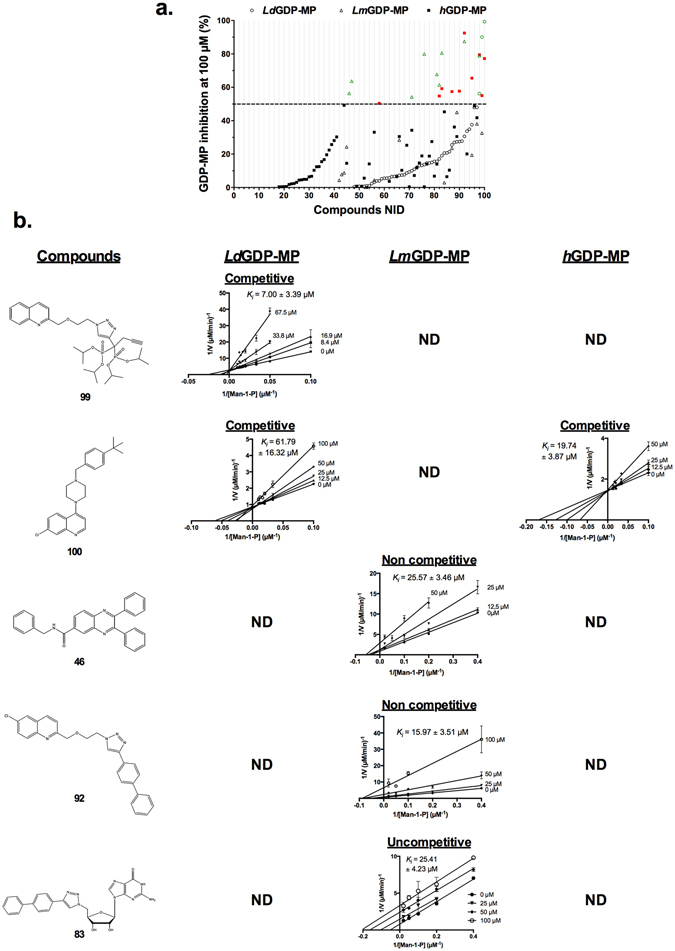
Leishmaniases are an ensemble of diseases caused by the protozoan parasite of the genus Leishmania. Current antileishmanial treatments are limited and present main issues of toxicity and drug resistance emergence. Therefore, the generation of new inhibitors specifically directed against a leishmanial target is an attractive strategy to expand the chemotherapeutic arsenal. GDP-Mannose Pyrophosphorylase (GDP-MP) is a prominent therapeutic target involved in host-parasite recognition which has been described to be essential for parasite survival. In this work, we produced and purified GDP-MPs from L. mexicana (LmGDP-MP), L. donovani (LdGDP-MP), and human (hGDP-MP), and compared their enzymatic properties. From a rationale design of 100 potential inhibitors, four compounds were identified having a promising and specific inhibitory effect on parasite GDP-MP and antileishmanial activities, one of them exhibits a competitive inhibition on LdGDP-MP and belongs to the 2-substituted quinoline series.


Conflict of interest statement

The authors declare that they have no competing interests.

Figures

Figure 1
Figure 1
Purification of LdGDP-MP, LmGDP-MP and hGDP-MP. (a,d,h) Coomassie blue stained SDS-PAGE of LdGDP-MP (a), LmGDP-MP (d), and hGDP-MP (h) purified from Ni-NTA column. Lane TOT: total, Lane SN: supernatant, Lane P: Pellet, Lane FT: flow through, Lane W: wash, Lanes E1, E2 and E4: elution fraction at 100 µM, 200 µM and 400 µM imidazole, respectively. The arrowheads show the proteins of interest. The protein ladders on the left are indicated in kDa. Full length gels are presented in Supplementary Fig. S7. (b,e,i) Size exclusion chromatography profiles of LdGDP-MP (b), LmGDP-MP (e), and hGDP-MP (i). In the inset, the filled triangles and open circles represent standard proteins and the peaks of proteins of interest, respectively. (c,f,j) Coomassie blue stained SDS-PAGE of fraction volumes of peaks 1 (49–51 mL), 2 (66–68 mL), 3 (75–78 mL) and 4 (84–86 mL) of LdGDP-MP (c), peaks 1 (12–13 mL) and 2 (16 mL) of LmGDP-MP (f) and peak 1 (77–82 mL) of hGDP-MP (j). Lo is the loading control. The arrowheads show the proteins of interest. The protein ladder on the left is indicated in kDa. Full length gels are presented in Supplementary Fig. S7. (g) Coomassie blue stained SDS-PAGE of a concentrated pool of fractions containing LmGDP-MP eluted from anion exchange chromatography (AEC). The arrowhead shows the protein of interest. The protein ladder on the left is indicated in kDa. Full length gel is presented in Supplementary Fig. S7.
Figure 2
Figure 2
Determination of LdGDP-MP, LmGDP-MP and hGDP-MP optimal conditions of enzymatic reaction and specificities. (a,b,c) Determination of LdGDP-MP, LmGDP-MP and hGDP-MP optimal temperatures (a), pH (b) and Mg2+ concentrations (c). For the determination of the optimal pH, the buffers used are 50 mM MES for pH 5.6 and 6.5, and 50 mM Tris-HCl for pH between 6.8 and 10. In each graph, enzyme activities were calculated relative to the maximal specific activity obtained with LmGDP-MP and are expressed in percent. The results correspond to the mean of three independent experiments ± SD. (d) Optimal parameters of LdGDP-MP, LmGDP-MP and hGDP-MP enzymatic reactions. All reactions were performed with the optimal reaction time of 20 min. (e,f,g) Determination of LdGDP-MP, LmGDP-MP and hGDP-MP specificities for cofactors (e), nucleotide triphosphates (f) or sugar monophosphates (g). All cofactors (Mg2+, Mn2+, Ca2+, Ni2+, Cu2+, Zn2+) were used at 5 mM. The NTPs (GTP, ATP, CTP, TTP, UTP and ITP) and sugar monophosphates (Man-1-P, Glu-1-P and Gal-1-P) were used at 100 µM. Enzyme activities were calculated relative to each enzyme specific activity obtained with Mg2+ (e), GTP (f) or Man-1-P (g), and are expressed in percent. The results correspond to the mean of three independent experiments ± SD.
Figure 3
Figure 3
Determination of LdGDP-MP, LmGDP-MP and hGDP-MP kinetic constants. (a,c,e) Lineweaver Burk double reciprocal plots 1/V = f(1/[Man-1-P]) of LdGDP-MP (a), LmGDP-MP (c) and hGDP-MP (e). GTP concentration was held constant at 150 µM for LdGDP-MP and 80 µM for LmGDP-MP and hGDP-MP. The results expressed correspond to the mean of three independent experiments ± SD. (b,d,f) Lineweaver Burk double reciprocal plots 1/V = f(1/[GTP]) of LdGDP-MP (b), LmGDP-MP (d) and hGDP-MP (f). Man-1-P concentration was held constant at 150 µM for LdGDP-MP and 80 µM for LmGDP-MP and hGDP-MP. The results expressed correspond to the mean of three independent experiments ± SD. (g) Kinetic constants (V m, K m, k cat, k cat/K m) of LdGDP-MP, LmGDP-MP and hGDP-MP for both substrates Man-1-P and GTP.
Figure 4
Figure 4
Determination of LdGDP-MP, LmGDP-MP and hGDP-MP mechanisms of reaction. (a and b) Lineweaver Burk plots 1/V = f(1/[Man-1-P]) (a) and 1/V = f(1/[GTP]) (b) of LdGDP-MP. Constant concentrations of GTP (a) and Man-1-P (b) were held at 10 µM, 20 µM, 40 µM and 150 µM. The results expressed correspond to the mean of three independent experiments ± SD. (c and d) Lineweaver Burk plots 1/V = f(1/[Man-1-P]) (c) and 1/V = f(1/[GTP]) (d) of LmGDP-MP. Constant concentrations of GTP (c) and Man-1-P (d) were held at 5 µM, 10 µM, 20 µM and 50 µM. The results expressed correspond to the mean of three independent experiments ± SD. (e and f) Lineweaver Burk plots 1/V = f(1/[Man-1-P]) (e) and 1/V = f(1/[GTP]) (f) of hGDP-MP. Constants concentrations of GTP (e) and Man-1-P (f) were held at 10 µM, 20 µM, 40 µM and 80 µM. The results expressed correspond to the mean of three independent experiments ± SD.
Figure 5
Figure 5
Evaluation of compound activities on LdGDP-MP, LmGDP-MP and hGDP-MP. (a) Percentage of LdGDP-MP (Ο), LmGDP-MP (Δ) and hGDP-MP (■) inhibition as a function of compounds numerical ID (NID). Each compound was used at 100 µM. Compounds showing inhibition above 50% are represented in green and red for leishmanial and human GDP-MPs, respectively. For a question of readability, all values at 0% inhibition were removed. The results expressed correspond to the mean of three independent experiments. (b) Lineweaver plots double reciprocal plots 1/V = f(1/[Man-1-P]) of compounds 99, 100, 46, 92 and 83 on LdGDP-MP, LmGDP-MP and hGDP-MP. Each compound was evaluated with a range of concentrations: 0–67.5 µM for compound 99 on LdGDP-MP, 0–50 µM for compound 46 on LmGDP-MP and compound 100 on hGDP-MP, and 0–100 µM for compounds 83 and 92 on LmGDP-MP and compound 100 on LdGDP-MP. Man-1-P was used at 10–100 µM for LdGDP-MP and hGDP-MP and 2.5–50 µM for LmGDP-MP. GTP was held constant at 100 µM for LdGDP-MP and hGDP-MP, and 50 µM for LmGDP-MP. The formula of each compound is represented on the left of the figure. The compounds K i are indicated on each plot, as well as the type of inhibition. ND: no K i could be determined since the double reciprocal plots did not fit with any (competitive, non-competitive, uncompetitive or mixed) inhibition model. The results expressed correspond to the mean of three independent experiments ± SD.
Figure 6
Figure 6
Docking analysis of compounds 99 and 100 on LdGDP-MP and hGDP-MP. (a) Percentage of “good” poses of GDP-Mannose, compound 99 and 100 over the hundred conformations generated by the ten docking calculations onto LdGDP-MP and hGDP-MP. For GDP-Mannose, a “good” pose is the one observed in the PDB crystallographic structure 2X5Z. For the two compounds 99 and 100, a “good” pose has the quinoline group located on the GDP-Mannose guanine. (b,c) Position and orientation of the lowest energy “good” pose of compound 99 in the catalytic site of LdGDP-MP (b) and hGDP-MP (c). The protein surface is colored as a function of the charge density (red, white and blue colors indicating negative, neutral and positive area, respectively). The amino-acids that make contacts with compound 99 are indicated by their one-letter code and number in the sequence.

Similar articles

Cited by

References

    1. World Health Organization. Investing to overcome the global impact of neglected tropical diseases. Third WHO report on neglected tropical diseases. WHO/HTM/NTD/2015.1 (2015).
    1. Sundar S, Singh A. Recent developments and future prospects in the treatment of visceral leishmaniasis. Ther. Adv. Infect. Dis. 2016;3:98–109. doi: 10.1177/2049936116646063. - DOI - PMC - PubMed
    1. Gutierrez V, Seabra AB, Reguera RM, Khandare J, Calderon M. New approaches from nanomedicine for treating leishmaniasis. Chem. Soc. Rev. 2016;45:152–168. doi: 10.1039/C5CS00674K. - DOI - PubMed
    1. Garami A, Ilg T. Disruption of mannose activation in Leishmania mexicana: GDP-mannose pyrophosphorylase is required for virulence, but not for viability. EMBO J. 2001;20:3657–3666. doi: 10.1093/emboj/20.14.3657. - DOI - PMC - PubMed
    1. Stewart J, et al. Characterization of Leishmania mexicana knockout lacking guanosine diphosphate-mannose pyrophosphorylase. Int. J. Parasitol. 2005;35:861–873. doi: 10.1016/j.ijpara.2005.03.008. - DOI - PubMed

Publication types

MeSH terms

LinkOut - more resources