

Comparative histological and immunohistochemical study of ameloblastomas and ameloblastic carcinomas
- PMID: 28390135
- PMCID: PMC5432081
- DOI: 10.4317/medoral.21901
Comparative histological and immunohistochemical study of ameloblastomas and ameloblastic carcinomas
Abstract
Background: This study aimed to compare the histological and immunohistochemical characteristics of ameloblastomas (AM) and ameloblastic carcinomas (AC).
Material and methods: Fifteen cases of AM and 9 AC were submitted to hematoxilin and eosin (H&E) and immunohistochemical analysis with the following antibodies: cytokeratins 5,7,8,14 and 19, Ki-67, p53, p63 and the cellular adhesion molecules CD138 (Syndecan-1), E-cadherin and β-catenin. The mean score of the expression of Ki-67 and p53 labelling index (LIs) were compared between the groups using the t test. A value of p<0.05 was considered to be statistically significant.
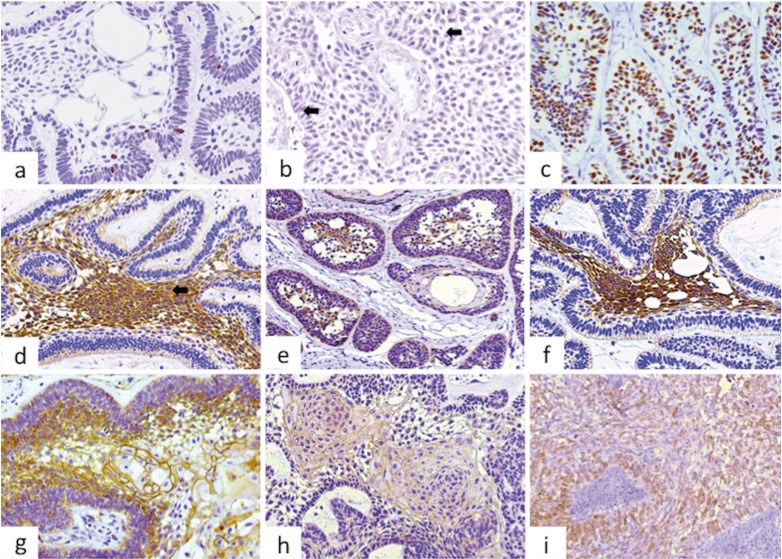
Results: All cases were positive for CKs 5, 14 and 19, but negative for CKs 7 and 8. CKs 5 and 19 were positive mainly in the central regions of the ameloblastic islands, while the expression in AC was variable in intensity and localization. CK14 was also variably expressed in both AM and AC. Ki-67 (P=.001) and p53 (P=.004) immunoexpression was higher in AC. All cases were positive for p63, but values were higher in AC. CD138 was mainly expressed in peripheral cells of AM, with a weak positivity in the central areas, while it was positive in most areas of ACs, except in less differentiated regions, where expression was decreased or lost. E-cadherin and β-catenin were weakly positive in both AM and AC.
Conclusions: These results shows that Ki-67, p53 and p63 expression was higher in AC as compared to AM, suggesting that these markers can be useful when considering diagnosis of malignancy, and perhaps could play a role in malignant transformation of AM. Pattern of expression of CKs 5 and 19 in AC were different to those found in AM, suggesting genetic alterations of these proteins in malignant cells. It was confirmed that CK19 is a good marker for benign odontogenic tumors, such as AM, but it is variably expressed in malignant cases.
Conflict of interest statement
Figures


Similar articles
-
Comparative expression of syndecan-1 and Ki-67 in peripheral and desmoplastic ameloblastomas and ameloblastic carcinoma.Pathol Int. 2009 Apr;59(4):229-33. doi: 10.1111/j.1440-1827.2009.02355.x. Pathol Int. 2009. PMID: 19351365
-
Comparative immunohistochemical study of ameloblastoma and ameloblastic carcinoma.Oral Surg Oral Med Oral Pathol Oral Radiol Endod. 2011 Dec;112(6):767-76. doi: 10.1016/j.tripleo.2011.06.036. Epub 2011 Oct 19. Oral Surg Oral Med Oral Pathol Oral Radiol Endod. 2011. PMID: 22014999
-
A immunohistochemical study of the peripheral ameloblastoma.Oral Dis. 2007 Nov;13(6):575-80. doi: 10.1111/j.1601-0825.2006.01340.x. Oral Dis. 2007. PMID: 17944675
-
Ameloblastic carcinoma, primary type: case report, immunohistochemical analysis and literature review.Anticancer Res. 2012 Apr;32(4):1515-25. Anticancer Res. 2012. PMID: 22493395 Review.
-
Differential Expression of Immunohistochemical Markers in Ameloblastoma & Ameloblastic Carcinoma: A Systematic Review and Meta-analysis of observational studies.F1000Res. 2024 May 31;13:557. doi: 10.12688/f1000research.149861.1. eCollection 2024. F1000Res. 2024. PMID: 39082057 Free PMC article.
Cited by
-
Clear-Cell Variant of Ameloblastic Carcinoma: Pathological Insights for a more Specific Categorization.J Microsc Ultrastruct. 2022 Aug 4;12(3):165-168. doi: 10.4103/jmau.jmau_34_22. eCollection 2024 Jul-Sep. J Microsc Ultrastruct. 2022. PMID: 39507648 Free PMC article.
-
Comparison of the IHC Markers CD138 and CD43 in Oral Leukoplakia: An Original Research.J Pharm Bioallied Sci. 2023 Jul;15(Suppl 1):S209-S212. doi: 10.4103/jpbs.jpbs_454_22. Epub 2023 Jul 5. J Pharm Bioallied Sci. 2023. PMID: 37654342 Free PMC article.
-
Expression Profile of Stemness Markers CD138, Nestin and Alpha-SMA in Ameloblastic Tumours.Int J Environ Res Public Health. 2021 Apr 8;18(8):3899. doi: 10.3390/ijerph18083899. Int J Environ Res Public Health. 2021. PMID: 33917771 Free PMC article.
-
Differences in E-Cadherin and Syndecan-1 Expression in Different Types of Ameloblastomas.Anal Cell Pathol (Amst). 2018 Apr 23;2018:9392632. doi: 10.1155/2018/9392632. eCollection 2018. Anal Cell Pathol (Amst). 2018. PMID: 29850393 Free PMC article.
-
Ameloblastic Carcinoma with Calcification: A Rare Case Report in the Mandible and Literature Review.Case Rep Dent. 2020 Oct 13;2020:4216489. doi: 10.1155/2020/4216489. eCollection 2020. Case Rep Dent. 2020. PMID: 33110663 Free PMC article.
References
-
- Martínez Martínez M, Mosqueda-Taylor A, Carlos R, Delgado-Azañero W, de Almeida OP. Malignant odontogenic tumors: a multicentric Latin American study of 25 cases. Oral Dis. 2014;20:380–5. - PubMed
-
- Yoon HJ, Hong SP, Lee JI, Lee SS, Hong SD. Ameloblastic carcinoma: an analysis of 6 cases with review of the literature. Oral Surg Oral Med Oral Pathol Oral Radiol Endod. 2009;108:904–13. - PubMed
-
- Barnes L, Eveson JW, Reichart PA. World Health Organization Classification Of Tumours: Pathology and Genetics of Tumours of the Head and Neck. Lyon: IARC; 2005.
-
- Lesot H, Brook AH. Epithelial histogenesis during tooth develop- ment. Arch Oral Biol. 2009;54:S25–33. - PubMed
-
- Costa P, Parsons M. New insights into the dynamics of cell adhesions. Int Rev Cell Mol Biol. 2010;283:57–91. - PubMed
Publication types
MeSH terms
Substances
LinkOut - more resources
Full Text Sources
Research Materials
Miscellaneous
