

Acute exposure of primary rat soleus muscle to zilpaterol HCl (β2 adrenergic agonist), TNFα, or IL-6 in culture increases glucose oxidation rates independent of the impact on insulin signaling or glucose uptake
- PMID: 28390265
- PMCID: PMC5483180
- DOI: 10.1016/j.cyto.2017.03.014
Acute exposure of primary rat soleus muscle to zilpaterol HCl (β2 adrenergic agonist), TNFα, or IL-6 in culture increases glucose oxidation rates independent of the impact on insulin signaling or glucose uptake
Abstract
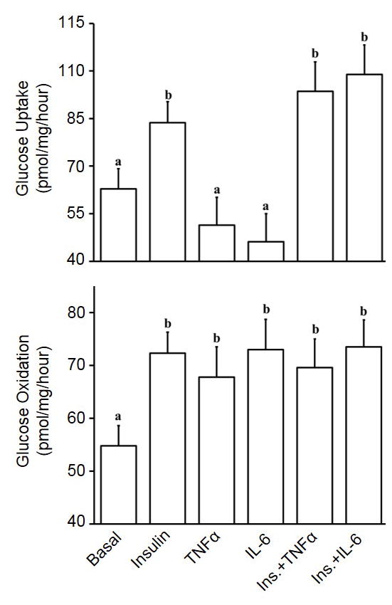
Recent studies show that adrenergic agonists and inflammatory cytokines can stimulate skeletal muscle glucose uptake, but it is unclear if glucose oxidation is similarly increased. Thus, the objective of this study was to determine the effects of ractopamine HCl (β1 agonist), zilpaterol HCl (β2 agonist), TNFα, and IL-6 on glucose uptake and oxidation rates in unstimulated and insulin-stimulated soleus muscle strips from adult Sprague-Dawley rats. Effects on phosphorylation of Akt (phospho-Akt), p38 MAPK (phospho-p38), and p44/42 MAPK (phospho-p44/42) was also determined. Incubation with insulin increased (P<0.05) glucose uptake by ∼47%, glucose oxidation by ∼32%, and phospho-Akt by ∼238%. Insulin also increased (P<0.05) phospho-p38, but only after 2h in incubation. Muscle incubated with β2 agonist alone exhibited ∼20% less (P<0.05) glucose uptake but ∼32% greater (P<0.05) glucose oxidation than unstimulated muscle. Moreover, co-incubation with insulin+β2 agonist increased (P<0.05) glucose oxidation and phospho-Akt compared to insulin alone. Conversely, β1 agonist did not appear to affect basal or insulin-stimulated glucose metabolism, and neither β agonist affected phospho-p44/42. TNFα and IL-6 increased (P<0.05) glucose oxidation by ∼23% and ∼33%, respectively, in the absence of insulin. This coincided with increased (P<0.05) phospho-p38 and phospho-p44/42 but not phospho-Akt. Furthermore, co-incubation of muscle with insulin+either cytokine yielded glucose oxidation rates that were similar to insulin alone, despite lower (P<0.05) phospho-Akt. Importantly, cytokine-mediated increases in glucose oxidation rates were not concomitant with greater glucose uptake. These results show that acute β2 adrenergic stimulation, but not β1 stimulation, directly increases fractional glucose oxidation in the absence of insulin and synergistically increases glucose oxidation when combined with insulin. The cytokines, TNFα and IL-6, likewise directly increased glucose oxidation in the absence of insulin, but were not additive in combination with insulin and in fact appeared to disrupt Akt-mediated insulin signaling. Rather, cytokines appear to be acting through MAPKs to elicit effects on glucose oxidation. Regardless, stimulation of glucose oxidation by these key stress factors did not rely upon greater glucose uptake, which may promote metabolic efficiency during acute stress by increasing fractional glucose oxidation without increasing total glucose consumption by muscle.
Keywords: Glucose oxidation; IL-6; Metabolic regulation; TNFα; β2 adrenergic agonist.
Copyright © 2017 Elsevier Ltd. All rights reserved.
Conflict of interest statement
The authors declare that there are no conflicts of interest regarding the publication of this paper.
Figures


Similar articles
-
Interleukin-6 directly increases glucose metabolism in resting human skeletal muscle.Diabetes. 2007 Jun;56(6):1630-7. doi: 10.2337/db06-1733. Epub 2007 Mar 15. Diabetes. 2007. PMID: 17363741
-
Cytokine regulation of skeletal muscle fatty acid metabolism: effect of interleukin-6 and tumor necrosis factor-alpha.Am J Physiol Endocrinol Metab. 2004 Oct;287(4):E616-21. doi: 10.1152/ajpendo.00150.2004. Epub 2004 May 18. Am J Physiol Endocrinol Metab. 2004. PMID: 15149950
-
In vivo stimulation of oestrogen receptor α increases insulin-stimulated skeletal muscle glucose uptake.J Physiol. 2011 Apr 15;589(Pt 8):2041-54. doi: 10.1113/jphysiol.2010.199018. Epub 2011 Feb 21. J Physiol. 2011. PMID: 21486807 Free PMC article.
-
Insulin signaling: a potential signaling pathway for the stimulatory effect of kaempferitrin on glucose uptake in skeletal muscle.Eur J Pharmacol. 2013 Jul 15;712(1-3):1-7. doi: 10.1016/j.ejphar.2013.02.029. Epub 2013 Mar 1. Eur J Pharmacol. 2013. PMID: 23458067 Review.
-
Effects of H2O2 on insulin signaling the glucose transport system in mammalian skeletal muscle.Methods Enzymol. 2013;528:269-78. doi: 10.1016/B978-0-12-405881-1.00016-1. Methods Enzymol. 2013. PMID: 23849871 Review.
Cited by
-
Maternofetal inflammation induced for 2 wk in late gestation reduced birth weight and impaired neonatal growth and skeletal muscle glucose metabolism in lambs.J Anim Sci. 2021 May 1;99(5):skab102. doi: 10.1093/jas/skab102. J Anim Sci. 2021. PMID: 33780540 Free PMC article.
-
Current Developments on the Role of α1-Adrenergic Receptors in Cognition, Cardioprotection, and Metabolism.Front Cell Dev Biol. 2021 May 25;9:652152. doi: 10.3389/fcell.2021.652152. eCollection 2021. Front Cell Dev Biol. 2021. PMID: 34113612 Free PMC article. Review.
-
Sustained heat stress elevated corneal and body surface temperatures and altered circulating leukocytes and metabolic indicators in wether lambs supplemented with ractopamine or zilpaterol.J Anim Sci. 2021 Sep 1;99(9):skab236. doi: 10.1093/jas/skab236. J Anim Sci. 2021. PMID: 34370018 Free PMC article.
-
Reducing Systemic Inflammation in IUGR-Born Neonatal Lambs via Daily Oral ω-3 PUFA Supplement Improved Skeletal Muscle Glucose Metabolism, Glucose-Stimulated Insulin Secretion, and Blood Pressure.Metabolites. 2025 May 22;15(6):346. doi: 10.3390/metabo15060346. Metabolites. 2025. PMID: 40559370 Free PMC article.
-
Impaired muscle stem cell function in cows with high concentrations of androstenedione in their follicular fluid.Transl Anim Sci. 2018 Sep;2(Suppl 1):S27-S30. doi: 10.1093/tas/txy050. Epub 2018 Sep 27. Transl Anim Sci. 2018. PMID: 30627703 Free PMC article. No abstract available.
References
-
- DeFronzo RA, Jacot E, Jequier E, Maeder E, Wahren J, Felber JP. The effect of insulin on the disposal of intravenous glucose. Results from indirect calorimetry and hepatic and femoral venous catheterization. Diabetes. 1981;30(12):1000–7. - PubMed
-
- Pillon NJ, Bilan PJ, Fink LN, Klip A. Cross-talk between skeletal muscle and immune cells: muscle-derived mediators and metabolic implications. Am J Physiol Endocrinol Metab. 2013;304(5):E453–65. - PubMed
-
- Glund S, Deshmukh A, Long YC, Moller T, Koistinen HA, Caidahl K, Zierath JR, Krook A. Interleukin-6 directly increases glucose metabolism in resting human skeletal muscle. Diabetes. 2007;56(6):1630–7. - PubMed
MeSH terms
Substances
Grants and funding
LinkOut - more resources
Full Text Sources
Other Literature Sources
Medical
