Does stress perfusion imaging improve the diagnostic accuracy of late gadolinium enhanced cardiac magnetic resonance for establishing the etiology of heart failure?
- PMID: 28390413
- PMCID: PMC5385076
- DOI: 10.1186/s12872-017-0529-y
Does stress perfusion imaging improve the diagnostic accuracy of late gadolinium enhanced cardiac magnetic resonance for establishing the etiology of heart failure?
Erratum in
-
Correction to: does stress perfusion imaging improve the diagnostic accuracy of late gadolinium enhanced cardiac magnetic resonance for establishing the etiology of heart failure?BMC Cardiovasc Disord. 2019 Jan 21;19(1):24. doi: 10.1186/s12872-019-1001-y. BMC Cardiovasc Disord. 2019. PMID: 30665364 Free PMC article.
Abstract
Background: Late gadolinium enhanced cardiovascular magnetic resonance (LGE-CMR) has excellent specificity, sensitivity and diagnostic accuracy for differentiating between ischemic cardiomyopathy (ICM) and non-ischemic dilated cardiomyopathy (NICM). CMR first-pass myocardial perfusion imaging (perfusion-CMR) may also play role in distinguishing heart failure of ischemic and non-ischemic origins, although the utility of additional of stress perfusion imaging in such patients is unclear. The aim of this retrospective study was to assess whether the addition of adenosine stress perfusion imaging to LGE-CMR is of incremental value for differentiating ICM and NICM in patients with severe left ventricular systolic dysfunction (LVSD) of uncertain etiology.
Methods: We retrospectively identified 100 consecutive adult patients (median age 69 years (IQR 59-73)) with severe LVSD (mean LV EF 26.6 ± 7.0%) referred for perfusion-CMR to establish the underlying etiology of heart failure. The cause of heart failure was first determined on examination of CMR cine and LGE images in isolation. Subsequent examination of complete adenosine stress perfusion-CMR studies (cine, LGE and perfusion images) was performed to identify whether this altered the initial diagnosis.
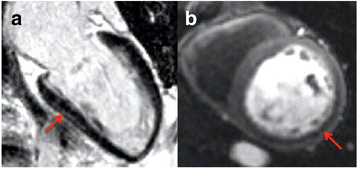
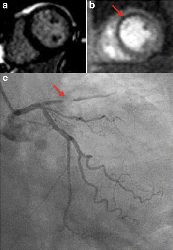
Results: On LGE-CMR, 38 patients were diagnosed with ICM, 46 with NICM and 16 with dual pathology. With perfusion-CMR, there were 39 ICM, 44 NICM and 17 dual pathology diagnoses. There was excellent agreement in diagnoses between LGE-CMR and perfusion-CMR (κ 0.968, p<0.001). The addition of adenosine stress perfusion images to LGE-CMR altered the diagnosis in only two of the 100 patients.
Conclusion: The addition of adenosine stress perfusion-CMR to cine and LGE-CMR provides minimal incremental diagnostic yield for determining the etiology of heart failure in patients with severe LVSD.
Keywords: Adenosine stress perfusion; Cardiovascular magnetic resonance; Heart failure; Late gadolinium enhancement; Non-ischemic cardiomyopathy.
Figures


References
-
- Ponikowski P, Voors AA, Anker SD, Bueno H, Cleland JG, Coats AJ, et al. 2016 ESC Guidelines for the diagnosis and treatment of acute and chronic heart failure: The Task Force for the diagnosis and treatment of acute and chronic heart failure of the European Society of Cardiology (ESC) Developed with the special contribution of the Heart Failure Association (HFA) of the ESC. Eur Heart J. 2016. - PubMed
-
- Cerrato E, D'Ascenzo F, Biondi-Zoccai G, Calcagno A, Frea S, Grosso Marra W, et al. Cardiac dysfunction in pauci symptomatic human immunodeficiency virus patients: a meta-analysis in the highly active antiretroviral therapy era. Eur Heart J. 2013;34(19):1432–1436. doi: 10.1093/eurheartj/ehs471. - DOI - PubMed
-
- Yancy CW, Jessup M, Bozkurt B, Butler J, Casey DE, Jr, Drazner MH, et al. 2013 ACCF/AHA guideline for the management of heart failure: executive summary: a report of the American College of Cardiology Foundation/American Heart Association task force on practice guidelines. Circulation. 2013;128(16):1810–1852. doi: 10.1161/CIR.0b013e31829e8807. - DOI - PubMed
MeSH terms
Substances
Grants and funding
LinkOut - more resources
Full Text Sources
Other Literature Sources
Medical

