Review
. 2017 Apr 8;18(1):54.
doi: 10.1186/s12931-017-0544-7.

The roles and regulation of the actin cytoskeleton, intermediate filaments and microtubules in smooth muscle cell migration

Affiliations
Review

The roles and regulation of the actin cytoskeleton, intermediate filaments and microtubules in smooth muscle cell migration

Dale D Tang et al. Respir Res. .

Abstract

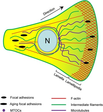
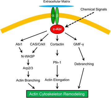
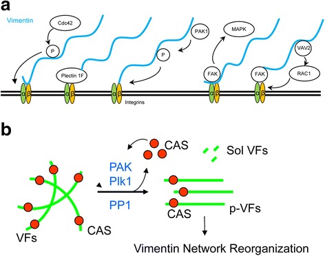
Smooth muscle cell migration has been implicated in the development of respiratory and cardiovascular systems; and airway/vascular remodeling. Cell migration is a polarized cellular process involving a protrusive cell front and a retracting trailing rear. There are three cytoskeletal systems in mammalian cells: the actin cytoskeleton, the intermediate filament network, and microtubules; all of which regulate all or part of the migrated process. The dynamic actin cytoskeleton spatially and temporally regulates protrusion, adhesions, contraction, and retraction from the cell front to the rear. c-Abl tyrosine kinase plays a critical role in regulating actin dynamics and migration of airway smooth muscle cells and nonmuscle cells. Recent studies suggest that intermediate filaments undergo reorganization during migration, which coordinates focal adhesion dynamics, cell contraction, and nucleus rigidity. In particular, vimentin intermediate filaments undergo phosphorylation and reorientation in smooth muscle cells, which may regulate cell contraction and focal adhesion assembly/disassembly. Motile cells are characterized by a front-rear polarization of the microtubule framework, which regulates all essential processes leading to cell migration through its role in cell mechanics, intracellular trafficking, and signaling. This review recapitulates our current knowledge how the three cytoskeletal systems spatially and temporally modulate the migratory properties of cells. We also summarize the potential role of migration-associated biomolecules in lung and vascular diseases.


Figures

Fig. 1
Fig. 1
A. Schematic illustration of major cytoskeletal components in motile cells. Lamellipodia and focal adhesions are located in the front of motile cells. The cross-hatched region represents the actin framework in lamellipodia. F-actin is present throughout the cell body, which interacts with myosin to generate traction force. Aging focal adhesions in the rear are disassembled to allow for cell retraction. Intermediate filaments surround the nucleus (N), some of which associate with focal adhesions in lamellipodia. Intermediate filaments modulate focal adhesion dynamics and cell contraction. Microtubules are polarized along the direction of migration and accumulate toward the front of the cell. Microtubule organizing centers (MOTCs) are localized in the front of the nucleus. Through their roles in mechanics, trafficking and signaling, polarized microtubules facilitate all important events leading to cell migration
Fig. 2
Fig. 2
Focal adhesion formation, actin dynamics and actomyosin activity in motile cells. Engagement of integrins with the extracellular matrix recruits structural proteins (talin, vinculin, ILK, PINCH, parvins, α-actinin, etc.) and signaling proteins (Cdc42, c-Abl, cortactin, FAK, paxillin, Abi1, etc.) to the near integrin region, which promotes focal adhesion formation (see detailed molecular interactions at focal adhesions in reference [2]). Signaling proteins activate N-WASP and the Arp2/3 complex, which induce actin filament branching (1). Activation of profilin-1, VASP and mDia promotes actin filament elongation (2). Activation of gelsolin and cofilin results in actin filament severing and depolymerization (3). GMF-γ promotes actin filament debranching (4). Myosin light chain phosphorylation triggers actomyosin activity and leads to cell contraction (5). Soluble cues activate receptors (e.g. growth factor receptors, cytokine receptors) and signaling proteins, which promote actin filament polymerization and focal adhesion assembly (See details in text). GFs, growth factors
Fig. 3
Fig. 3
Regulation of airway smooth muscle cell migration by c-Abl tyrosine kinase. c-Abl is recruited to the leading edge by integrin β1, which activates the downstream pathways and regulates actin cytoskeletal remodeling in lamellipodia. Abi1, Abl interactor 1; Arp2/3, Actin-related protein 2/3; CAS, CrkII-associated substrate; GMF-γ, glia maturation factor-γ; N-WASP, neuronal Wiskott-Aldrich Syndrome Protein; Pfn-1, profilin-1
Fig. 4
Fig. 4
Vimentin intermediate filaments, focal adhesions and cell migration. a Vimentin filaments may directly bind to integrins β1 or β3, or indirectly to integrins via FAK or plectin 1F. Cdc42 or PAK induces vimentin phosphorylation, which activates integrins. Vimentin recruits VAV2 to focal adhesions to promote FAK activation. Interactions of vimentin filaments with focal adhesions can activate the MAPK pathway. b PAK1 and Plk1 are able to induce vimentin phosphorylation at Ser-56 in smooth muscle whereas protein phosphatase 1 (PP1) dephosphorylates vimentin. Vimentin phosphorylation induces vimentin disassembly and spatial reorientation, which regulates cell contraction and focal adhesion dynamics. Vimentin disassembly also releases CAS to affect actin dynamics. VFs, vimentin filaments; p-VFs, phospho-vimentin filaments; Sol-VFs, soluble vimentin filaments; CAS, Crk-associated substrate
Fig. 5
Fig. 5
Cell migration regulated by microtubule-associated processes. Microtubule dynamics is regulated by plus-end trafficking proteins (+TIPs),+TIP stabilizing protein (e.g. adenomatous polyposis coli, APC), and cytoplasmic linker associated proteins (CLAPs). Microtubules regulate cell migration through their roles in mechanics, trafficking and signaling. GEF, guanine nucleotide exchange factor; MAP4K4, mitogen-activated protein kinase kinase kinase kinase 4.

References

    1. Gerthoffer WT. Migration of airway smooth muscle cells. Proc Am Thorac Soc. 2008;5:97–105. doi: 10.1513/pats.200704-051VS. - DOI - PMC - PubMed
    1. Tang DD. Critical role of actin-associated proteins in smooth muscle contraction, cell proliferation, airway hyperresponsiveness and airway remodeling. Respir Res. 2015;16:134. doi: 10.1186/s12931-015-0296-1. - DOI - PMC - PubMed
    1. Cleary RA, Wang R, Waqar O, Singer HA, Tang DD. Role of c-Abl tyrosine kinase in smooth muscle cell migration. Am J Physiol Cell Physiol. 2014;306:C753–61. doi: 10.1152/ajpcell.00327.2013. - DOI - PMC - PubMed
    1. Pollard TD, Cooper JA. Actin, a central player in cell shape and movement. Science. 2009;326:1208–12. doi: 10.1126/science.1175862. - DOI - PMC - PubMed
    1. Krause M, Gautreau A. Steering cell migration: lamellipodium dynamics and the regulation of directional persistence. Nat Rev Mol Cell Biol. 2014;15:577–90. doi: 10.1038/nrm3861. - DOI - PubMed

Publication types

LinkOut - more resources