Review
. 2017 May;40(5):260-275.
doi: 10.1016/j.tins.2017.03.005. Epub 2017 Apr 5.

Deciphering Neural Codes of Memory during Sleep

Affiliations
Review

Deciphering Neural Codes of Memory during Sleep

Zhe Chen et al. Trends Neurosci. 2017 May.

Abstract

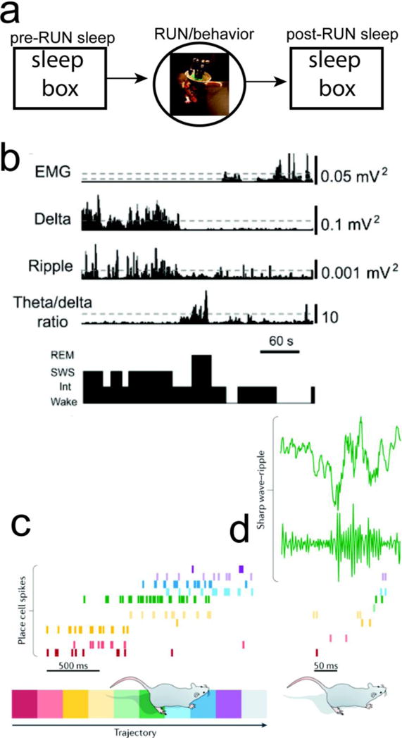
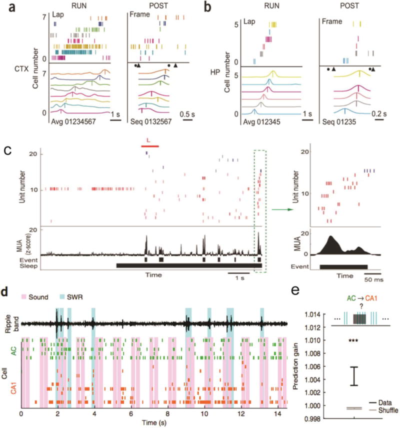
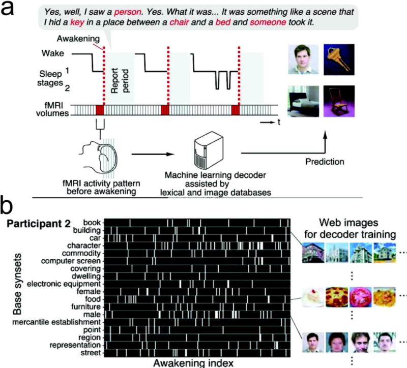
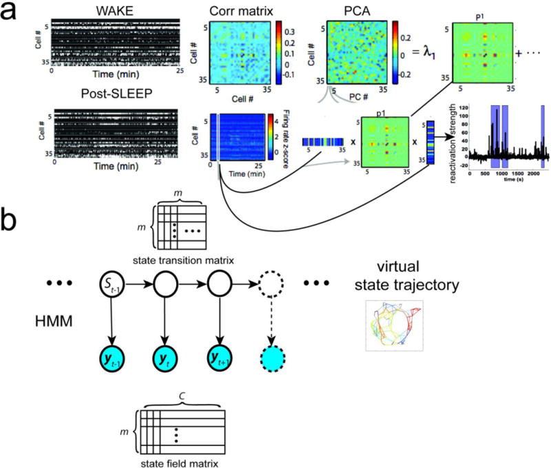
Memories of experiences are stored in the cerebral cortex. Sleep is critical for the consolidation of hippocampal memory of wake experiences into the neocortex. Understanding representations of neural codes of hippocampal-neocortical networks during sleep would reveal important circuit mechanisms in memory consolidation and provide novel insights into memory and dreams. Although sleep-associated ensemble spike activity has been investigated, identifying the content of memory in sleep remains challenging. Here we revisit important experimental findings on sleep-associated memory (i.e., neural activity patterns in sleep that reflect memory processing) and review computational approaches to the analysis of sleep-associated neural codes (SANCs). We focus on two analysis paradigms for sleep-associated memory and propose a new unsupervised learning framework ('memory first, meaning later') for unbiased assessment of SANCs.

Keywords: functional imaging; memory consolidation; memory replay; neural representation; population decoding; sleep-associated memory.


Figures

FIGURE 1
FIGURE 1. Study of Rodent Hippocampal Memory and Sleep
(a) A standard study paradigm for rodent hippocampal memory consists of pre-RUN sleep, RUN/behavior, and post-RUN sleep. (b) Classification of sleep stages from EMG, cortical LFP (Delta power), hippocampal ripple power, and cortical theta/delta power ratio [21]. (c) Rodent hippocampal population spike activity during RUN on a linear track. (d) Rodent hippocampal LFP and SWRs during post-RUN SWS, and the associated spatiotemporal spike pattern that shows a similar temporal order (“replay”) (reproduced with permission [18]).
FIGURE 2
FIGURE 2. Dissection of Hippocampal-Neocortical Memories during Sleep
(a,b) Neuronal firing sequences in rat V1 (a) and hippocampus (b) during RUN and POST-RUN SWS episodes. Lap: population neuronal firing pattern during a single running lap on the left-to-right trajectory. Each row represents a cell and each tick represents a spike. Avg: template firing sequence obtained by averaging over all laps on the trajectory. Each curve represents the average firing rate of a cell. Cells were assigned to numbers 0, 1, etc. and then arranged (01234567) from bottom to top according to the order of their firing peaks (vertical lines). Frame: the same population firing patterns in a POST-RUN SWS episode. Triangles and circles denote the onset of UP and DOWN states, respectively. Seq: firing sequence in the frame. Spike trains were convolved with a Gaussian window and cells were ordered (0132567) according to the peaks (vertical lines) of the resulted curves [36]. (c) Auditory sound (L, in red, indicating a left turn) biased the hippocampal reactivation during SWS [37]. In the raster plot, spikes from place cells with place fields on the right side of the track are blue, and left-sided place fields in red. Place fields are ordered from top to bottom by their location on the track (right → left side). Prior to sleep onset, the rat was resting in the sleep chamber. The reactivation event in the green dashed box is shown to the right. (d) Sound-biased auditory cortical neuronal ensembles (green) predict reactivations of hippocampal neurons (orange) during SWRs. Pink bars indicate sounds; cyan bars indicate detected SWRs. Top black trace is ripple-filtered LFP in hippocampal CA1 [38]. (e) Quantification of prediction gain of using sound-based pre-SWR auditory cortical (AC) ensemble spike patterns to predict hippocampal CA1 firing. Data is significantly different from the shuffled statistics (n=96) [38]. All figures are reproduced with permission.
FIGURE 3
FIGURE 3. Decoding the Content of Visual Imagery during Human REM Sleep
(a) fMRI data were acquired from sleeping participants simultaneously with polysomnography. Participants were awakened during sleep stage 1 or 2 (red dashed line) and verbally reported their visual experience during sleep. The fMRI data immediately before awakening (9 s) were used as the input for main decoding analyses (sliding time windows were used for time course analyses). Words describing visual objects or scenes (red letters) were extracted. The visual contents were predicted using machine-learning decoders trained on fMRI responses to natural images. (b) During the training phase, words describing visual objects or scenes were first mapped onto synsets of the WordNet tree [a dictionary of nouns, verbs, adverbs, adjectives, and their lexical relations]. Synsets were grouped into “base synsets” located higher in the tree. Visual reports (participant 2) are represented by visual content vectors, in which the presence or absence of the base synsets in the report at each awakening is indicated by white or black, respectively. Examples of images used for decoder training are shown for some of the base synsets. During the testing phase, a pairwise or multi-label decoder is applied to awakening event for predicting the visual object label (reproduced with permission [56]).
Box 2 FIGURE I
Box 2 FIGURE I. Unbiased Assessment of Sleep-Associated Neuronal Population Codes
(a) Principal component analysis (PCA) for computing the similarity of two templates of correlation matrices from population spike counts (WAKE and SLEEP) and assessing the reactivation strength during sleep (reproduced with permission, [43]). In WAKE, {λ,1,p1} are associated with the dominant principal component (PC) extracted from PCA. In SLEEP, time-varying reactivation strength is computed. (b) Unsupervised population decoding using a finite-state hidden Markov model (HMM). Specifically, the spatial environment is represented by a finite discrete state space. Trajectories across spatial locations (“states”) are associated with consistent hippocampal ensemble spike patterns, which are characterized by a state transition matrix. From the state transition matrix, a topology graph that defines the connectivity in the state space is inferred [69]. In these two methods, no assumption is made about neuronal RF, and the bin size in Post-SLEEP is independent on the bin size used in WAKE. Since the order of WAKE and SLEEP can be switched, and one can apply these methods to SLEEP data first and then examine their meanings in the WAKE behavior; therefore they both fall into the new paradigm (“memory first, meaning later”).

References

    1. Andersen P, et al. The Hippocampus Book. Oxford University Press; 2006.
    1. Stickgold R. Sleep-dependent memory consolidation. Nature. 2005;437:1272–1278. - PubMed
    1. Marshall L, Born J. Contribution of sleep to hippocampus-dependent memory consolidation. Trends Cog Sci. 2007;11:442–450. - PubMed
    1. Buzsaki G. Memory consolidation during sleep: a neurophysiology perspective. J Sleep Res. 1998;7:17–23. - PubMed
    1. Breton J, Robertson EM. Memory processing: The critical role of neuronal replay during sleep. Curr Biol. 2013;23:R836–R838. - PubMed

Publication types