Review
. 2017 Jun:184:68-76.
doi: 10.1016/j.trsl.2017.03.003. Epub 2017 Mar 23.

Skin microbiome and mast cells

Affiliations
Review

Skin microbiome and mast cells

Satomi Igawa et al. Transl Res. 2017 Jun.

Abstract

Microbiotas in the skin have high levels of diversity at the species level, but low phylum-level diversity. The human skin microbiota is composed predominantly of Gram-positive bacteria especially Actinobacteria, which are the dominant bacterial phylum on the skin. Lipoteichoic acid (LTA) is a major constituent of the cell wall of Gram-positive bacteria and is therefore abundant in the skin microbiome. Recent studies have shown that LTA, and other bacterial products, permeates the whole skin and comes into contact with epidermal and dermal cells, including mast cells (MCs), with the potential of stimulating MC toll-like receptors (TLRs). MCs express a variety of pattern recognition receptors, including TLRs, on their cell surface in order to detect bacteria. Recent publications suggest that the skin microbiome has influence on MC migration, localization and maturation in the skin. Germ free (no microbiome) animals possess an underdeveloped immune system and immature MCs. Despite much research done on skin microbiota and many papers describing skin interaction with "the good microbiota", there is still controversy regarding how mast cells, communicate with surface bacteria. The present review intends to quell the controversy by illuminating the communication mechanism between bacteria and MCs.


Figures

Figure 1
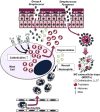
Figure 1. The two roles of mast cells (MCs) in bacterial infection
In group A Streptococcus skin infection, MC cathelicidin (LL-37) expression is increased by IL-4 and ligands of TLR2 and -4. The cathelicidins derived from MCs (LL-37) can not only kill bacteria directly, but also facilitate recruitment of neutrophils. On the other hand, MCs can show different antimicrobial activity against Streptococcus pyogenes, when they are exposed to a large number of bacteria, by emitting extracellular structures called MC extracellular traps (MCETs). IL-4: interleukin 4, LPS: lipopolysaccharide, LTA: lipoteichoic acid, TLR: Toll-like receptor
Figure 2
Figure 2. Skin microbiome-derived LTA reinforces the clearance of vaccinia virus (VV) with increasing antimicrobial peptide (AMP) expression in mast cells (MCs)
LTA derived from the commensal microbiome on the skin surface increases the expression of cathelicidins in MC granules via induction of Cnlp. TLR2 mediates this LTA-Cnlp response. VV challenge to MCs can also induce weak Cnlp expression in MCs, but this pathway is not mediated by TLR2. When VV enter MCs by membrane fusion, this event induces S1P formation and S1PR2 activation, leading to MC degranulation. Cnlp: cathelicidin-like protein, LTA: lipoteichoic acid, S1P: sphingosine-1-phosphate, S1PR2: S1P receptor 2, TLR: Tolllike receptor
Figure 3
Figure 3. Commensal skin microbiome promotes mast cell (MC) maturation
In a normal mouse, LTA from the skin microbiome induces keratinocytes to produce sufficient SCF, which results in proper MC maturation. In germ-free mice, MCs present lower expressions of the c-Kit and IgE receptors because SCF is insufficient. c-kit (CD117): mast/stem cell growth factor receptor, LTA: lipoteichoic acid, SCF: stem cell factor

References

    1. Supajatura V, Ushio H, Nakao A, Akira S, Okumura K, Ra C, et al. Differential responses of mast cell Toll-like receptors 2 and 4 in allergy and innate immunity. J Clin Invest. 2002;109:1351–9. - PMC - PubMed
    1. Vliagoftis H, Befus AD. Rapidly changing perspectives about mast cells at mucosal surfaces. Immunol Rev. 2005;206:190–203. - PubMed
    1. Forsythe P. Microbes taming mast cells: Implications for allergic inflammation and beyond. Eur J Pharmacol. 2016;778:169–75. - PubMed
    1. Gurish MF, Austen KF. Developmental origin and functional specialization of mast cell subsets. Immunity. 2012;37:25–33. - PubMed
    1. Gurish MF, Ghildyal N, McNeil HP, Austen KF, Gillis S, Stevens RL. Differential expression of secretory granule proteases in mouse mast cells exposed to interleukin 3 and c-kit ligand. J Exp Med. 1992;175:1003–12. - PMC - PubMed

MeSH terms