

The role of epidermal growth factor receptor (EGFR) signaling in SARS coronavirus-induced pulmonary fibrosis
- PMID: 28390872
- PMCID: PMC5507769
- DOI: 10.1016/j.antiviral.2017.03.022
The role of epidermal growth factor receptor (EGFR) signaling in SARS coronavirus-induced pulmonary fibrosis
Abstract
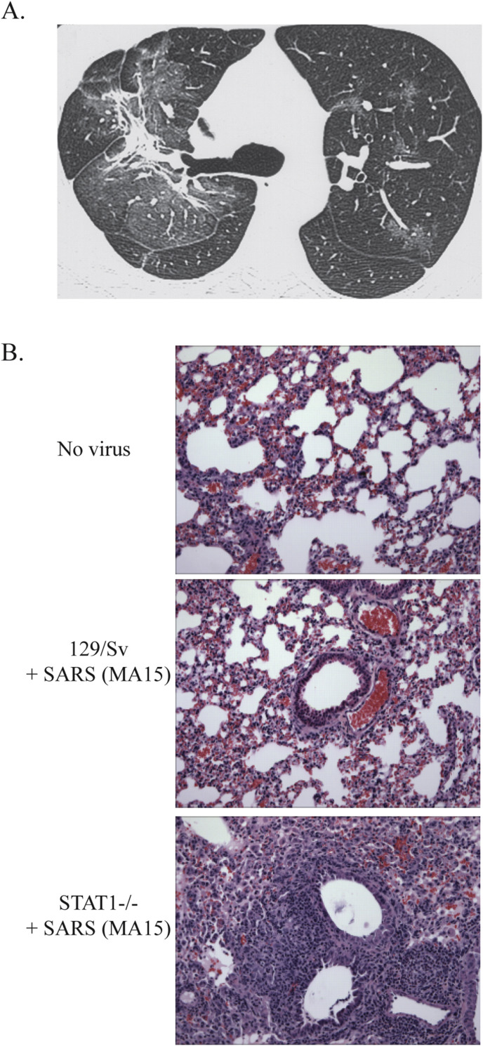
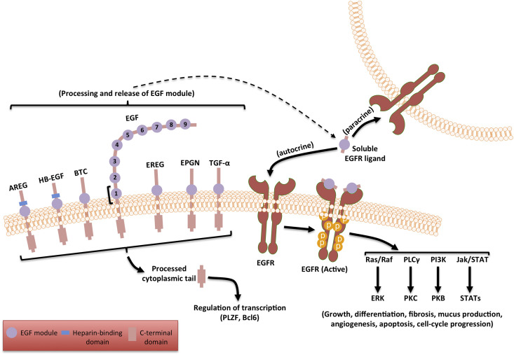
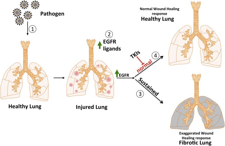
Many survivors of the 2003 outbreak of severe acute respiratory syndrome (SARS) developed residual pulmonary fibrosis with increased severity seen in older patients. Autopsies of patients that died from SARS also showed fibrosis to varying extents. Pulmonary fibrosis can be occasionally seen as a consequence to several respiratory viral infections but is much more common after a SARS coronavirus (SARS-CoV) infection. Given the threat of future outbreaks of severe coronavirus disease, including Middle East respiratory syndrome (MERS), it is important to understand the mechanisms responsible for pulmonary fibrosis, so as to support the development of therapeutic countermeasures and mitigate sequelae of infection. In this article, we summarize pulmonary fibrotic changes observed after a SARS-CoV infection, discuss the extent to which other respiratory viruses induce fibrosis, describe available animal models to study the development of SARS-CoV induced fibrosis and review evidence that pulmonary fibrosis is caused by a hyperactive host response to lung injury mediated by epidermal growth factor receptor (EGFR) signaling. We summarize work from our group and others indicating that inhibiting EGFR signaling may prevent an excessive fibrotic response to SARS-CoV and other respiratory viral infections and propose directions for future research.
Keywords: EGFR; Fibrosis; SARS-CoV; Wound healing.
Copyright © 2017 Elsevier B.V. All rights reserved.
Figures
References
-
- Antonio G.E., Wong K.T., Hui D.S., Wu A., Lee N., Yuen E.H., Leung C.B., Rainer T.H., Cameron P., Chung S.S., Sung J.J., Ahuja A.T. Thin-section CT in patients with severe acute respiratory syndrome following hospital discharge: preliminary experience. Radiology. 2003 Sep;228(3):810–815. - PubMed
-
- Baas T., Taubenberger J.K., Chong P.Y., Chui P., Katze M.G. SARS-CoV virus-host interactions and comparative etiologies of acute respiratory distress syndrome as determined by transcriptional and cytokine profiling of formalin-fixed paraffin-embedded tissues. J. Interferon Cytokine Res. Off. J. Int. Soc. Interferon Cytokine Res. 2006;26:309–317. - PMC - PubMed
Publication types
MeSH terms
Substances
Grants and funding
LinkOut - more resources
Full Text Sources
Other Literature Sources
Medical
Research Materials
Miscellaneous
