. 2017 Aug:12:625-633.
doi: 10.1016/j.redox.2017.04.003. Epub 2017 Apr 4.

Artemisinin protects PC12 cells against β-amyloid-induced apoptosis through activation of the ERK1/2 signaling pathway

Affiliations

Artemisinin protects PC12 cells against β-amyloid-induced apoptosis through activation of the ERK1/2 signaling pathway

Zhiwen Zeng et al. Redox Biol. 2017 Aug.

Abstract

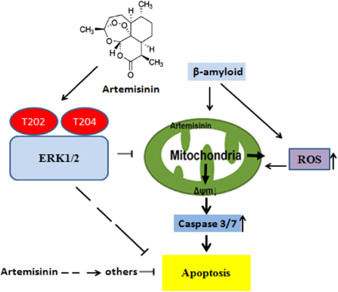
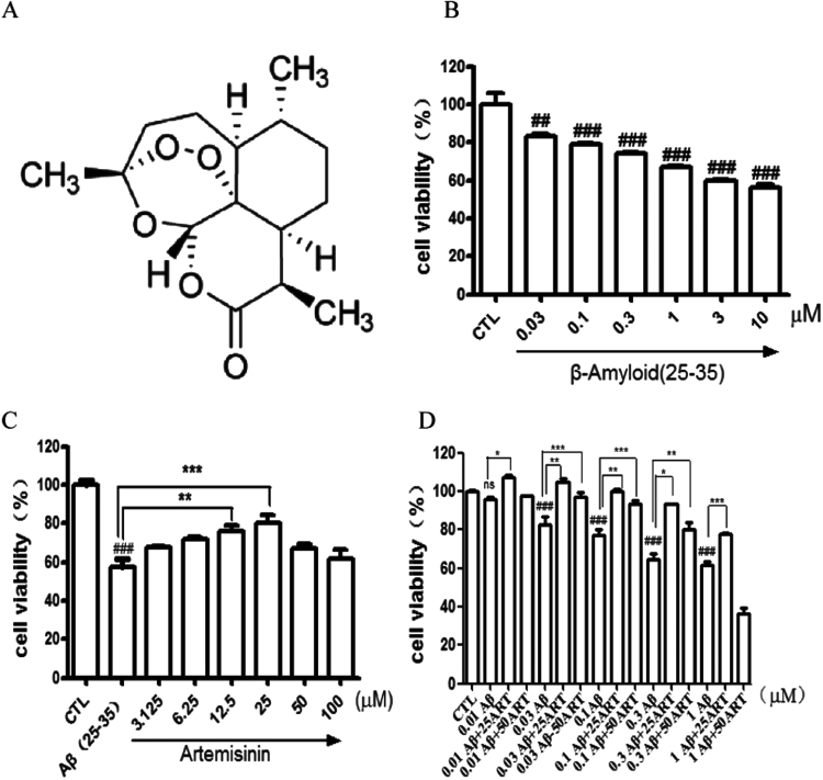
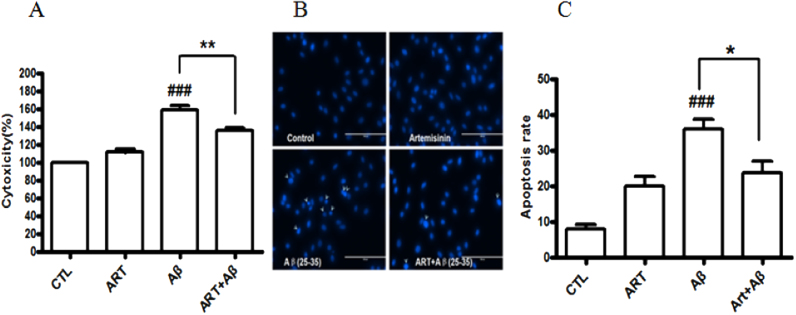
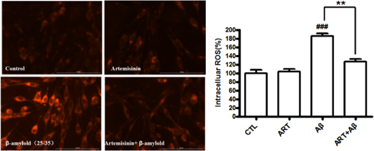
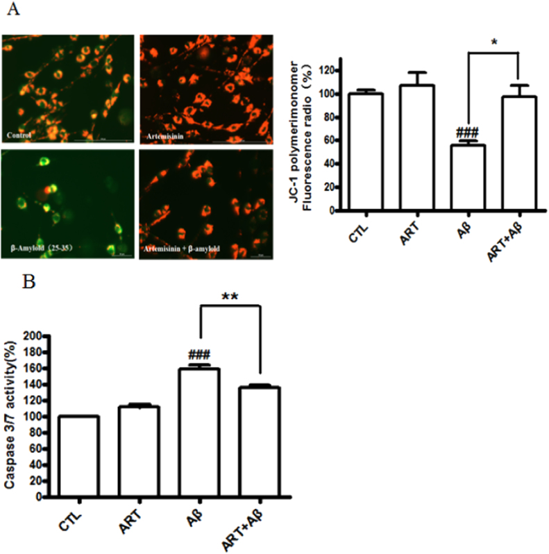
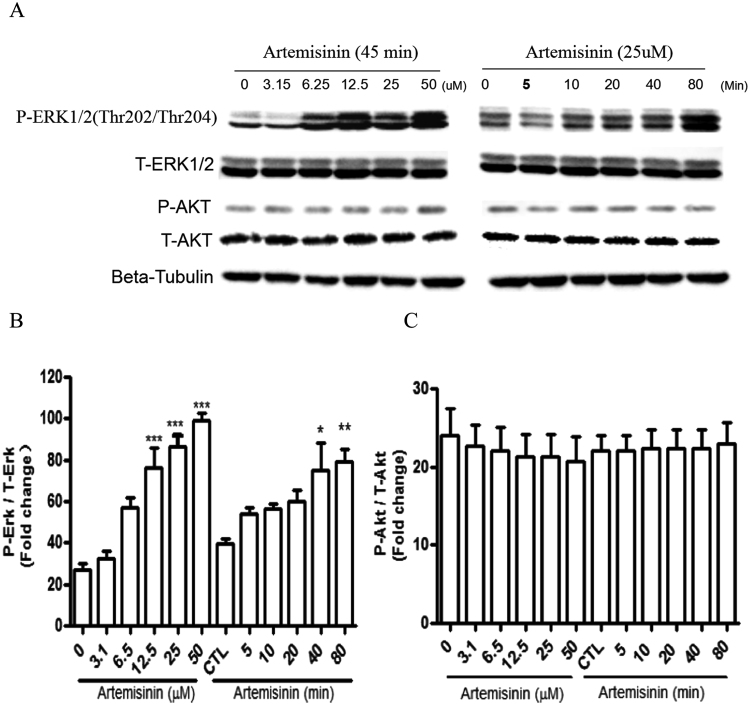
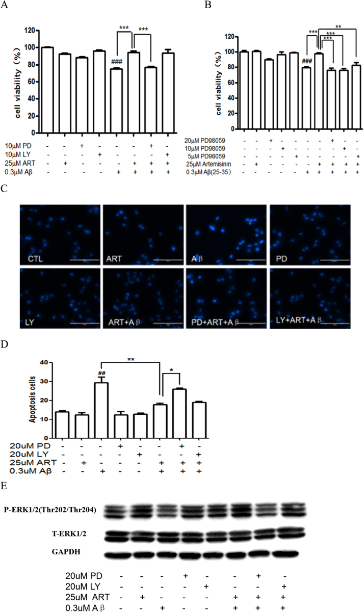
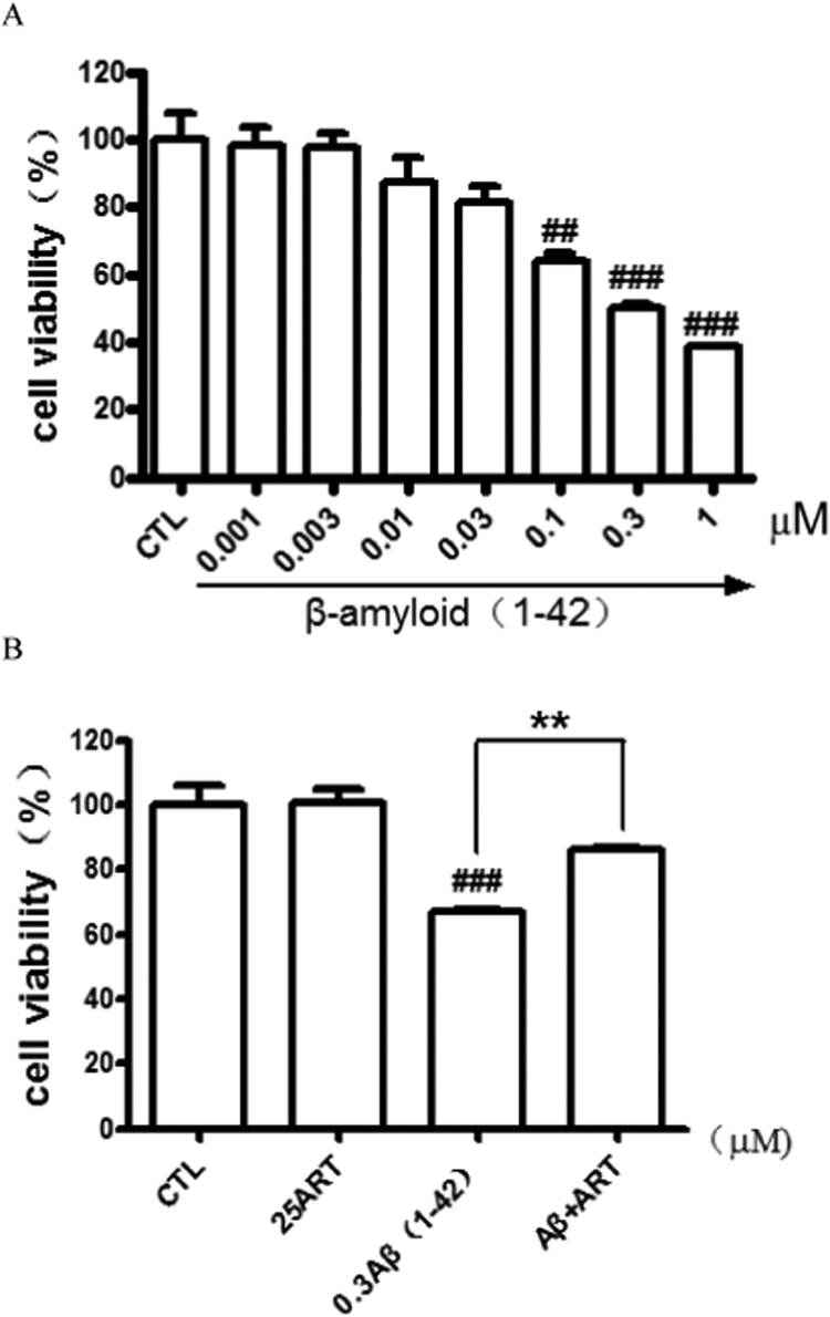
Accumulating evidence displays that an abnormal deposition of amyloid beta-peptide (Aβ) is the primary cause of the pathogenesis of Alzheimer's disease (AD). And therefore the elimination of Aβ is regarded as an important strategy for AD treatment. The discovery of drug candidates using culture neuronal cells against Aβ peptide toxicity is believed to be an effective approach to develop drug for the treatment of AD patients. We have previously showed that artemisinin, a FDA-approved anti-malaria drug, has neuroprotective effects recently. In the present study, we aimed to investigate the effects and potential mechanism of artemisinin in protecting neuronal PC12 cells from toxicity of β amyloid peptide. Our studies revealed that artemisinin, in clinical relevant concentration, protected and rescued PC12 cells from Aβ25-35-induced cell death. Further study showed that artemisinin significantly ameliorated cell death due to Aβ25-35 insult by restoring abnormal changes in nuclear morphology, lactate dehydrogenase, intracellular ROS, mitochondrial membrane potential and activity of apoptotic caspase. Western blotting analysis demonstrated that artemisinin activated extracellular regulated kinase ERK1/2 but not Akt survival signaling. Consistent with the role of ERK1/2, preincubation of cells with ERK1/2 pathway inhibitor PD98059 blocked the effect of artemisinin while PI3K inhibitor LY294002 has no effect. Moreover, Aβ1-42 also caused cells death of PC12 cells while artemisinin suppressed Aβ1-42 cytotoxicity in PC12 cells. Taken together, these results, at the first time, suggest that artemisinin is a potential protectant against β amyloid insult through activation of the ERK1/2 pathway. Our finding provides a potential application of artemisinin in prevention and treatment of AD.

Keywords: Alzheimer's disease; Artemisinin; Aβ25–35; ERK1/2; PC12 cells.


Figures

fx1
Graphical abstract
Fig. 1
Fig. 1
Artemisinin concentration-dependently suppressed Aβ25-35-induced cell viability lose in PC12 cells. (A) The structure of artemisinin. (B) Cells were treated with Aβ25-35 (0.03–10 μM) or 0.1% DMSO (vehicle control) for 24 h and cell viability was measured using the MTT assay(N=3). (C) Cells were pre-treated with artemisinin (3,125–100 μM) or 0.1% DMSO (vehicle control) for 1 h and then incubated with or without 0.3 μM Aβ25-35 for a further 24 h and cell viability were measured by MTT assay (N=3). (D) PC12 cells were incubated with 0.1, 0.3, 1 μM Aβ25-35 for 30 min and post-treated with artemisinin (25 or 50 μM) for 24 h and cell viability were measured by MTT assay (N=3). *P<0.05, **P<0.01,***P<0.005 versus the control or Aβ25-35 treated group as indicated; ###P<0.005 versus control group was considered statistically significant differences.
Fig. 2
Fig. 2
Artemisinin suppressed Aβ25-35-induced LDH release and apoptosis in PC12 cells. After pre-treatment with 25 μM artemisinin or 0.1% DMSO (vehicle control) for 1 h, PC12 cells were incubated with or without 0.3 μM Aβ25-35 for another 24 h the release of LDH (A) was measured by LDH assay and the apoptosis cells (N=3) (B) was detected by staining with Hoechst 33342 and visualized by fluorescence microscopy. The number of apoptotic nuclei with condensed chromatin (C) was counted from the photomicrographs and presented as a percentage of the total number of nuclei (N=3). ###P<0.005 versus control group; *P<0.05, **P<0.005 versus the Aβ-treated group were considered statistically significant differences.
Fig. 3
Fig. 3
Artemisinin reduced the increase of Aβ25-35-induced oxidative stress in PC12 cells. After pre-treatment with 25 μM artemisinin or 0.1% DMSO (vehicle control) for 1 h, PC12 cells were incubated with or without 0.3 μM Aβ25-35 for another 24 h. Intracellular ROS level was determined by the CellROXs Deep Red Reagent (N=3). ###P<0.005 versus control group; **P<0.01 versus Aβ-treated group was considered significantly different.
Fig. 4
Fig. 4
Artemisinin attenuated Aβ-induced mitochondrial membrane potential (△ψm) loss and caspase 3/7 activity increase in PC12 cells. After pre-treatment with 25 μM artemisinin or 0.1% DMSO (vehicle control) for 1 h, PC12 cells were incubated with or without 0.3 μM Aβ25-35 for another 24 h, △ψm was determined by the JC-1 assay (N=3) (Fig. 4A) and quantification of caspase 3/7 activity was determined by caspase 3/7 activity assay (N=3) (Fig. 4B). ###P<0.005 versus control group; *P<0.05, **P<0.01 versus Aβ-treated group were considered significantly different.
Fig. 5
Fig. 5
Artemisinin stimulated the phosphorylation of ERK1/2 in a time- and concentration-dependent manner in PC12 cells. (A) The PC12 cells were collected with artemisinin treatment for different times (0, 5, 10, 20, 40 and 80 min) at 25 μM, or at different concentrations (3.15, 6.25, 12.5, 25 and 50 μM) for 45 min. The expression of phosphorylated ERK1/2, total ERK1/2, phosphorylated Akt, total Akt and beta-tubulin (A) were detected by Western blotting with specific antibodies. Quantification of representative protein band from Western blotting (B and C N=3). *P<0.05, **P<0.01, ***P<0.005 versus the control group was considered significantly different.
Fig. 6
Fig. 6
ERK 1/2 pathway mediated the protect effects of artemisinin in PC12 cells. PC12 cells were pre-treated with10 μM PD98059 or 10 μM LY294002 (A), or 5, 10, 20 μM PD98059 (B) for 30 min, and treated with 25 μM artemisinin for 1 h, then incubated with or without 0.3 μM Aβ25-35 for a further 24 h, and the cell viability was determined by MTT assay (N=3). PC12 cells were pre-treated with the ERK1/2 inhibitor PD98059 (20 μM) or PI3K inhibitor LY294002 (20 μM) for 30 min, and afterward treated with 25 μM artemisinin for 1 h, then incubated with or without 0.3 μM Aβ25-35 for a further 24 h; the apoptosis cells was detected by staining with Hoechst 33342 and visualized by fluorescence microscopy (C). The number of apoptotic nuclei with condensed chromatin was counted from the photomicrographs and presented as a percentage of the total number of nuclei (N=3) (D). PC12 cells were pre-treated with the ERK1/2 inhibitor PD98059 (20 μM) or PI3K inhibitor LY294002 (20 μM) for 30 min, and afterward treated with 25uM artemisinin for 1 h, then incubated with or without 0.3 μM Aβ25-35, The expression of phosphorylated ERK1/2, total ERK1/2; and GAPDH were detected by Western blotting with specific antibodies (N=3). ###P<0.005 versus control group; **P<0.01, ***P<0.005 versus the artemisinin or artemisinin plus Aβ25-35-treated group as indicated were considered significantly different.
Fig. 7
Fig. 7
Artemisinin suppressed Aβ1-42-induced cell viability lose in PC12 cells. (A) Cells were treated with Aβ1-42 (0.001–1 μM) or 0.1% DMSO (vehicle control) for 24 h and cell viability was measured using the MTT assay. (B) Cells were pre-treated with artemisinin(25 μM) or 0.1% DMSO (vehicle control) for 1 h and then incubated with or without 0.3 μM Aβ1-42 for a further 24 h and cell viability were measured by MTT assay (N=3). ###P<0.005 versus control group; **P<0.01 versus the Aβ1-42 treated group was considered statistically differences.

References

    1. Vila M., Przedborski S. Targeting programmed cell death in neurodegenerative diseases. Nat. Rev. Neurosci. 2003;4(5):365–375. - PubMed
    1. Forman M.S., Trojanowski J.Q., Lee V.M. Neurodegenerative diseases: a decade of discoveries paves the way for therapeutic breakthroughs. Nat. Med. 2004;10(10):1055–1063. - PubMed
    1. Taylor J.P., Hardy J., Fischbeck K.H. Toxic proteins in neurodegenerative disease. Science. 2002;296(5575):1991–1995. - PubMed
    1. Colucci L. Alzheimer's disease costs: what we know and what we should take into account. J. Alzheimers Dis. 2014;42(4):1311–1324. - PubMed
    1. Selkoe D.J. Alzheimer's disease is a synaptic failure. Science. 2002;298(5594):789–791. - PubMed

MeSH terms

LinkOut - more resources