. 2017 Mar 1;9(3):398-413.
doi: 10.1093/gbe/evx001.

Evolutionary Thrift: Mycobacteria Repurpose Plasmid Diversity during Adaptation of Type VII Secretion Systems

Affiliations

Evolutionary Thrift: Mycobacteria Repurpose Plasmid Diversity during Adaptation of Type VII Secretion Systems

Tatum D Mortimer et al. Genome Biol Evol. .

Abstract

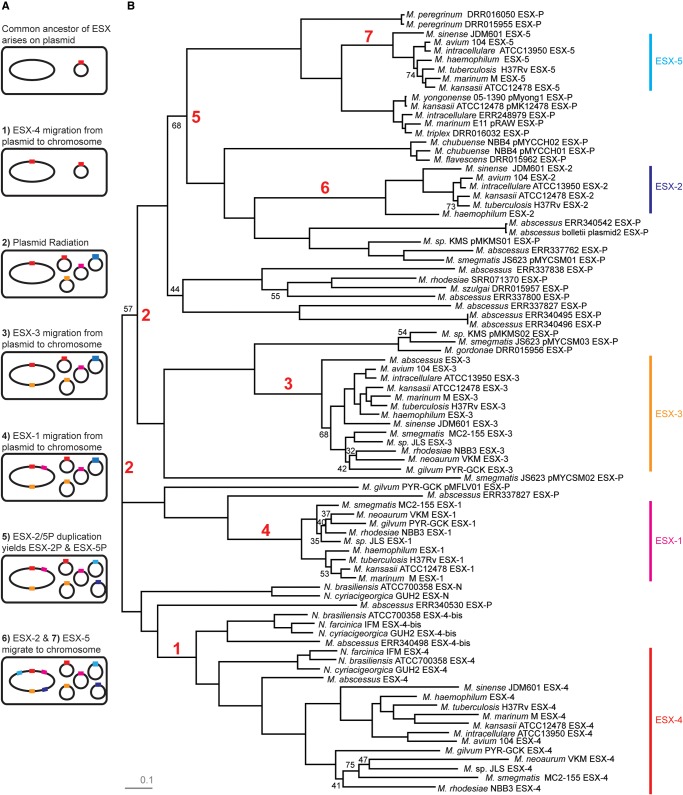
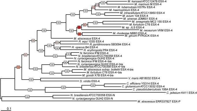
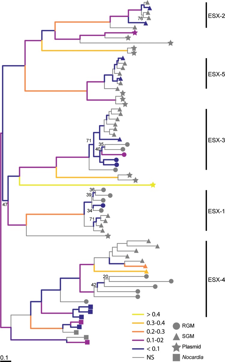
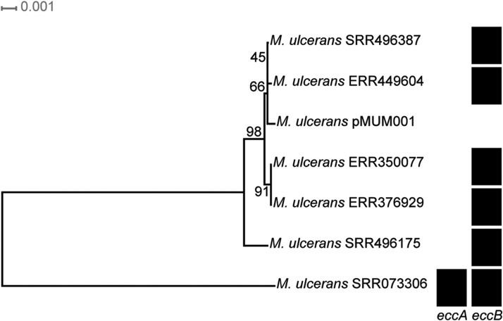
Mycobacteria have a distinct secretion system, termed type VII (T7SS), which is encoded by paralogous chromosomal loci (ESX) and associated with pathogenesis, conjugation, and metal homeostasis. Evolution of paralogous gene families is of interest because duplication is an important mechanism by which novel genes evolve, but there are potential conflicts between adaptive forces that stabilize duplications and those that enable evolution of new functions. Our objective was to delineate the adaptive forces underlying diversification of T7SS. Plasmid-borne ESX were described recently, and we found evidence that the initial duplication and divergence of ESX systems occurred on plasmids and was driven by selection for advantageous mutations. Plasmid conjugation has been linked to T7SS and type IV secretion systems (T4SS) in mycobacteria, and we discovered that T7SS and T4SS genes evolved in concert on the plasmids. We hypothesize that differentiation of plasmid ESX helps to prevent conjugation among cells harboring incompatible plasmids. Plasmid ESX appear to have been repurposed following migration to the chromosome, and there is evidence of positive selection driving further differentiation of chromosomal ESX. We hypothesize that ESX loci were initially stabilized on the chromosome by mediating their own transfer. These results emphasize the diverse adaptive paths underlying evolution of novelty, which in this case involved plasmid duplications, selection for advantageous mutations in the mobile and core genomes, migration of the loci between plasmids and chromosomes, and lateral transfer among chromosomes. We discuss further implications for the choice of model organism to study ESX functions in Mycobacterium tuberculosis.

Keywords: ESX; gene duplication; mycobacteria; plasmid; selection; type VII secretion system.


Figures

F<sc>ig</sc>. 1.—
Fig. 1.—
Mycobacterial chromosomal ESX loci. Core gene content in the ESX loci are colored as follows: eccA- red, eccB- dark blue, eccC- green, eccD- light blue, eccE- orange, mycP- pink, esxA/B- purple, PE/PPE- yellow. Other variable genes in the loci are black. Orthologs and paralogs are based on OrthoMCL (Li et al. 2003) output. Locus diagrams were made using GenomeTools (Gremme et al. 2013). Each locus has a distinct structure, which developed during adaptation on mycobacterial plasmids and chromosomes (see text for details).
F<sc>ig</sc>. 2.—
Fig. 2.—
Maximum likelihood phylogeny of Actinobacteria with presence/absence matrix of type VII secretion system loci. RAxML was used for phylogenetic inference of the Actinobacteria core genome alignment (concatenated amino acid alignments of genes (n = 171) present in all genomes without duplications). The phylogeny is midpoint rooted, and branches without labels have a boostrap value of 100. Presence of ESX loci is indicated with black boxes. We have abbreviated the genus Mycobacterium in the tip labels. Some M. tuberculosis complex (MTBC) species have characteristic deletions located in ESX loci. Partially deleted ESX loci are represented by black triangles. M. caprae has a deletion in ESX-2 spanning PE/PPE, esxC, espG2, Rv3888c, eccD2, and mycP2. M. pinnipedii has a deletion in ESX-1 spanning PE/PPE and a portion of eccC1b. Patterns of ESX presence/absence are consistent with an initial emergence of the FtsK/WXG100 gene cluster, followed by ESX-4 bis and ESX-4, with subsequent duplications giving rise to ESX-3, ESX-1, ESX-2, and ESX-5.
F<sc>ig</sc>. 3.—
Fig. 3.—
Network of ESX loci in mycobacteria, Nocardia, and mycobacterial plasmids. The network was created in SplitsTree4 from a concatenated alignment of eccA, eccB, eccC, eccD, eccE, and mycP. Light blue dots correspond to ESX loci from RGM, light purple dots correspond to ESX loci from SGM, magenta dots correspond to ESX loci from mycobacterial plasmids, and black dots correspond to ESX loci from Nocardia chromosomes. The earliest branching lineages are all plasmid-associated, suggesting that the ancestral ESX locus was plasmid-borne (putative location of migration events to the chromosome marked “M” on the network). The PHI test was insignificant (p = 1.0) for this alignment, indicating that there was no evidence for intralocus recombination. A version of this figure with tip labels is available in the supplementary fig. S1, Supplementary Material online.
F<sc>ig</sc>. 4.—
Fig. 4.—
Network of plasmid-borne ESX loci. The network was created in SplitsTree4 from a concatenated alignment of eccA, eccB, eccC, eccD, eccE, and mycP from ESX loci encoded on mycobacterial plasmids. The star-like appearance of the network is consistent with rapid diversification of this gene family on the plasmids. Some bacterial strains harbored multiple plasmids, and these are indicated with colored branches. The phylogenetic relationships among plasmid ESX loci do not follow the core genome phylogeny, and plasmids with divergent ESX loci can be found within the same host species or even the same cell. This suggests that plasmid ESX diversification has not been shaped by adaptation to bacterial host species. We did not find evidence of intra-locus recombination in this alignment with the PHI test (P = 1.0).
F<sc>ig</sc>. 5.—
Fig. 5.—
ESX plasmid-mediated duplication and migration to the chromosome. (A) Simplified schematic of major steps in the evolutionary history of mycobacterial ESX loci. ESX loci are colored as follows: Ancestral/ESX-4: red, ESX-3: orange, ESX-1: pink, ESX-2: light blue, ESX-5: dark blue. (B) Maximum likelihood phylogeny of ESX loci (eccA, eccB, eccC, eccD, eccE, and mycP) in mycobacteria, Nocardia, and mycobacterial plasmids. Branches without black labels have a bootstrap value greater than 75. Red labels correspond to events presented in the schematic. ESX-N, found on the chromosomes of some Nocardia species, appear to have been recently transferred from a plasmid (see text). ESX-N and other plasmid-associated ESX are basal to chromosomal ESX 1–5. This suggests that their common ancestor was plasmid-borne and that extant chromosomal loci trace to migrations from plasmid to chromosome. The model outlined here is highly simplified: for example, there were likely several migrations of ESX-4 like loci to the chromosome (step 1 in the schematic) and the chromosomal loci show a mixture of vertical and horizontal inheritance (details in text).
F<sc>ig</sc>. 6.—
Fig. 6.—
Maximum likelihood phylogeny of ESX-4 in Actinobacteria. The phylogeny is rooted using ESX-N and a basal plasmid-borne ESX locus. Bootstrap values are colored based on support (white = 100, red = lowest support). The location of chromosomal Corynebacterium ESX-4 and M. goodii ESX-4-bis are in conflict with the core genome phylogeny (fig. 2). In the core genome phylogeny, Corynebacterium is more closely related to Nocardia and Rhodococcus than Verrucosispora or Saccharamonaspora. However, in the ESX-4 phylogeny, this relationship is reversed. Based on the relationship of Mycobacterium species in the core genome phylogeny, we would expect M. goodii ESX-4-bis and M. fortuitum ESX-4-bis to be more closely related to one another than either is to M. abscessus (the most basal Mycobacterium species). These conflicts suggest that chromosomal ESX-4 like loci have been laterally transferred among species. Genus names have been abbreviated, but full length names can be found in the core genome phylogeny (fig. 2).
F<sc>ig</sc>. 7.—
Fig. 7.—
Episodic directional selection during the evolution of ESX loci. Maximum likelihood phylogeny inferred using RAxML from a concatenated alignment of eccA, eccB, eccC, eccD, eccE, and mycP. In order to minimize potential effects of misalignment on inference of selection, only data from finished genomes were included in this analysis: see figures 3 and 5B for network and phylogenetic analyses of the complete dataset. Plasmid associated taxa for ESX-4, ESX-2 and ESX-5 are not shown on this phylogeny for this reason. Branches in this phylogeny without labels have a bootstrap value >75. We used the aBSREL test implemented in HyPhy to identify branches with significant evidence (P < 0.05) of episodic directional selection; these branches are colored based on the proportion of sites affected by positive selection (ω > 1). Circles correspond to ESX loci from RGM chromosomes, triangles correspond to ESX loci from SGM chromosomes, stars correspond to ESX loci from mycobacterial plasmids, and squares correspond to ESX loci from Nocardia chromosomes. A version of this figure is included in the supplementary figure S4, Supplementary Material online that shows the tips labeled with species names. There is evidence of directional (positive) selection at each duplication event (short internal branches), as well as on the branches leading to the extant chromosomal loci.
F<sc>ig</sc>. 8.—
Fig. 8.—
Congruence of tree topologies of plasmid encoded secretion systems. Bayesian phylogenetic analysis was performed in MrBayes using amino acid alignments of EccA, EccB, EccC, EccD, EccE, MycP, VirB4, VirD, TcpC, EspI, NrdH, and a hypothetical protein (Hyp1) encoded proximal to known T4SS genes. (A) We used TreeScape to calculate the Kendell Colijn metric between pairs of trees and perform multidimensional scaling (MDS). Clusters of trees are visualized as a scatterplot of the first and third principal components from the MDS. The inset bar chart is a scree plot showing the eigenvalues for the principal components. The T4SS and T7SS gene trees overlap in the MDS, whereas topologies of NrdH gene trees are incongruent with those of T4SS and T7SS. This suggests that plasmid-encoded T4SS and T7SS have co-diverged during their evolutionary history and that they have evolved independently of other gene content on the plasmids. (B) Kendell Colijn distances among secretion system gene trees and between secretion system and NrdH gene trees. The means of these distributions are significantly different (P < 2.2 × 10⁻¹⁶) according to a Mann–Whitney U test.
F<sc>ig</sc>. 9.—
Fig. 9.—
Core gene phylogeny of Mycobacterium ulcerans plasmids and presence/absence of T7SS genes. RAxML was used for phylogenetic inference from a core gene alignment (concatenated amino acid alignments of genes (n = 21) present in all Mycobacterium ulcerans plasmids without duplications). The phylogeny is midpoint rooted. Presence of ESX genes is indicated with black boxes. ESX genes found on M. ulcerans plasmids are most closely related to ESX-2. The most basal M. ulcerans plasmid encodes both eccA and eccB. Most M. ulcerans plasmids only encode eccB, and some have lost all ESX genes (e.g. pMUM001). The locus shows other signs of degradation (discussed in the text). We hypothesize that selection to maintain the conjugation machinery of these plasmids has been relaxed as a result of host selection for its other gene content, likely mycolactone.

Similar articles

Cited by

References

    1. Abascal F, Zardoya R, Telford MJ. 2010. TranslatorX: multiple alignment of nucleotide sequences guided by amino acid translations. Nucleic Acids Res. 38:W7–W13. - PMC - PubMed
    1. Abdallah AM, et al. 2009. PPE and PE_PGRS proteins of Mycobacterium marinum are transported via the type VII secretion system ESX-5. Mol Microbiol. 73:329–340. doi: 10.1111/j.1365-2958.2009.06783.x. - PubMed
    1. Adler M, Anjum M, Berg OG, Andersson DI, Sandegren L. 2014. High fitness costs and instability of gene duplications reduce rates of evolution of new genes by duplication-divergence mechanisms. Mol Biol Evol. 31:1526–1535. - PubMed
    1. Andersson DI, Hughes D. 2009. Gene amplification and adaptive evolution in bacteria. Annu. Rev. Genet. 43:167–195. - PubMed
    1. Antipov D, et al. 2016. plasmidSPAdes: Assembling Plasmids from Whole Genome Sequencing Data. bioRxiv. 48942. doi: 10.1101/048942. - PubMed

Publication types

MeSH terms

Substances

LinkOut - more resources