Review
. 2017 Jan 1;32(suppl_1):i71-i79.
doi: 10.1093/ndt/gfw300.

Biomarkers of lupus nephritis histology and flare: deciphering the relevant amidst the noise

Affiliations
Review

Biomarkers of lupus nephritis histology and flare: deciphering the relevant amidst the noise

Daniel J Birmingham et al. Nephrol Dial Transplant. .

Abstract

Biomarker development in lupus nephritis (LN) has traditionally relied on comparing the characteristics of candidate markers to clinical findings in patients and controls from cross-sectional cohorts. In this work, two additional strategies for LN biomarker development that are gaining ground will be discussed. One approach compares analytes directly to kidney histology. The second strategy utilizes longitudinal measurements of biomarker levels at regular intervals as patients move from disease quiescence to disease flare. These approaches have begun to empower biomarkers as diagnostic and prognostic tools in LN and have revealed novel and sometimes unexpected roles for these biomarkers in the pathogenesis and prediction of LN disease activity.

Keywords: discovery and validation; longitudinal cohort; urine analyte.


Figures

FIGURE 1:
FIGURE 1:
Biomarkers of interstitial injury in LN. These composite biomarkers of interstitial injury were derived using linear discriminant analysis to combine urine and serum analytes into equations that gave the highest sum of sensitivity and specificity for distinguishing between none–mild and moderate–severe interstitial fibrosis and interstitial inflammation.
FIGURE 2:
FIGURE 2:
Studies of the complement system in the OSS. All figures reproduced in modified form from previous reports [27, 32, 36], with permission. (A) Risk curves, according to factor H 402Y->H genotype, for renal flare as plasma C3 levels decrease. The vertical arrows show the median plasma C3 levels at the ‘no-flare’ and ‘at-flare’ follow-up visits. (B) Mean E-CR1 levels in nonrenal and LN patients at baseline, 2 months before (−2), at the time of (0) and 2 months after (+2) a nonrenal or renal flare. **P < 0.001 for a decrease in detectable E-CR1 level at nonrenal flare compared with nonrenal flare baseline in LN. (C) Anti-C1q IgG levels at 8, 6, 4 and 2 months before (−8, −6, −4, −2) and at (0) renal flare in patients who were anti-C3b negative or anti-C3b-positive. *P = 0.02 for an increase at renal flare for anti-C3b positive patients.
FIGURE 3:
FIGURE 3:
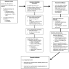
A strategy for biomarker development in LN: from context to validation. The phrase ‘LN biomarker’ is vague. The purpose of a biomarker in managing LN should be precisely defined. Some important unmet needs in the care of patients with LN include how to predict who will flare before they do so and who is likely to respond to therapy or not respond and develop chronic kidney disease (CKD) or end-stage renal disease (ESRD). Disease activity is generally assessed clinically, but clinical impressions do not necessarily reflect histologic activity. Understanding intrarenal histologic activity in a noninvasive way would improve LN management. Biomarkers that predict these outcomes or are surrogates of a kidney biopsy will be of high clinical value. Candidate LN biomarkers are identified in discovery cohorts of LN patients. Such cohorts are generally small but well-characterized clinically. LN outcomes are known for each patient. For example, to identify biomarkers of histologic activity the comparator for potential candidates should be the kidney biopsy. Biomarker candidates can be identified by a variety of techniques. Screening with omic techniques is time efficient. Candidates identified in the discovery phase of biomarker development need to be verified before proceeding with validation. This is especially important for candidates derived using omic techniques. Depending on how large omic data sets are analyzed, candidates found to be significant may not be differentially expressed when measured by an alternate technique specific for that analyte. Alternatively, differentially expressed transcripts may not translate to changes in protein levels, and would not be further developed. It is also important to understand how well the biomarker candidates reflect the clinical outcomes of interest in a cohort that is larger and possibly more diverse than the discovery cohort. By studying a larger cohort, the diagnostic metrics of a biomarker candidate can be determined. This will be important in deciding whether a candidate is superior to existing clinical diagnostics, and thus should be taken forward, and whether a candidate can stand on its own, or would be more robust if part of a composite biomarker (a biomarker panel). Finally, biomarkers need to be validated using independent cohorts by other investigators.

Similar articles

Cited by

  • Urine proteomic signatures of histological class, activity, chronicity, and treatment response in lupus nephritis.
    Fava A, Buyon J, Magder L, Hodgin J, Rosenberg A, Demeke DS, Rao DA, Arazi A, Celia AI, Putterman C, Anolik JH, Barnas J, Dall'Era M, Wofsy D, Furie R, Kamen D, Kalunian K, James JA, Guthridge J, Atta MG, Monroy Trujillo J, Fine D, Clancy R, Belmont HM, Izmirly P, Apruzzese W, Goldman D, Berthier CC, Hoover P, Hacohen N, Raychaudhuri S, Davidson A, Diamond B; Accelerating Medicines Partnership in RA/SLE network; Petri M. Fava A, et al. JCI Insight. 2024 Jan 23;9(2):e172569. doi: 10.1172/jci.insight.172569. JCI Insight. 2024. PMID: 38258904 Free PMC article.
  • Alternative pathway diagnostics.
    Thurman JM, Fremeaux-Bacchi V. Thurman JM, et al. Immunol Rev. 2023 Jan;313(1):225-238. doi: 10.1111/imr.13156. Epub 2022 Oct 28. Immunol Rev. 2023. PMID: 36305168 Free PMC article. Review.
  • Precision medicine in lupus nephritis: can biomarkers get us there?
    Caster DJ, Merchant ML, Klein JB, Powell DW. Caster DJ, et al. Transl Res. 2018 Nov;201:26-39. doi: 10.1016/j.trsl.2018.08.002. Epub 2018 Aug 10. Transl Res. 2018. PMID: 30179587 Free PMC article. Review.
  • Adhesion molecules: a way to understand lupus.
    Nowak K, Gumkowska-Sroka O, Kotyla P. Nowak K, et al. Reumatologia. 2022;60(2):133-141. doi: 10.5114/reum.2022.115664. Epub 2022 May 18. Reumatologia. 2022. PMID: 35782027 Free PMC article. Review.
  • [Lupus nephritis].
    Melderis S, Wiech T, Iking-Konert C, Steinmetz OM. Melderis S, et al. Z Rheumatol. 2018 Sep;77(7):593-608. doi: 10.1007/s00393-018-0496-4. Z Rheumatol. 2018. PMID: 29955955 German.

References

    1. Malvar A, Pirruccio P, Alberton V, et al. Histologic versus clinical remission in proliferative lupus nephritis. Nephrol Dial Transplant 2015; doi: 10.1093/ndt/gfv296doi:10.1093/ndt/gfv296 - DOI - PMC - PubMed
    1. Hsieh C, Chang A, Brandt D, et al. Predicting outcomes of lupus nephritis with tubulointerstitial inflammation and scarring. Arthritis Care Res (Hoboken) 2011; 63: 865–874 - PMC - PubMed
    1. Rovin BH, Song H, Birmingham DJ, et al. Urine chemokines as biomarkers of human systemic lupus erythematosus activity. J Am Soc Nephrol 2005; 16: 467–473 - PubMed
    1. Merchant ML, Perkins BA, Boratyn GM, et al. Urinary peptidome may predict renal function decline in type 1 diabetes and microalbuminuria. J Am Soc Nephrol 2009; 20: 2065–2074 - PMC - PubMed
    1. Triantafyllopoulou A, Franzke CW, Seshan SV, et al. Proliferative lesions and metalloproteinase activity in murine lupus nephritis mediated by type I interferons and macrophages. Proc Natl Acad Sci USA 2010; 107: 3012–3017 - PMC - PubMed

LinkOut - more resources