Review
. 2017 Dec 1:122:31-64.
doi: 10.1016/j.addr.2017.04.001. Epub 2017 Apr 6.

Nanoparticles for drug delivery to the anterior segment of the eye

Affiliations
Review

Nanoparticles for drug delivery to the anterior segment of the eye

Dileep R Janagam et al. Adv Drug Deliv Rev. .

Abstract

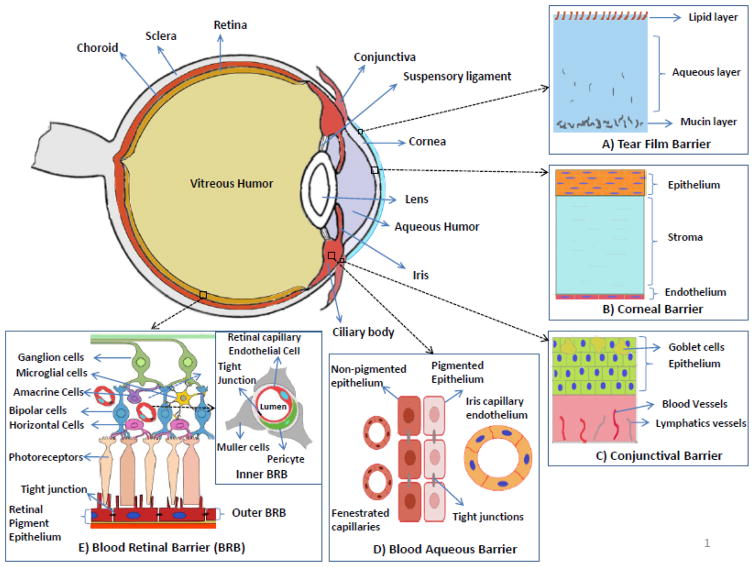
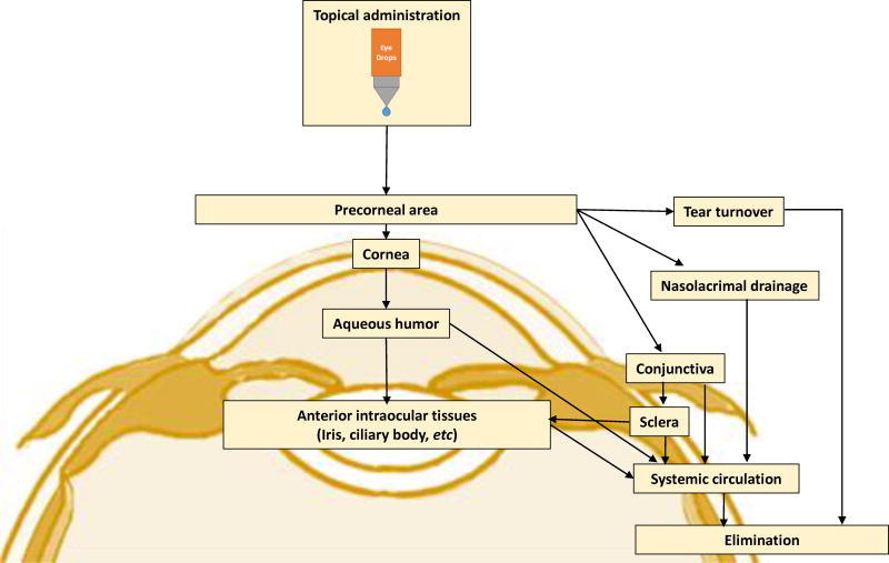
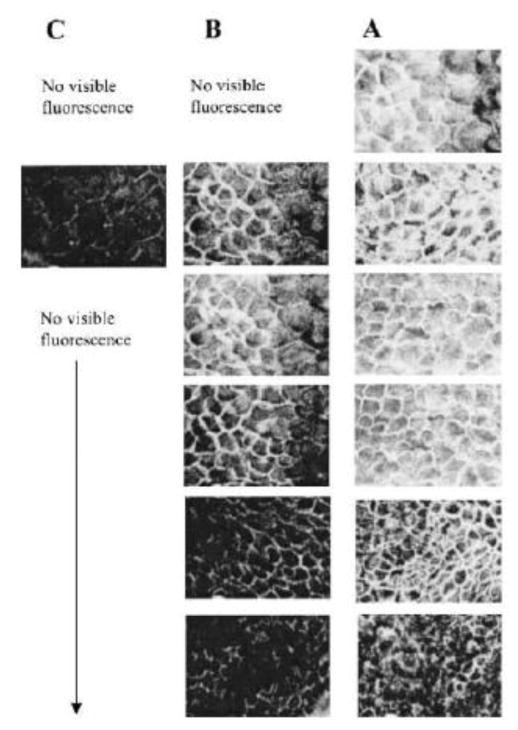
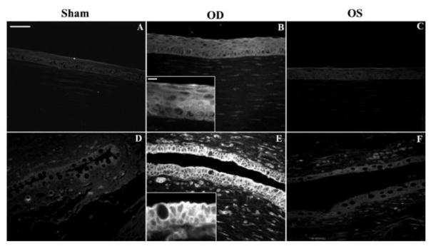
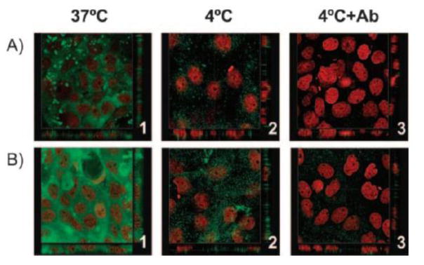
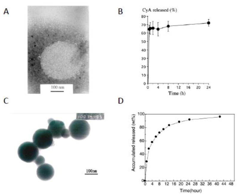
Commercially available ocular drug delivery systems are effective but less efficacious to manage diseases/disorders of the anterior segment of the eye. Recent advances in nanotechnology and molecular biology offer a great opportunity for efficacious ocular drug delivery for the treatments of anterior segment diseases/disorders. Nanoparticles have been designed for preparing eye drops or injectable solutions to surmount ocular obstacles faced after administration. Better drug pharmacokinetics, pharmacodynamics, non-specific toxicity, immunogenicity, and biorecognition can be achieved to improve drug efficacy when drugs are loaded in the nanoparticles. Despite the fact that a number of review articles have been published at various points in the past regarding nanoparticles for drug delivery, there is not a review yet focusing on the development of nanoparticles for ocular drug delivery to the anterior segment of the eye. This review fills in the gap and summarizes the development of nanoparticles as drug carriers for improving the penetration and bioavailability of drugs to the anterior segment of the eye.

Keywords: Anterior segment of the eye; Contact lenses; Dendrimer; EUDRAGIT®; Lipid; Nanoparticles; Ocular barriers; Poly(alkyl cyanoacrylate); Polyester; Polysaccharide.


Figures

Fig. 1
Fig. 1
Structure of the anterior segment of the eye and ocular barriers for drug delivery. A) Tear film barrier: Main components of the tear film include mucins, water and lipid, and acts a defensive barrier to the foreign-object access to the cornea and conjunctiva. B) Corneal barrier: avascular and comprised of three major layers which are epithelium (multiple layers stacked on each other), stroma and endothelium (single layer). Acts as a barrier preventing the drug absorption from the lacrimal fluid into the anterior chamber after the topical administration. C) Conjunctival barrier: mucous membrane consisting of conjunctival epithelium (2–3 layers thick), and an underlying vascularized connective tissue. Acts a barrier to the topically administered drugs and relatively in-efficient compared to the corneal barrier. D) Blood-aqueous barrier: located in the anterior segment of the eye. Formed by the capillary endothelium in the iris, and the ciliary epithelium which both contain tight junctions. The barrier is relatively inefficient compared to the blood retinal barrier and small molecules can reach the aqueous humor by permeation through fenestrated capillaries in the ciliary processes. E) Blood-retinal barrier (BRB): located in the poster segment of the eye. Formed by the retinal pigment epithelium (outer BRB) and the endothelial membrane of the retinal blood vessels (inner BRB), both contain tight junctions. The tight junctions restrict the entry of the drugs from the blood (systemic) into the retina/aqueous humor.
Fig. 2
Fig. 2
Flow chart presenting the drug pathway into the eye after topical application.
Fig. 3
Fig. 3
Scanning electron microphotographs of Chang cells exposed to culture medium, chitosan nanoparticles, and benzalkonium chloride for 24 h. (A) (Negative control, culture medium) Cells showing abundant microvilli and intact membrane details. (B) (Chitosan nanoparticles, 0.25 mg/ml) cells showing well-preserved morphology, an intact cell surface, and abundant microvilli, as expected for an epithelial cell. (C) (Chitosan nanoparticles, 1 mg/ml). (D) (Positive control, benzalkonium chloride) Cells were flat and showed absence of microvilli and broken membrane. Magnification ×750 (bar =15 μm). [Reprinted from “Pharmaceutical research, Chitosan nanoparticles as new ocular drug delivery systems: in vitro stability, in vivo fate, and cellular toxicity, 21.5 (2004): 803–810. De Campos, A. M., Diebold, Y., Carvalho, E. L., Sánchez, A., & José Alonso, M. c 2004 Plenum Publishing Corporation” With permission of Springer.]
Fig. 4
Fig. 4
Ocular surface structures of CSNP-treated (OD) and control (OS) rabbit eyes. Rabbits were exposed to CSNPs for 24 hours. Representative conjunctival impression cytology (A, B) and conjunctival (C, D) and corneal (E, F) sections are shown. Conjunctival and corneal epithelia from both control and treated eyes displayed normal cell layers and morphology. No signs of tissue edema were observed in any structure studied after exposure to CSNPs compared with controls. Scale bar, 100 μm. [Reprinted from De Salamanca, Amalia Enríquez, et al. “Chitosan nanoparticles as a potential drug delivery system for the ocular surface: toxicity, uptake mechanism and in vivo tolerance.” Investigative ophthalmology & visual science 47.4 (2006): 1416–1425. Copyright © Association for Research in Vision and Ophthalmology]
Fig. 5
Fig. 5
Confocal fluorescence images at different levels from the rabbit corneal epithelium (5 μm sequential cross sections from the corneal surface) at 1 h postinstillation of (C) Chitosan-fluorescein nanoparticles, (B) Chitosan-fluorescein solution. (A) image of cross section of a nontreated cornea. Round fluorescent spots corresponding to the chitosan-fluorescein nanoparticles were observed inside the cells. [Reprinted from “Pharmaceutical research, Chitosan nanoparticles as new ocular drug delivery systems: in vitro stability, in vivo fate, and cellular toxicity, 21.5 (2004): 803–810. De Campos, A. M., Diebold, Y., Carvalho, E. L., Sánchez, A., & José Alonso, M. c 2004 Plenum Publishing Corporation” With permission of Springer.]
Fig. 6
Fig. 6
Chitosan nanoparticle in vivo uptake. Fluorescence microscopy of ocular surface structures of sham-treated (A, D), CSNP-treated (B, E), and contralateral control (C, F) rabbit eyes. Representative corneal (A–C) and conjunctival (D–F) sections are shown. No fluorescence was detected in sham control corneas (A) or conjunctivas (D). (B) Corneal epithelial cells of CSNP-treated rabbits were uniformly fluorescent. (B, inset): enlargement showing a detail of corneal epithelial fluorescence pattern. (E) Fluorescence in conjunctival epithelial cells was intense in apical cell membranes and positive along the basolateral cell membrane. (E, inset) Enlargement showing the basolateral membrane fluorescence staining in goblet and non–goblet cells. (C, F) Some fluorescence was detected in corneal and conjunctival epithelial cells from contralateral control eye (OS), although much less intense than in the treated (OD) eye. Scale bar (A–F) 50 μm; insets: 10 μm). The in vivo uptake by conjunctival and corneal epithelia was confirmed from these fluorescence microscopy images of eyeball and lids sections confirmed. [Reprinted from De Salamanca, Amalia Enrique, et al. “Chitosan nanoparticles as a potential drug delivery system for the ocular surface: toxicity, uptake mechanism and in vivo tolerance.” Investigative ophthalmology & visual science 47.4 (2006): 1416–1425. Copyright © Association for Research in Vision and Ophthalmology.]
Fig. 7
Fig. 7
Confocal images showing the internalization of (A) Hyaluronic acid: Chitosan oligomer (mass ratio, 1:2), and (B) Hyaluronic acid: Chitosan (mass ratio, 2:1) NP in HCE cells. The NPs were incubated at 37°C (1), 4°C (2), and 4°C after blocking of the CD44 receptor with the monoclonal antibody Hermes-1 (3). The images show cross sections in the x–y and x–z axes of the series. HA was labeled with fluoresceinamine (green), and the cell nuclei were stained with propidium iodide (red). Magnification, 63. An extensive internalization of nanoparticles was observed at 37°C evidencing endocytic uptake of the nanoparticles into the cells (B1). The observed minor cell association at 4°C could be attributed to the receptor-mediated uptake of the particles (B2). However, when the study was repeated at 4°C in presence of CD44 receptor blocker (monoclonal antibody Hermes-1), a negligible uptake of nanoparticles was observed (B3), thus concluding the internationalization mechanism as CD44 receptor-mediated endocytosis. [Reprinted from de la Fuente, Maria, Begona Seijo, and Maria J. Alonso. “Novel hyaluronic acid-chitosan nanoparticles for ocular gene therapy.” Investigative ophthalmology & visual science 49.5 (2008): 2016–2024. Copyright © Association for Research in Vision and Ophthalmology]
Fig. 8
Fig. 8
Transmission electron micrograph of the CsA-loaded CS nanoparticles (prepared by ionotropic gelation method) (A). In vitro CsA release profile from CyA-loaded CS nanoparticles (B) [Reprinted from International journal of pharmaceutics, 224(1), (2001). De Campos, A. M., Sánchez, A., & Alonso, M. J. Chitosan nanoparticles: a new vehicle for the improvement of the delivery of drugs to the ocular surface. Application to cyclosporine A. 159–168. Copyright © 2001 Elsevier Science B.V., with permission from Elsevier]. TEM micrographs of cholesterol modified chitosan nanoparticles (formed by self-aggregation) (C). In vitro CsA release from CS-CH self-aggregated nanoparticles (D) [Reprinted from Carbohydrate Polymers 65.3 (2006). Yuan, X. B., Li, H., & Yuan, Y. B. Preparation of cholesterol-modified chitosan self-aggregated nanoparticles for delivery of drugs to ocular surface. 337–345 Copyright © 2006 Elsevier Ltd, with permission from Elsevier.]
Fig. 9
Fig. 9
Uptake of FITC labeled Chitosan-Dextran sulfate Nanoparticles (FCDNs) occurs by a clathrin-dependent mechanism. (A, D) Images taken incubation with of cells FCDNs for 30 min at 37 C. (B, E) Cells are stained with clathrin (red). (C, F) Merged image showing the colocalization of FCDNs and clathrin (i.e., yellow spots). Magnification: (A–C) ×20; (D–F) ×63. Scale bar: (A–C) = 50 μm; (D–F) = 20 μm. Images shown are typical results after 3 independent trials. [Reprinted from Colloids and Surfaces B: Biointerfaces 149 (2017). Chaiyasan, W., Praputbut, S., Kompella, U. B., Srinivas, S. P., & Tiyaboonchai, W. Penetration of mucoadhesive chitosan-dextran sulfate nanoparticles into the porcine cornea. 288–296. © 2016 Elsevier B.V. with permission from Elsevier.]
Fig. 10
Fig. 10
Cross section of the porcine cornea after its exposure to FCDNs for 3 h. (A) Intact corneal epithelium. (B) After removal of the corneal epithelium. Scale bar = 250 μm. Images shown are typical results after 3 independent trials. [Reprinted from Colloids and Surfaces B: Biointerfaces 149 (2017). Chaiyasan, W., Praputbut, S., Kompella, U. B., Srinivas, S. P., & Tiyaboonchai, W. Penetration of mucoadhesive chitosan-dextran sulfate nanoparticles into the porcine cornea. 288–296. © 2016 Elsevier B.V. with permission from Elsevier.]
Fig. 11
Fig. 11
TEM images, size and zeta potential distribution curves for brimonidine-loaded nanoparticles. TEM image (a), size distribution curve (b) and zeta potential distribution curve (c) of PCL-nanoparticles, TEM image (d), size distribution curve (e) and zeta potential distribution curve (f) of PLA-nanoparticles, TEM image (g), size distribution curve (h) and zeta potential distribution curve (i) of PLGA-nanoparticles. [Reprinted permission “Pharmaceutical research. Novel topical ophthalmic formulations for management of glaucoma. 30.11 (2013): 2818–2831. Ibrahim, M. M., Abd-Elgawad, A. E. H., Soliman, O. A., & Jablonski, M. M. © Springer Science+Business Media New York 2013.” With permission of Springer]
Fig. 12
Fig. 12
Histopathology analysis of ocular tissues of mice after seven different treatment types: (a) healthy, experimental dry eye treated with (b) Saline (x1/week), (c) Saline (x2/week), (d) Blank nanoparticles, (e) nanoparticle+ CsA (x1/week), (f) nanoparticle+CsA (x2/week), and (g) RESTASIS (x3/day). The scale bars (black) are 300 μmin length. The arrows (red) represent some of the inflammatory infiltrates such as lymphocytes, polymorphonuclears and eosinophils observed. [Reprinted from “Pharmaceutical research, Phenylboronic acid modified mucoadhesive nanoparticle drug carriers facilitate weekly treatment of experimentally induced dry eye syndrome, 8(2), (2015) 621-635. Liu, S., Chang, C. N., Verma, M. S., Hileeto, D., Muntz, A., Stahl, U., Woods, J., Jones, W., Gu, F. X. © Tsinghua University Press and Springer-Verlag Berlin Heidelberg 2014” With permission of Springer.]
Fig. 13
Fig. 13
Time-dependent nanoparticles uptake by L929 (mouse fibroblast) cells. Photographs of representative series of cells exposed to 0.5 mg/mL NPs for 30, 120 and 360 min. Original magnification 400× (A). Cellular uptake efficiency (%) of Nile red labeled nanoparticles (B). [Reprinted from Journal of controlled release 151.3 (2011). Aksungur, P., Demirbilek, M., Denkbaş, E. B., Vandervoort, J., Ludwig, A., & Unlu, N. Development and characterization of Cyclosporine A loaded nanoparticles for ocular drug delivery: Cellular toxicity, uptake, and kinetic studies. 286–294. Copyright © 2011 Elsevier B.V. with permission from Elsevier.]
Fig. 14
Fig. 14
Images of histopathology of isolated cornea after treatment with (a) Control (0.9% NaCl), (b) Irritant (0.1N NaOH) and (c) SLN. Irritant treated cornea showed a severely injured epithelial layer, which was separated from the Bowman’s layer at various regions of the cornea, and showed a significant corneal swelling. The control and SLN-treated cornea displayed minimal or insignificant swelling. [Reprinted form Kumar, Rakesh, and Vivek Ranjan Sinha. “Solid lipid nanoparticle: an efficient carrier for improved ocular permeation of voriconazole.” Drug development and industrial pharmacy 42.12 (2016): 1956–1967. © 2016 Informa UK Limited, trading as Taylor & Francis Group. With permission from Taylor & Francis Ltd (www.tandfonline.com)]
Fig. 15
Fig. 15
Images of HETCAM assay after treatment with (a) Control (0.9% NaCl), (b) Irritant (0.1N NaOH) and (c) SLN. Irritant treated showed a significant lysis, hemorrhage, and coagulation while the control and SLN-treated displayed non-irritant property. [Reprinted from Kumar, Rakesh, and Vivek Ranjan Sinha. “Solid lipid nanoparticle: an efficient carrier for improved ocular permeation of voriconazole.” Drug development and industrial pharmacy 42.12 (2016): 1956–1967. © 2016 Informa UK Limited, trading as Taylor & Francis Group. With permission from Taylor & Francis Ltd (www.tandfonline.com)]

References

    1. Cunha-Vaz JG. The blood-ocular barriers: Past, present, and future. Documenta Ophthalmologica. 1997;93:149–157. - PubMed
    1. Wadhwa S, Paliwal R, Vyas SP. Nanocarriers in ocular drug delivery: An update review. Current Pharmaceutical Design. 2009;15:2724–2750. - PubMed
    1. Gaudana R, Ananthula HK, Parenky A, Mitra AK. Ocular drug delivery. The AAPS journal. 2010;12:348–360. - PMC - PubMed
    1. Bucolo C, Drago F, Salomone S. Ocular drug delivery: a clue from nanotechnology. Frontiers in pharmacology. 2012;3:188. - PMC - PubMed
    1. Ghate D, Edelhauser HF. Ocular drug delivery. Expert Opinion on Drug Delivery. 2006;3:275–287. - PubMed

Publication types

MeSH terms