. 2017 Jun;19(6):668-679.
doi: 10.1016/j.jcyt.2017.03.002. Epub 2017 Apr 6.

Persistence and proliferation of human mesenchymal stromal cells in the right ventricular myocardium after intracoronary injection in a large animal model of pulmonary hypertension

Affiliations

Persistence and proliferation of human mesenchymal stromal cells in the right ventricular myocardium after intracoronary injection in a large animal model of pulmonary hypertension

Roza Badr Eslam et al. Cytotherapy. 2017 Jun.

Abstract

Background aims: In this study, we demonstrate long-term persistence of human mesenchymal stromal cells (hMSCs) after intracoronary injection in a large animal model of pulmonary hypertension (PH).

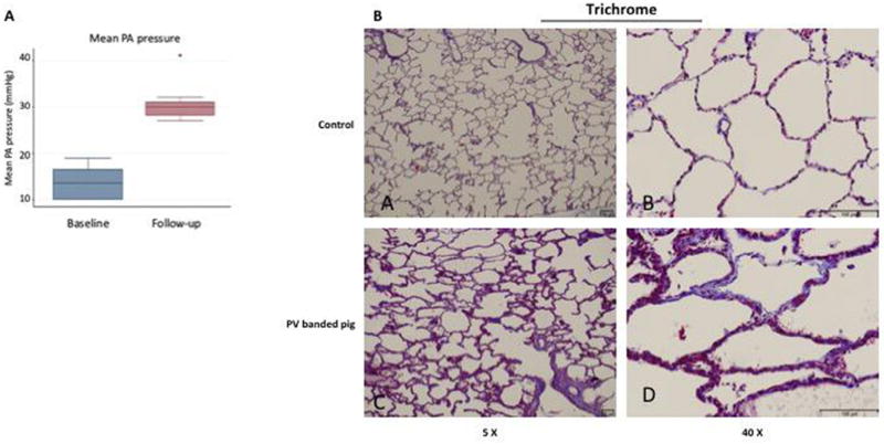
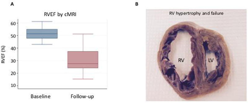
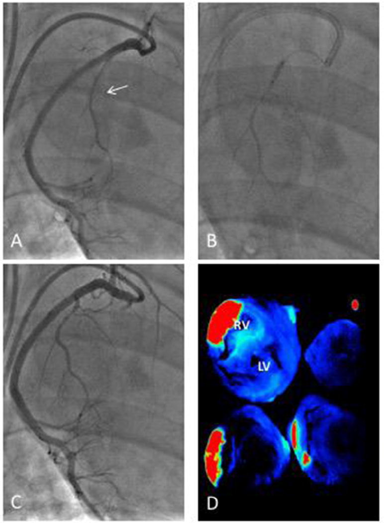
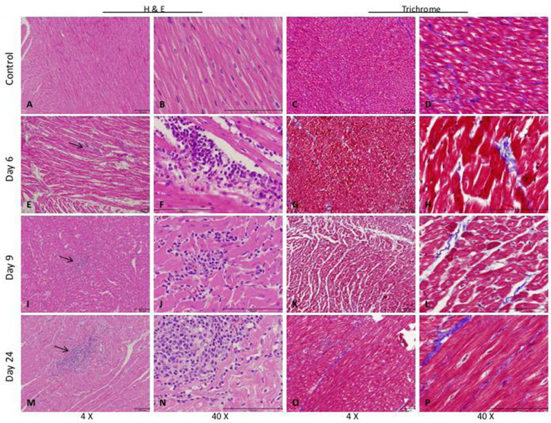
Methods: Commercially available placenta-derived hMSCs were used. Experiments were conducted on 14 female Yorkshire swine. Four animals served as controls, and 10 underwent pulmonary vein (PV) banding. After 12 ± 2 weeks, PH and PV dysfunction were confirmed by right heart catheterization and cardiac magnetic resonance imaging. hMSCs were injected in the marginal branch of the right coronary artery. Tissues were harvested 6, 9 or 24 days after infusion.

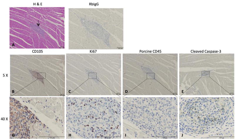
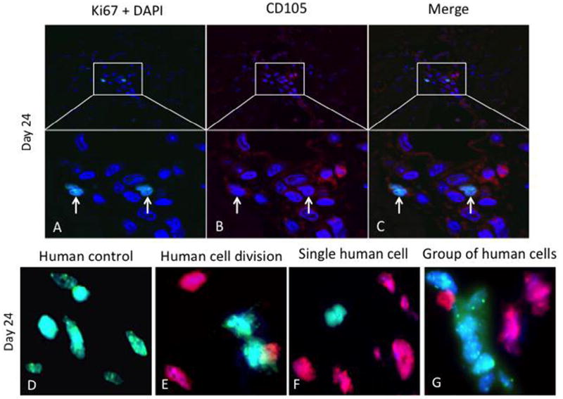
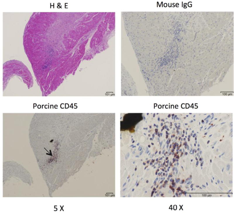
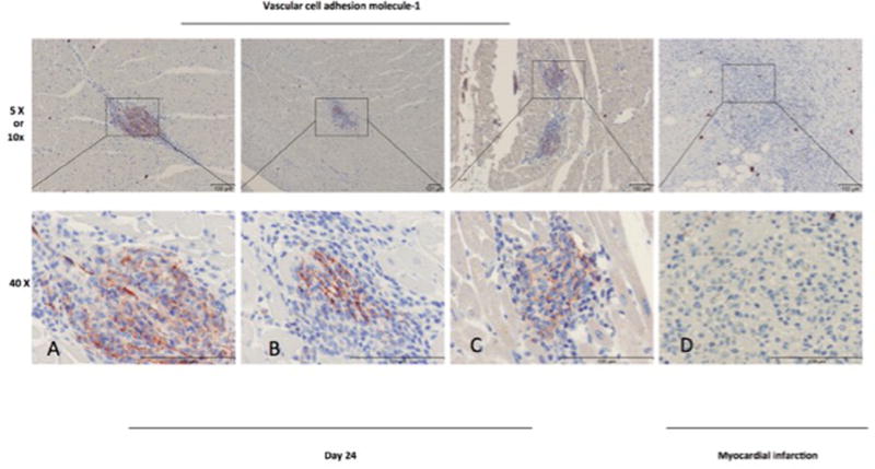
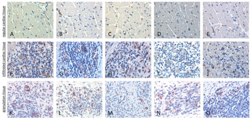
Results: After 12 ± 2 weeks after PV banding, all subjects had increased mean pulmonary artery pressure (13.6 ± 3.6 versus 30.8 ± 4.5 mm Hg, P < 0.007) and a decrease in right ventricular ejection fraction from 51.7 ± 5.7% versus 30.5 ± 11.3% (P = 0.003). Intracoronary injection of hMSCs was well tolerated. Up to 24 days after hMSC injection, immunohistochemistry revealed extravascular viable human CD105+ mononuclear cells in the right ventricle (RV) that were Ki67+. This was confirmed by fluorescence in situ hybridization. CD45+ porcine inflammatory cells were identified, commonly seen adjacent to areas of healing microscopic infarction that likely dated to the time of the original hMSC injection. Anti-CD31 staining produced strong signals in areas of injected hMSCs. Immunohistochemistry staining for vascular cell adhesion molecule-1 showed upregulation in the clusters, where mononuclear cells were located.

Conclusions: hMSCs injected via intracoronary infusion survived up to 24 days and demonstrated proliferative capacity. hMSCs can persist long term in the RV and are potential cell source for tissue repair in RV dysfunction.

Keywords: differentiation; human mesenchymal stromal cells; intracoronary injection; pulmonary hypertension; right ventricular remodeling.


Figures

Figure 1
Figure 1
PH and vascular remodeling. (A) Mean pulmonary artery (PA) pressures were measured by right heart catheterization at the time of pulmonary vein banding (baseline, n = 10) and 12 ± 2 weeks later just before cell injection (follow-up, n = 8). *P = 0.007. (B) Pulmonary arteriole remodeling and collagen deposition was assessed by Masson’s trichrome staining in control non-diseased pigs (A,B) and PV-banded pigs with PH (C,D).
Figure 2
Figure 2
RV remodeling. (A) RV ejection fraction (RVEF) was evaluated by cMRI at the time of PV banding (baseline, n = 10) and 12 ± 2 weeks later just before cell injection (follow-up, n = 8). *P = 0.003. (B) Cross-section through the heart of a PV-banded pig at week 16 demonstrating RV hypertrophy and dilation consistent with RV failure. RV, right ventricle; LV, left ventricle.
Figure 3
Figure 3
Intracoronary cell injection technique via the RCA marginal branch. (A) Intracoronary cell delivery was performed after initial coronary angiography identified the largest right coronary artery (RCA) marginal branch (arrow). (B) The marginal branch was wired with a 0.014-inch coronary guide wire and a balloon inflated to occlude blood flow for cell delivery. (C) After cell delivery, blood flow was normal in the coronary artery. (D) Selective delivery of cells to the right ventricle (RV) using this technique was confirmed by intracoronary injection of fluorescent microbeads in the marginal and using ex vivo fluorescence reflectance imaging of RV sections. Red and green indicate higher fluorescence while blue is low fluorescence. A representative image is shown. LV, left ventricle.
Figure 4
Figure 4
Cell collections in the right ventricle (RV) after hMSC delivery. (A–D) RV section from a control pig injected with physiological saline as a negative control. RV section from pigs with PH 6 days after hMSC injection (E–H), 9 days hMSC injection (I–L) or 24 days post-hMSC injection (M–P). Arrows indicate areas of cell collections/cell islands. Representative images are shown.
Figure 5
Figure 5
Cell islands in the right ventricle (RV) 24 days after delivery of hMSCs. (A) Hematoxylin and eosin–stained sections of the RV; arrows indicate cell islands 24 days post-hMSC delivery. (B,G) Cell collection of CD105+ mononuclear cells. (C,H) Immunohistochemistry (IHC) with antihuman Ki67 proliferative marker. (D,I) Negative IHC staining for porcine inflammatory cells in area of cell collection using CD45 cell marker. (E,J) IHC using caspase-3 antibody showing positive cytoplasmic localization. Representative images are shown.
Figure 6
Figure 6
hMSCs are retained and proliferate in the right ventricle (RV). Double immunofluorescence staining of RV sections for (A) 4′-6-diamidino-2-phenylindole (DAPI) for nuclear staining (blue) and Ki67 (green), (B) CD105 (red) from PV-banded pigs 24 days after intracoronary injection of hMSCs. (C) Merged sections show co-localization of CD105 and Ki67, indicating proliferation of hMSCs. Arrows show positive immunostaining. Representative images are shown. FISH was performed on RV sections to confirm the presence of human cells. (E) Human control hybridized with combined green (human) and red (pig) species-specific Cot-1 FISH probe. (F) Two adjacent human (daughter) cells in the final stages of cell division (telophase/cytokinesis). (G) An individual human cell (green) and (H) a group of human cells (green) are seen in the RV. Representative images are shown.
Figure 7
Figure 7
Inflammatory cells in the right ventricle (RV). Porcine CD45+ inflammatory cells (arrow) were identified adjacent to areas of microscopic infarction by immunohistochemistry of the RV. Representative images are shown. H&E, hematoxylin and eosin.
Figure 8
Figure 8
VCAM-1: immunohistochemistry staining of paraffin-embedded porcine right ventricle tissue 24 days post-hMSC injection (A–C) and infarcted porcine right ventricle tissue (D).
Figure 9
Figure 9
CD31: comparison of vessel cross-sections from porcine right ventricle in a PH-diseased pig 9 days after hMSC injection. (A– E) Cardiac tissue without hMSC infiltration; (F–J) cardiac tissue with hMSC infiltration; (K–O) infarction/granulation tissue.

Similar articles

Cited by

References

    1. Zhu Y, Song X, Wang J, Li Y, Yang Y, Yang T, et al. Placental mesenchymal stem cells of fetal origin deposit epigenetic alterations during long-term culture under serum-free condition. Expert Opin Biol Ther. 2015;15(2):163–80. - PubMed
    1. Zhu Y, Yang Y, Zhang Y, Hao G, Liu T, Wang L, et al. Placental mesenchymal stem cells of fetal and maternal origins demonstrate different therapeutic potentials. Stem Cell Res Ther. 2014;5(2):48. - PMC - PubMed
    1. Clifford DM, Fisher SA, Brunskill SJ, Doree C, Mathur A, Watt S, et al. Stem cell treatment for acute myocardial infarction. Cochrane Database Syst Rev. 2012;(2):CD006536. - PubMed
    1. Martin-Rendon E, Snowden JA, Watt SM. Stem cell-related therapies for vascular diseases. Transfus Med. 2009;19(4):159–71. - PubMed
    1. Harvey E, Fisher SA, Doree C, Taggart DP, Martin-Rendon E. Current evidence of the efficacy of cell-based therapies in heart failure. Circ J. 2015;79(2):229–36. - PubMed

Substances