. 2017 Aug:64:330-343.
doi: 10.1016/j.bbi.2017.04.003. Epub 2017 Apr 6.

Modulation of experimental arthritis by vagal sensory and central brain stimulation

Affiliations

Modulation of experimental arthritis by vagal sensory and central brain stimulation

Gabriel Shimizu Bassi et al. Brain Behav Immun. 2017 Aug.

Abstract

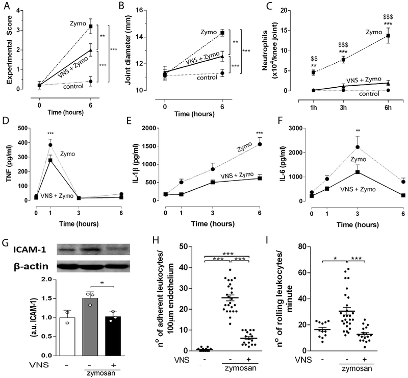
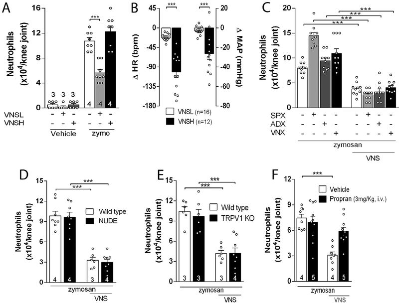
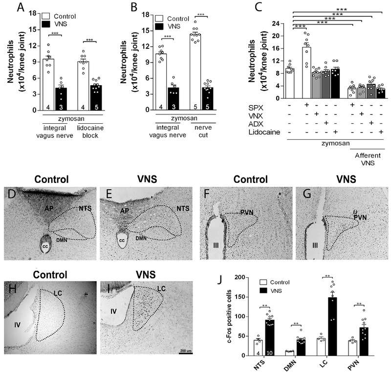
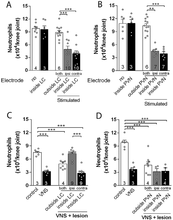
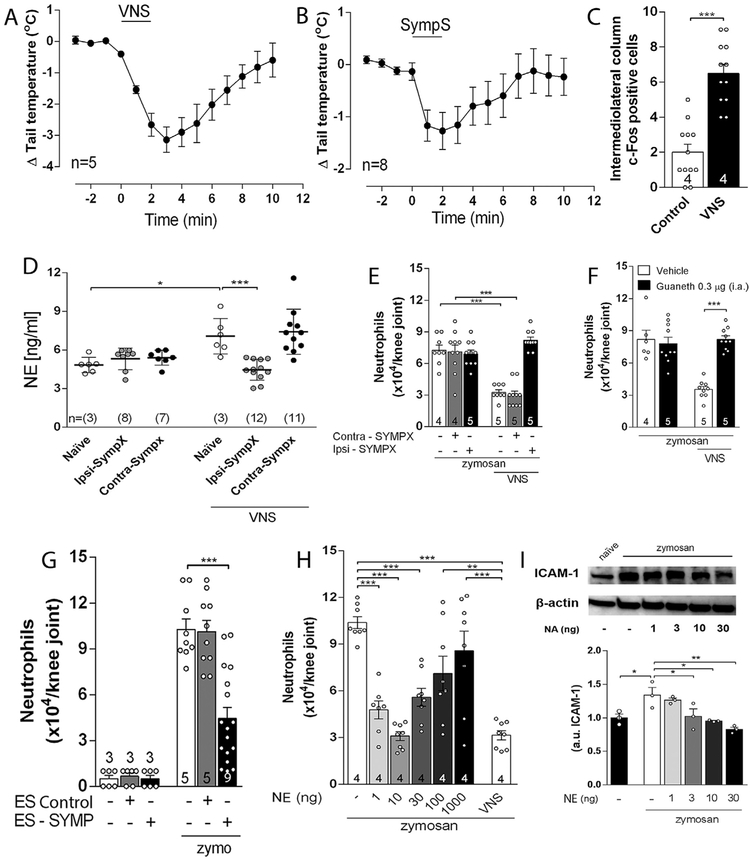
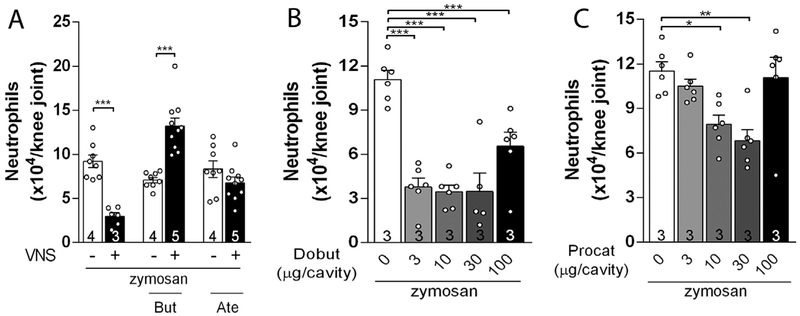
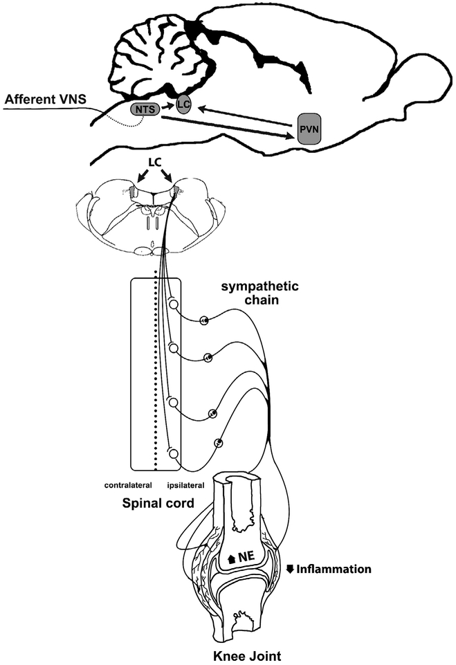
Articular inflammation is a major clinical burden in multiple inflammatory diseases, especially in rheumatoid arthritis. Biological anti-rheumatic drug therapies are expensive and increase the risk of systemic immunosuppression, infections, and malignancies. Here, we report that vagus nerve stimulation controls arthritic joint inflammation by inducing local regulation of innate immune response. Most of the previous studies of neuromodulation focused on vagal regulation of inflammation via the efferent peripheral pathway toward the viscera. Here, we report that vagal stimulation modulates arthritic joint inflammation through a novel "afferent" pathway mediated by the locus coeruleus (LC) of the central nervous system. Afferent vagal stimulation activates two sympatho-excitatory brain areas: the paraventricular hypothalamic nucleus (PVN) and the LC. The integrity of the LC, but not that of the PVN, is critical for vagal control of arthritic joint inflammation. Afferent vagal stimulation suppresses articular inflammation in the ipsilateral, but not in the contralateral knee to the hemispheric LC lesion. Central stimulation is followed by subsequent activation of joint sympathetic nerve terminals inducing articular norepinephrine release. Selective adrenergic beta-blockers prevent the effects of articular norepinephrine and thereby abrogate vagal control of arthritic joint inflammation. These results reveals a novel neuro-immune brain map with afferent vagal signals controlling side-specific articular inflammation through specific inflammatory-processing brain centers and joint sympathetic innervations.

Keywords: Arthritis; Neuroimmune interactions; Neuroimmunomodulation; Neutrophil migration; Sympathetic nervous system; Vagus nerve.


Conflict of interest statement

Disclosures

The authors have no financial conflicts of interest.

Figures

Fig. 1.
Fig. 1.
VNS controls local inflammation via the sympathetic activity modulation. VNS improved knee (A) experimental score, (B)articular edema, (C) neutrophil count (n = 5 for each group), and synovial levels of (D) TNF, (E) IL-1 β, and (F) IL-6 at 0, 1, 3 and 6 h (0 h, n = 2; 1 h, n = 4; 3 h, n = 3; 6 h, n = 4) after intra-articular zymosan. Data were analyzed using two-way ANOVA followed by a Bonferroni’s multiple comparison test comparing individual time-point. (G) Synovial ICAM-1 expression observed 6 h after vagal stimulation. Number of adherent (H) and rolling (I) leukocytes observed 1 h after VNS in the synovial endothelium in zymosan-injected knee-joints. Data were analyzed using one-way ANOVA followed by Tukey’s multiple comparison test. **p < 0.01; ***p < 0.001 versus control, $$p < 0.01; $$$p < 0.001 versus VNS + Zymo.
Fig. 2.
Fig. 2.
Low intensity VNS reduces articular inflammation by a new neuroimmune pathway. (A) Effects of VNS of high (VNSH: 5 Hz, 0.1 ms, 3 V) or low (VNSL: 5 Hz, 0.1 ms, 1 V) intensity on neutrophil migration and on (B) heart rate (HR) and mean arterial pressure (MAP); The anti-inflammatory effect of VNS is maintained after (C) splenectomy (SPX), adrenalectomy (ADX), or subdiaphragmatic vagotomy (sVNX) (n = 5 for each group), and in (D) NUDE or (E) TRPV1 KO mice, but not in (F) propranolol (Propan) treated-animals. Data were analyzed using one-way ANOVA followed by Tukey’s multiple comparison test; The number of animals used for each group is displayed at the bottom of the corresponding bar. ***p < 0.001 versus control.
Fig. 3.
Fig. 3.
Afferent VNS reduces articular inflammation and activates brain structures. Afferent VNS via (A) intra-nerve administration of lidocaine or (B) via surgical cut reduces knee inflammation. Afferent vagal stimulation reduces peripheral inflammation in (C) splenectomized (SPX), subdiaphragmatic vagotomized (sVNX) or adrenalectomized (ADX) rats (n = 4 for each group). Data were analyzed using one-way ANOVA followed by Tukey’s multiple comparison test; VNS increases the expression of c-Fos in (D-E) the nucleus of the tractus solitarius (NTS), dorsal motor nucleus (DMN), (F-G) hypothalamic paraventricular nucleus (PVN), and (H-I) locus coeruleus (LC) as compared to the control (non-stimulated) group (D-J). Unpaired Student t-test was applied to this comparison. The number of animals used for each group is displayed at the bottom of the corresponding bar. **p < 0.01; ***p < 0.001.
Fig. 4.
Fig. 4.
Central structures activation is essential to the vagal anti-inflammatory effect. Electrical stimulation of the (A) LC or the (B) PVN decreases neutrophil migration into both knee joints. Temporary deactivation by prior injection of CoCL2 into the (C) LC, but not (D) PVN, abrogated the vagal anti-inflammatory effect only in the ipsilateral, but not in the contralateral, joint to the hemispheric LC deactivation. Both: both knees; Ipsi: ipsilateral knee to the injected brain hemisphere; Contra: contralateral knee to the injected brain hemisphere. Data were analyzed using one-way ANOVA followed by Tukey’s multiple comparison test. The number of animals used for each group is displayed at the bottom of the corresponding bar. **p < 0.01; ***p < 0.001.
Fig. 5.
Fig. 5.
Vagal anti-inflammatory signaling depends on local sympathetic system to evoke its anti-inflammatory effect. (A) Intact vagus nerve stimulation (VNS) or (B) sympathetic chain stimulation (SympS) decreases rat tail temperature; (C) VNS increases c-Fos expression in both sides of the intermediolateral column of the spinal cord; (D) Measurement of norepinephrine (NE) 5 min after VNS in the ipsilateral (Ipsi–SYMPX) and contralateral (Contra – SYMPX) joint to surgical sympathectomy (ELISA); (E) VNS anti-inflammatory effect is blocked in the ipsilateral (Ipsi–SYMPX), but not in the contralateral (Contra – SYMPX), joint by surgical sympathectomy; (F) VNS effect is blocked by prior intra-articular administration of guanethidine (Guaneth), the contralateral joint received vehicle solution; (G) Stimulation of the sympathetic chain (ES – SYMP), but not sham stimulation (ES Control), decreases neutrophil migration to the inflamed joints; (H-I) Intra-articular administration of NA decreases synovial ICAM-1 expression and neutrophil migration to the inflamed joint. Data were analyzed using one-way ANOVA followed by Tukey’s multiple comparison test. The number of animals used for each group is displayed at the bottom of the corresponding bar. *p < 0.05; **p < 0.01; ***p < 0.001.
Fig. 6.
Fig. 6.
The activation of joint β-adrenergic receptors is mandatory for the central vagal anti-inflammatory signaling. Intra-articular administration of (A) β2 (Butoxamine – But) or β1 (Atenolol – Ate) antagonists abrogate the anti-inflammatory effects of VNS; Intra-articular injection of (B) β1 (Dobutamine – Dobut) or (C) β2 (Procaterol – Procat) agonists decrease knee inflammation. Data were analyzed using one-way ANOVA followed by Tukey’s multiple comparison test. The number of animals used for each group is displayed at the bottom of the corresponding bar. *p < 0.05; **p < 0.01; ***p < 0.001.
Fig. 7.
Fig. 7.
Comprehensive illustration of the central inflammatory processing centers (LC and PVN) activated through afferent vagal stimulation. Afferent VNS (green) controls arthritic joint inflammation by activating specific sympatho-excitatory brain areas (blue) and sympathetic nerve fibers to increase synovial norepinephrine (NE) levels to reduce articular inflammation. (For interpretation of the references to colour in this figure legend, the reader is referred to the web version of this article.)

References

    1. Ardell JL, Rajendran PS, Nier HA, KenKnight BH, Armour JA, 2015. Central-peripheral neural network interactions evoked by vagus nerve stimulation: functional consequences on control of cardiac function. Am. J. Physiol. Heart Circ. Physiol 309, 1740–1752. - PMC - PubMed
    1. Armour JA, Randall WC, 1975. Functional anatomy of canine cardiac nerves. Acta Anat. (Basel) 91, 510–528. - PubMed
    1. Bassi GS, Kanashiro A, Santin FM, de Souza GE, Nobre MJ, Coimbra NC, 2012. Lipopolysaccharide-induced sickness behaviour evaluated in different models of anxiety and innate fear in rats. Basic Clin. Pharmacol. Toxicol 110, 359–369. 10.1111/j.1742-7843.2011.00824.x. - DOI - PubMed
    1. Bassi GS, Brognara F, Castania JA, Talbot J, Cunha TM, Cunha FQ, Ulloa L, Kanashiro A, Dias DP, Salgado HC, 2015. Baroreflex activation in conscious rats modulates the joint inflammatory response via sympathetic function. Brain Behav. Immun 49, 140–147. 10.1016/j.bbi.2015.05.002. - DOI - PMC - PubMed
    1. Bellinger DL, Nance DM, Lorton D, 2013. Innervation of the Immune System In: The Wiley-Blackwell Handbook of Psychoneuroimmunology. Wiley-Blackwell, pp. 24–72.

MeSH terms