Review
. 2017 Jun;18(6):345-361.
doi: 10.1038/nrg.2017.15. Epub 2017 Apr 10.

Scaling by shrinking: empowering single-cell 'omics' with microfluidic devices

Affiliations
Review

Scaling by shrinking: empowering single-cell 'omics' with microfluidic devices

Sanjay M Prakadan et al. Nat Rev Genet. 2017 Jun.

Abstract

Recent advances in cellular profiling have demonstrated substantial heterogeneity in the behaviour of cells once deemed 'identical', challenging fundamental notions of cell 'type' and 'state'. Not surprisingly, these findings have elicited substantial interest in deeply characterizing the diversity, interrelationships and plasticity among cellular phenotypes. To explore these questions, experimental platforms are needed that can extensively and controllably profile many individual cells. Here, microfluidic structures - whether valve-, droplet- or nanowell-based - have an important role because they can facilitate easy capture and processing of single cells and their components, reducing labour and costs relative to conventional plate-based methods while also improving consistency. In this article, we review the current state-of-the-art methodologies with respect to microfluidics for mammalian single-cell 'omics' and discuss challenges and future opportunities.


Conflict of interest statement

Competing interests statement

The authors declare competing interests: see Web version for details.

Figures

Figure 1
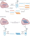
Figure 1. Technical and biological noise in single-cell measurements
a | Technical errors in cellular processing (‘technical noise’), such as failure to reverse transcribe an mRNA transcript or over-amplification during the ensuing PCR, can dramatically affect the utility of the measured value of any single gene in a single-cell experiment. b | Similarly, the physical, spatial and temporal processes governing biological phenomena (‘intrinsic noise’), such as the ‘burstiness’ of mRNA transcription, can limit the information content in any single instantaneous end-point measurement.
Figure 2
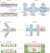
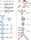
Figure 2. Overview of the major microfluidic device types: valves, droplets and nanowells
Basic microfluidic structures for processing cells (left) and example implementations (right). a | Valve-based microfluidics operate by aligning two perpendicular channels, one for cell (or cellular component) and solution flow and another for control, separated by a thin membrane. To isolate a chamber, pressure is applied to the control channel, deflecting a membrane into the flow channel to block it, and hence trap its contents. These channels can be arrayed, multiplexed and coupled to external computer-controlled pressure regulators to reliably execute complex workflows such as whole-transcriptome amplification (see, for example, REF. ). b | In many implementations, droplet-based strategies rely on flowing single cells (or cellular components) in aqueous medium into a co-flowed oil phase. Due to pressure-driven effects, small aqueous droplets that can contain cells (or their components) are formed through encapsulation with oil, isolating each individual cell or cellular component from its peers. During or after this initial encapsulation, additional reagents, such as barcoded oligo(dT) mRNA capture beads (see, for example, REF. ), can be incorporated or removed to enable more complex operations. c | Nanowells confine cells by gravity, and can subsequently be sealed with a membrane or glass slide to isolate single cells and their components. These cells or components can then be picked out of the wells for further processing (see, for example, REF. ) or characterized in-well using, for example, a functionalized seal. PDMS, polydimethylsiloxane; RT, reverse transcription.
Figure 3
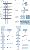
Figure 3. Selected examples of microfluidic devices used to measure single-cell genomes and epigenomes
a | A diagram (top view) of the valve-based microfluidic device used by Wang et al. for amplification of genomic material from single cells. In the implementation, individual cells were captured and lysed, genomic DNA was then stabilized and amplified and, finally, cellular products were collected independently to ensure single-cell resolution of information. b | Emulsion multiple-displacement amplification (eMDA), as performed by Fu et al. (top view). Here, genomic material from single-cell lysates was encapsulated in droplets, within which MDA was performed, enabling more uniform coverage of the chromosomal profile (relative to MDA performed en masse) of the individual cell after the droplets have been broken. c | A modified diagram representing the valve-based microfluidic device implemented by Cheow et al. to profile the DNA methylome and transcriptome (only the methylome is shown here) of single cells (top view). d | Single-cell assay for transposase-accessible chromatin using sequencing (ATAC-seq) by Buenrostro et al., which uses a Tn5 transposase to identify regions of open chromatin and was performed using a commercial C1 valve-based integrated fluidic circuit from Fluidigm (top view). Part a is adapted with permission from REF. , Elsevier. Part c is adapted with permission from REF. , Macmillan Publishers. Part d is adapted with permission from REF. , Macmillan Publishers.
Figure 4
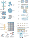
Figure 4. Selected examples of microfluidic devices used to measure single-cell transcriptomes and proteomes
a | InDrop, developed by Klein et al., is a droplet-based single-cell transcriptomics method that works by co-confining single cells with hydrogel beads containing uniquely barcoded primers (top view). On UV-light-mediated release of those primers, reverse transcription (RT) and barcoding is performed in droplet, and the resulting complementary DNA (cDNA) is collected after the droplets have been broken for subsequent processing. b | Seq-Well, a massively parallel single-cell RNA sequencing (scRNA-Seq) method from Gierahn et al., combines early bead-based barcoding with nanowells to generate thousands of single-cell libraries (the top row shows a top view; below this are side views). Single cells are gravity-loaded onto an array that has been preloaded with uniquely barcoded oligo(dT) capture beads, sealed with a membrane (which permits buffer exchange but not mRNA escape) and lysed; mRNAs are then captured by oligo(dT) primers bound to the surface of the barcoded beads, and the beads are removed for off-chip RT, amplification, library preparation and sequencing. c | The single-cell barcode chip (SCBC) developed by Ma et al. uses a valve-based strategy to isolate single cells and interrogate their secreted factors (top view). In each enclosed chamber, individual cells are exposed to antibodies that are specific for various extracellular protein targets. Following incubation of cells in the chip, streptavidin-bound fluorophores and biotinylated secondary antibodies are flowed into the channels, enabling a sandwich enzyme-linked immunosorbent assay (ELISA)-based fluorescent readout of protein abundance. d | Nanowells were coupled with capture-antibody-coated glass coverslips by Love et al. to measure the abundance of several secreted factors from single cells, such as active hybridomas in a reverse ELISA format (side view). PDMS, polydimethylsiloxane; T7 RNAP, T7 RNA polymerase; UMI, unique molecular identifier. Part a is adapted with permission from REF. , Elsevier. Part b is adapted with permission from REF. , Macmillan Publishers. Part c is adapted with permission from REF. , Macmillan Publishers. Part d is adapted with permission from REF. , Macmillan Publishers.
Figure 5
Figure 5. Future directions and extensions for microfluidic devices
a | Multiple cells can be deposited onto well-based profiling platforms, enabling the examination of multicellular and cooperative events, such as the killing of target cells by natural killer cells by Yamanaka et al. (side view). b | The Fluidigm C1 valve-based integrated fluidic circuit (IFC; top view) was used to simultaneously detect several transcripts and proteins from the same single cell in a single series of reactions. During lysis, targeted primers and complementary DNA-tagged antibodies were introduced, and reverse transcription and DNA extension reactions were carried out on both species to yield uniquely detectable, PCR-amplifiable DNAs for both cellular analytes. c | The directed evolution of enzymes in single cells can be probed with an activity assay in droplets. Here, horseradish peroxidase (HRP) surface proteins from mutated vectors were interrogated for substrate turnover with single-cell resolution (top view). Following capture of the most active cells, the interrogation was repeated to study their evolution over time. Part a is adapted from REF. with permission of The Royal Society of Chemistry. Part b is adapted with permission from REF. , Macmillan Publishers Limited. Part c is adapted with permission from REF. (Agresti, J. J. et al. Ultrahigh-throughput screening in drop-based microfluidics for directed evolution. Proc. Natl Acad. Sci. USA 107, 4004–4009; 2010).

Similar articles

Cited by

References

    1. Tirosh I, et al. Dissecting the multicellular ecosystem of metastatic melanoma by single-cell RNA-seq. Science. 2016;352:189–196. - PMC - PubMed
    1. Patel AP, et al. Single-cell RNA-seq highlights intratumoral heterogeneity in primary glioblastoma. Science. 2014;344:1396–1401. - PMC - PubMed
    1. Shalek AK, et al. Single-cell RNA-seq reveals dynamic paracrine control of cellular variation. Nature. 2014;510:363–369. - PMC - PubMed
    1. Shalek AK, et al. Single-cell transcriptomics reveals bimodality in expression and splicing in immune cells. Nature. 2013;498:236–240. A demonstration of how covariation in gene expression in scRNA-Seq data can be used to identify cellular circuits and molecular drivers of behaviour in seemingly identical immune cells. It also nicely demonstrates the use of microfluidic devices for targeted single-cell mRNA measurements (STA). - PMC - PubMed
    1. Lohr JG, et al. Whole-exome sequencing of circulating tumor cells provides a window into metastatic prostate cancer. Nat Biotechnol. 2014;32:479–484. - PMC - PubMed

Publication types