

Rosacea Subtypes Visually and Optically Distinct When Viewed with Parallel-Polarized Imaging Technique
- PMID: 28392643
- PMCID: PMC5383741
- DOI: 10.5021/ad.2017.29.2.167
Rosacea Subtypes Visually and Optically Distinct When Viewed with Parallel-Polarized Imaging Technique
Abstract
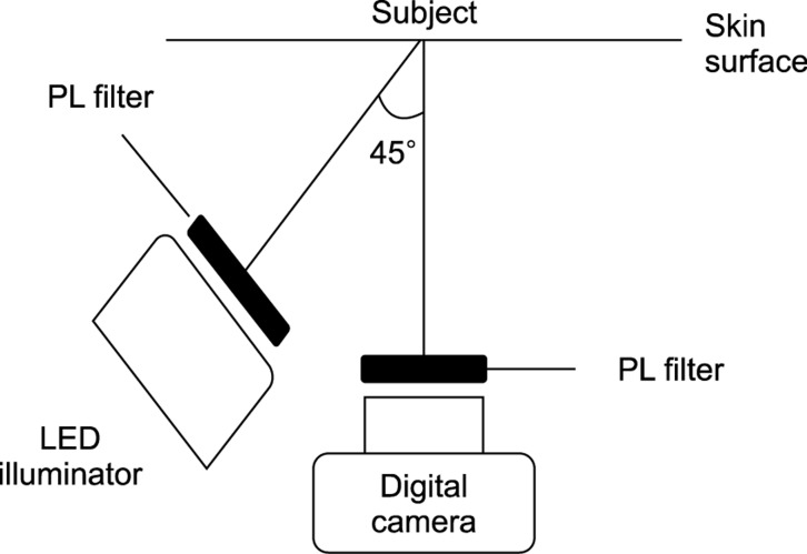
Background: Parallel-polarized light (PPL) photography evaluates skin characteristics by analyzing light reflections from the skin surface.
Objective: The aim of this study was to determine the significance of quantitative analysis of PPL images in rosacea patients, and to provide a new objective evaluation method for use in clinical research and practice.
Methods: A total of 49 rosacea patients were enrolled. PPL images using green and white light emitting diodes (LEDs) were taken of the lesion and an adjacent normal area. The values from the PPL images were converted to CIELAB coordinates: L* corresponding to the brightness, a* to the red and green intensities, and b* to the yellow and blue intensities.
Results: A standard grading system showed negative correlations with L* (r=-0.67862, p=0.0108) and b* (r=-0.67862, p=0.0108), and a positive correlation with a* (r=0.64194, p=0.0180) with the green LEDs for papulopustular rosacea (PPR) types. The xerosis severity scale showed a positive correlation with L* (r=0.36709, p=0.0276) and a negative correlation with b* (r=-0.33068, p=0.0489) with the white LEDs for erythematotelangiectatic rosacea (ETR) types. In the ETR types, there was brighter lesional and normal skin with white LEDs and a higher score on the xerosis severity scale than the PPR types.
Conclusion: This technique using PPL images is applicable to the quantitative and objective assessment of rosacea in clinical settings. In addition, the two main subtypes of ETR and PPR are distinct entities visually and optically.
Keywords: Optics and photonics; Rosacea.
Conflict of interest statement
CONFLICTS OF INTEREST: The authors have nothing to disclose.
Figures
References
-
- Kim DH, Choi JE, Ryu HJ, Seo SH, Kye YC, Ahn HH. Analytic parallel-polarized light imaging technique using various light-emitting diodes: a comparison with skin conductance values. Skin Res Technol. 2015;21:158–163. - PubMed
-
- Latreille J, Gardinier S, Ambroisine L, Mauger E, Tenenhaus M, Guéhenneux S, et al. Influence of skin colour on the detection of cutaneous erythema and tanning phenomena using reflectance spectrophotometry. Skin Res Technol. 2007;13:236–241. - PubMed
-
- Rogers RS, 3rd, Callen J, Wehr R, Krochmal L. Comparative efficacy of 12% ammonium lactate lotion and 5% lactic acid lotion in the treatment of moderate to severe xerosis. J Am Acad Dermatol. 1989;21:714–716. - PubMed
-
- Wilkin J, Dahl M, Detmar M, Drake L, Liang MH, Odom R, et al. Standard grading system for rosacea: report of the national rosacea society expert committee on the classification and staging of rosacea. J Am Acad Dermatol. 2004;50:907–912. - PubMed
LinkOut - more resources
Full Text Sources
Other Literature Sources
