. 2017 Mar 24:8:488.
doi: 10.3389/fmicb.2017.00488. eCollection 2017.

Anti-Biofilm Activity of a Self-Aggregating Peptide against Streptococcus mutans

Affiliations

Anti-Biofilm Activity of a Self-Aggregating Peptide against Streptococcus mutans

Juliana M Ansari et al. Front Microbiol. .

Abstract

Streptococcus mutans is the primary agent of dental cavities, in large part due to its ability to adhere to teeth and create a molecular scaffold of glucan polysaccharides on the tooth surface. Disrupting the architecture of S. mutans biofilms could help undermine the establishment of biofilm communities that cause cavities and tooth decay. Here we present a synthetic peptide P1, derived from a tick antifreeze protein, which significantly reduces S. mutans biofilm formation. Incubating cells with this peptide decreased biofilm biomass by approximately 75% in both a crystal violet microplate assay and an in vitro tooth model using saliva-coated hydroxyapatite discs. Bacteria treated with peptide P1 formed irregular biofilms with disconnected aggregates of cells and exopolymeric matrix that readily detached from surfaces. Peptide P1 can bind directly to S. mutans cells but does not possess bactericidal activity. Anti-biofilm activity was correlated with peptide aggregation and β-sheet formation in solution, and alternative synthetic peptides of different lengths or charge distribution did not inhibit biofilms. This anti-biofilm peptide interferes with S. mutans biofilm formation and architecture, and may have future applications in preventing bacterial buildup on teeth.

Keywords: Gram-positive cocci; Streptococcus mutans; aggregation; anti-biofilm; biofilm; synthetic peptide.


Figures

FIGURE 1
FIGURE 1
Anti-biofilm effect of peptide P1 in microplate assay and on sHA discs. (A) Peptide P1 reduces biofilm formation by Streptococcus mutans and oral streptococci in microplate assay. Cultures were incubated with peptide P1 and control peptide sP1 at 0.1 mg/ml final concentration. Biofilm biomass in microplate wells was stained with crystal violet (images below graph). Results are combined from three replicate experiments, and error bars indicate standard error of the mean. **p < 0.01, ****p < 0.0001, Student’s t-test. (B) Determination of minimum biofilm inhibitory concentration of peptide P1 against S. mutans. Biofilm inhibitory activity of P1 was measured by testing decreasing peptide concentrations in a microplate assay. Results were combined from three experiments, with biofilm biomass normalized to a percentage of the control. (C) Viability of S. mutans in the presence of peptide P1. Total S. mutans biomass (combined planktonic and biofilm cells from well) of cultures grown overnight in Brain Heart Infusion + 1% sucrose (BHI-S) without peptide or with 0.1 mg/ml P1 or sP1 was collected, diluted and plated, and cfu/ml was quantified for each condition. (D) Peptide P1 reduces Streptococcus mutans biofilm formation on saliva-coated hydroxyapatite (sHA) discs. Biofilm cfu/sHA disc showing combined results from three replicate experiments (total n = 9 discs/treatment). Peptide P1 and control peptide sP1 were used at 0.1 mg/ml final concentration. Error bars show standard error of the mean. *p < 0.05, using Student’s t-test. (E) Custom-made wire holder holding the hydroxyapatite disc vertically in the well of a 96-well plate.
FIGURE 2
FIGURE 2
Immunoblot assay of S. mutans binding to His-P1 peptide or His-sP1 control peptide. (A) Peptides were detected using a monoclonal His-tagged antibody (α-His). Cell wall bound fraction (Associated) versus unbound (Supernatant) bacterial fractions were distinguished by using a polyclonal anti-Wheat Germ Agglutinin antibody (α-WGA). (B) Quantification of bound antiserum of His-tagged peptide in the cell-associated fraction. The Relative Signal Intensity is the signal of “associated” fraction as a percentage of total signal detected for each peptide [associated/(supernatant + associated)]*(100). Lower dotted line indicates the limit of detection by the LICOR Odyssey system. *p < 0.05, using Student’s t-test.
FIGURE 3
FIGURE 3
Peptide P1 activity against biofilms of Gram-negative bacteria. Biofilm biomass in microplate wells was stained with crystal violet (images below graph). Results are combined from three replicate experiments with cultures grown in TSB + 1% glucose, using peptides P1 and sP1 at 0.1 mg/ml final concentration. Error bars indicate standard error of the mean.
FIGURE 4
FIGURE 4
Clumping and detachment of exopolymeric matrix in the presence of P1. (A) S. mutans cultures in well of 12-well plate after overnight incubation with peptide P1 or sP1 at 0.1 mg/ml. Detached chunks of floating matrix material are visible in P1-treated sample (no magnification). (B) Cells and exopolymeric matrix (same samples from A) viewed through 10× objective with phase contrast microscopy. Bar = 100 μm. Cells and matrix appear dark, clear areas show lack of biofilm attached to well. (C) Matrix clumping in wells of 24-well plate. (D) Biofilm eDNA (same samples from C) stained with fluorescent Miami Orange probe and viewed under fluorescent light with 20× objective. Bar = 100 μm.
FIGURE 5
FIGURE 5
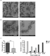
Scanning electron microscopy of S. mutans biofilm. (A) Reduced adherent material in biofilms formed in the presence of 0.1 mg/ml peptide P1; final magnification 500×, bar = 100 μm. (B) Biofilms in P1-treated sample display shorter streptococcal chain lengths; final magnification 3084×, bar = 10 μm. (C) Average streptococcal chain length under each treatment, measured with ImageJ software. ****p < 0.0001, Student’s t-test. (D) Distribution of streptococcal chain lengths under each treatment.
FIGURE 6
FIGURE 6
Effect of alternative peptide sequences on biofilm formation. (A) Biofilm biomass of S. mutans and S. aureus, with results combined from three replicate experiments. All peptides were used at 0.1 mg/ml final concentration. Error bars indicate standard error of the mean. **p < 0.01, ***p < 0.001, ****p < 0.0001. (B) Amino acid sequences of synthetic peptides with color-coded amino acid properties.
FIGURE 7
FIGURE 7
Aggregation of peptides in solution. Images were taken with phase contrast microscopy through the bottom of a 24-well tissue culture plate. All peptides were added at 0.1 mg/ml final concentration in BHI+1% sucrose (BHI-S). (A) Peptides in solution after 6 h incubation; bar = 50 μm. (B) Peptides in solution after 24 h incubation; bar = 100 μm. (C) Peptides in solution with S. mutans biofilm after 6 h incubation; bar = 50 μm. (D) Peptides in solution with S. mutans biofilm after 24 h incubation; bar = 100 μm. Arrow = visible aggregated peptide.
FIGURE 8
FIGURE 8
Secondary structure of peptides in solution using FTIR. Fourier transform infrared spectroscopy (FTIR) spectra of peptides P1, P17, and negative control sP1 at 1 mg/ml, with peaks at 1621 cm-1 indicative of β-sheet formation.

References

    1. Abraham N. M., Liu L., Jutras B. L., Yadav A. K., Narasimhan S., Gopalakrishnan V., et al. (2017). Pathogen-mediated manipulation of arthropod microbiota to promote infection. Proc. Natl. Acad. Sci. U.S.A. 114 E781–E790. 10.1073/pnas.1613422114 - DOI - PMC - PubMed
    1. Basak S., Rajurkar M., Attal R., Mallick S. (2013). “Biofilms: a challenge to medical fraternity in infection control,” in Infection Control ed. Basak S. (Rijeka: InTech; ), 57–74. 10.5772/56704 - DOI
    1. Besingi R. N., Wenderska I. B., Senadheera D. B., Cvitkovitch D. G., Long J. R., Wen Z. T., et al. (2017). Functional Amyloids in Streptococcus mutans, their use as targets of biofilm inhibition and initial characterization of SMU_63c. Microbiology 10.1099/mic.0.000443 [Epub ahead of print]. - DOI - PMC - PubMed
    1. Bjarnsholt T., Ciofu O., Molin S., Givskov M., Høiby N. (2013). Applying insights from biofilm biology to drug development–can a new approach be developed? Nat. Rev. Drug Discov. 12 791–808. 10.1038/nrd4000 - DOI - PubMed
    1. Blanco L. P., Evans M. L., Smith D. R., Badtke M. P., Chapman M. R. (2012). Diversity, biogenesis and function of microbial amyloids. Trends Microbiol. 20 66–73. 10.1016/j.tim.2011.11.005 - DOI - PMC - PubMed

LinkOut - more resources