. 2017 May;50(5):1555-1566.
doi: 10.3892/ijo.2017.3952. Epub 2017 Apr 5.

Overexpression of Srcin1 contributes to the growth and metastasis of colorectal cancer

Affiliations

Overexpression of Srcin1 contributes to the growth and metastasis of colorectal cancer

Mengnan Zhang et al. Int J Oncol. 2017 May.

Abstract

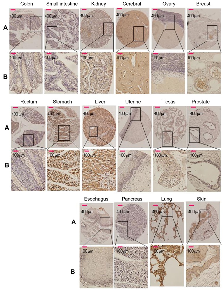
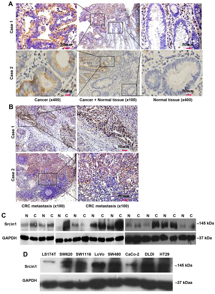
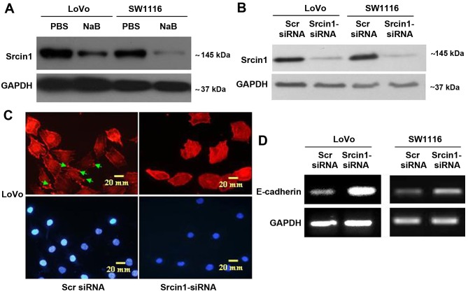
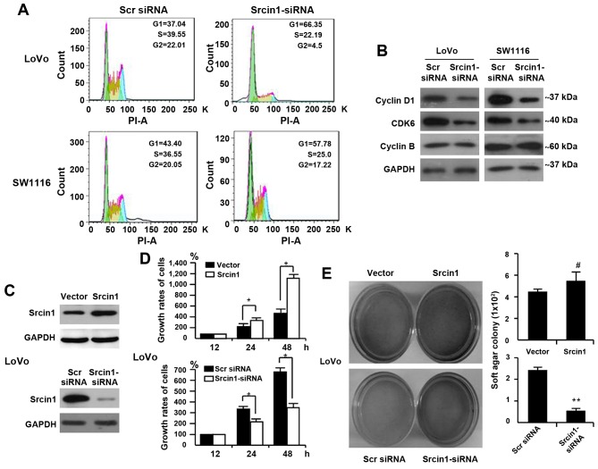
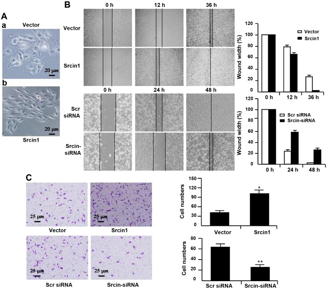
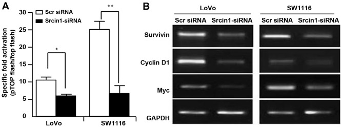
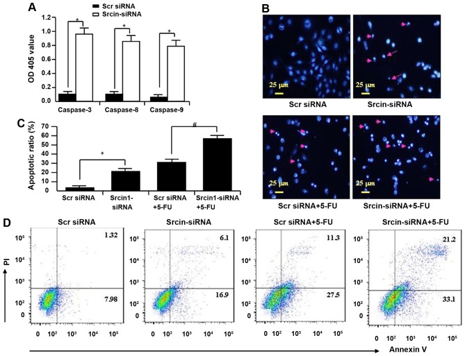
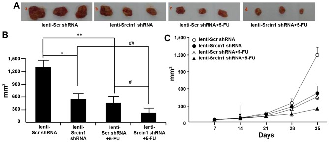
The adaptor protein Srcin1 is a novel Src-binding protein that regulates Src activation through C-terminal Src kinase (Csk). Srcin1 behaves as a tumour suppressor in breast cancer, but the role of Srcin1 in the development of colorectal cancer (CRC) remains unknown. In the present study, Srcin1 expression in normal tissue was examined by tissue microarray and assessed by immunohistochemistry in 10 patients. In addition, the biological impact of Srcin1 knockdown on CRC cells was investigated in vitro and in vivo. The results showed that Srcin1 was expressed in different types of normal human tissues, whereas its expression was increased in human CRC tissues. Srcin1 expression also correlated with tumour progression. The suppression of Srcin1 induced cell differentiation and G0/G1 cell cycle arrest. Furthermore, Srcin1 increased cell growth as well as the capacity of migration and invasion in CRC cells. Srcin1 induced the activation of the Wnt/β-catenin signalling pathway. Moreover, Srcin1 suppression sensitized cancer cells to 5-fluorouracil (5-FU)-induced apoptosis in vitro and in vivo. Together, these results demonstrate that Srcin1 contributes to CRC carcinogenesis, invasion and metastasis. These findings provide a rationale for a mechanistic approach to CRC treatment based on the development of Srcin1-targeted therapies.


Figures

Figure 1
Figure 1
Srcin1 expression in 16 normal human tissues was detected by immunohistochemistry. Breast, brain, colon, oesophagus, kidney, liver, lung, ovary, pancreas, prostate, skin, small intestine, stomach, testis, uterine and rectum, of 16 normal human tissues stained with anti-Srcin1 antibody by means of TMA. Original magnification, A, ×100 and B, ×400.
Figure 2
Figure 2
CRC cells express higher levels of Srcin1 than normal cells. (A) Expression of Srcin1 in normal and malignant human colorectal tissues were detected by IHC. (B) Expression of Srcin1 in metastatic lymph nodes were detected by IHC. (C) Srcin1 expression in colon cancer (C) tissues and matched normal colon (N) tissues as detected by western blot analysis. GAPDH was used as the internal control for western blot analysis. (D) Expression of Srcin1 in colorectal cancer cell lines. All of these experiments were repeated 3 times with identical findings.
Figure 3
Figure 3
Suppression with siRNA induces cell differentiation in CRC cells. (A) Srcin1 expression in LoVo and SW1116 cells treated without or with sodium butyrate (NaB) for 48 h, as detected by western blot analysis. (B) LoVo and SW1116 cells were transfected with Srcin1-siRNA and Scr-siRNA (scramble siRNA) for 48 h following by western blot analysis. (C) LoVo cells transfected with Srcin1-siRNA or Src-siRNA were stained with rhodamine-phallotoxin for 48 h, with F-actin filaments being visualized under fluorescent microscopy. (D) Expression of E-cadherin in LoVo and SW1116 cells transfected with Srcin1-siRNA and Src-siRNA for 48 h, as detected by RT-PCR. These pictures are representatives of 3 independent experiments with identical results.
Figure 4
Figure 4
Srcin1 expression modulates cell cycle and cell proliferation in human CRC cells. (A) Cell cycle analysis was examined by FACScan using LoVo and SW1116 cells. RNAi-mediated repression of Srcin1 modulated G0/G1 checkpoint. (B) Expression of cyclins and cyclin-dependent kinase (CDKs) proteins after treatment with Srcin1-shRNA. All these experiments were repeated two times with identical findings. (C) LoVo/vector and LoVo/Srcin1 or Srcin1-siRNA and Src-siRNA were detected by western blot analysis with GAPDH as the internal control. (D) Cells seeded in 96-well tissue culture plates in triplicate were collected 12, 24 and 48 h by WST-1 assay. The growth rates of the cells are expressed as means ± SEM (n=3; *P<0.05). (E) Cells were plated in a tissue culture dish with complete culture medium containing 0.35% agar on top and 0.5% agar at the bottom. After 14 days, cell colonies were visualized after staining with 0.005% crystal violet. Colonies containing >50 cells were considered viable. The results are expressed as means ± SD. #P>0.05 and **P<0.05.
Figure 5
Figure 5
Srcin1 expression modulates cell migration and invasive of LoVo cells. (A) LoVo cells stably expressing Srcin1 show spindle-like, fibroblastic morphology under phase-contrast microscope; (magnification, ×20). (B) Images of the wound closure of pooled monolayer of stable transfectants or knockdown of Srcin1 of LoVo cells; magnification, ×10. (C) Invasive potential of pooled stable transfectants or RNAi-mediated repression of Srcin1 of CRC cells; *P<0.05; **P<0.01. The experiments were repeated three times with identical findings.
Figure 6
Figure 6
Knockdown of Srcin1 decreases β-catenin transcriptional activity. (A) Transcription activities of pTOPFLASH and pFOPFLASH cotransfected with Scr siRNA or Srcin1-siRNA in LoVo and SW1116 cells. The results expressed relative luciferase unit ratios between pTOPFLASH and pFOPFLASH; *P<0.05; **P<0.01. (B) RNA expression of survivin, cyclin D1 and Myc were detected in cells with transfection of Scr siRNA or Srcin1-siRNA for 24 h by RT-PCR.
Figure 7
Figure 7
Suppression of Srcin1-siRNA increased cell susceptibility to apoptotic stimuli. (A) The activity of caspase-3, -8 and -9 in LoVo after Srcin1-siRNA for 72 h. OD405 nm of transfected cells and parental cells was obtained and calculated individually in each group (*P<0.05, comparing to Scr-siRNA). (B) LoVo cells were treated with Scr-siRNA, Srcin1-siRNA, Scr-siRNA + 5-FU or Srcin1-siRNA + 5-FU. Nuclei were stained with Hoechst 33258 and visualized under a fluorescent microscope (arrow indicates cells with nuclear fragmentation and condensed chromatin). Scale bars, 25 µm. (C) Illustration of apoptotic cells after staining with Hoechst 33258. The values are expressed as the means ± SEM from 3 separate experiments. *P<0.05; #P<0.05 between the 2 transfectants. (D) Cells with Scr siRNA or Srcin1-siRNA were treated with 5-FU for 48 h, double stained with Annexin V-FITC and PI, followed by flow cytometric analysis to determine apoptosis. These figures are representatives of two independent experiments with same findings.
Figure 8
Figure 8
Suppression of Srcin1 with shRNA sensitized cancer cells to 5-FU-induced apoptosis in vivo. (A) LoVo cells (5×10⁶) were injected subcutaneously in the right flanks of the nude mice. When the tumor nodules became visible, 5-FU (50 µg/kg, once per two days, 4 times) or NS were administered by intraperitoneal injection. Images shown were taken on day 35. (B) Tumor growth was monitored in 3 dimensions and expressed as tumor volume in cubic millimeters. Data are the pooled average ± standard error of mean of the tumor volumes for each of 3 animals per group. *P<0.05, lenti-Scr shRNA vs. lenti-Srcin1 shRNA, #P<0.05, lenti-Scr shRNA + 5-FU vs. lenti-Srcin1 shRNA+ 5-FU, **P<0.05, lenti-Scr shRNA vs. lenti-Scr shRNA + 5-FU, ##P<0.05, lenti-Srcin1 shRNA vs. lenti-Srcin1 shRNA+ 5-FU. (C) Tumor size was measured weekly after tumor cell inoculation in each group. The arrow indicates the time when intratumoral and intravenous injections were performed.

References

    1. Gill S, Sinicrope FA. Colorectal cancer prevention: Is an ounce of prevention worth a pound of cure? Semin Oncol. 2005;32:24–34. doi: 10.1053/j.seminoncol.2004.09.038. - DOI - PubMed
    1. Chinery R, Beauchamp RD, Shyr Y, Kirkland SC, Coffey RJ, Morrow JD. Antioxidants reduce cyclooxygenase-2 expression, prostaglandin production, and proliferation in colorectal cancer cells. Cancer Res. 1998;58:2323–2327. - PubMed
    1. Wang J, Yang Y, Xia HH, Gu Q, Lin MC, Jiang B, Peng Y, Li G, An X, Zhang Y, et al. Suppression of FHL2 expression induces cell differentiation and inhibits gastric and colon carcinogenesis. Gastroenterology. 2007;132:1066–1076. doi: 10.1053/j.gastro.2006.12.004. - DOI - PubMed
    1. Gu Q, Wang JD, Xia HH, Lin MC, He H, Zou B, Tu SP, Yang Y, Liu XG, Lam SK, et al. Activation of the caspase-8/Bid and Bax pathways in aspirin-induced apoptosis in gastric cancer. Carcinogenesis. 2005;26:541–546. doi: 10.1093/carcin/bgh345. - DOI - PubMed
    1. He H, Xia HH, Wang JD, Gu Q, Lin MC, Zou B, Lam SK, Chan AO, Yuen MF, Kung HF, et al. Inhibition of human telomerase reverse transcriptase by nonsteroidal antiinflammatory drugs in colon carcinoma. Cancer. 2006;106:1243–1249. doi: 10.1002/cncr.21694. - DOI - PubMed

MeSH terms