. 2017 May;37(5):2695-2701.
doi: 10.3892/or.2017.5538. Epub 2017 Mar 31.

The downstream of tyrosine kinase 7 is reduced in lung cancer and is associated with poor survival of patients with lung cancer

Affiliations

The downstream of tyrosine kinase 7 is reduced in lung cancer and is associated with poor survival of patients with lung cancer

Gang Chen et al. Oncol Rep. 2017 May.

Abstract

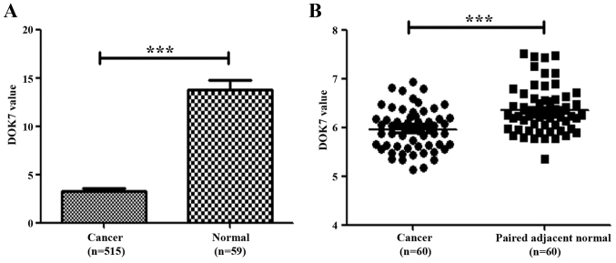
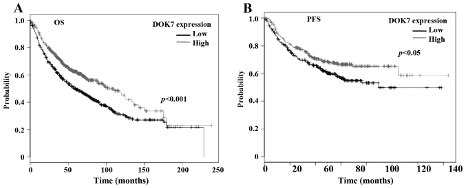
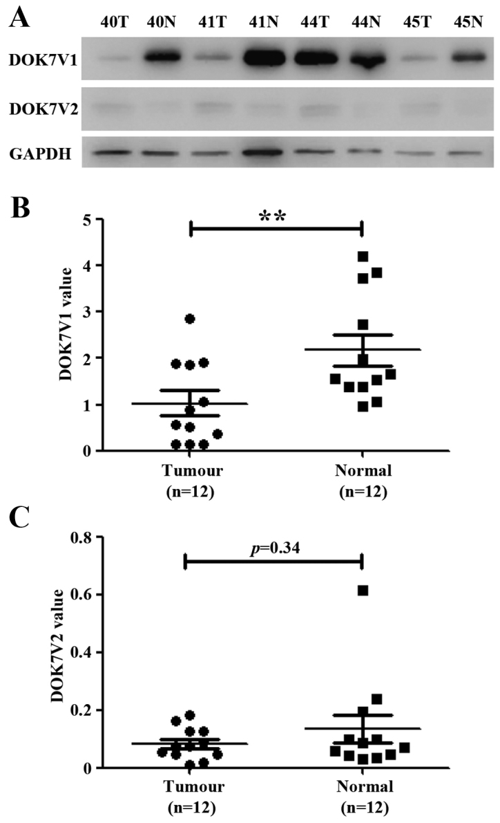
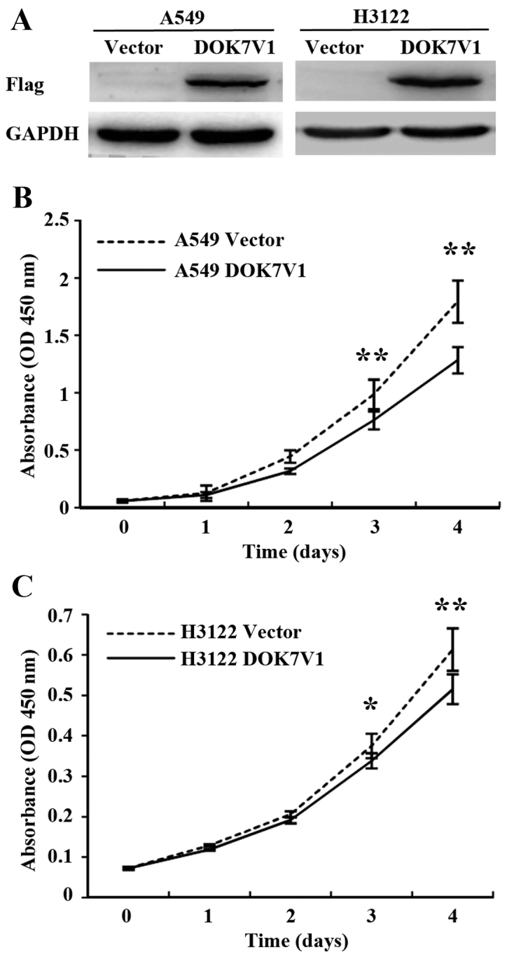
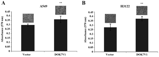
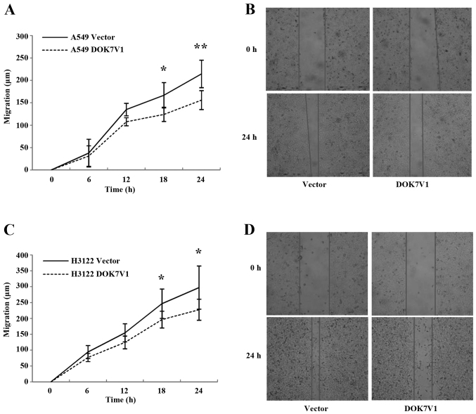
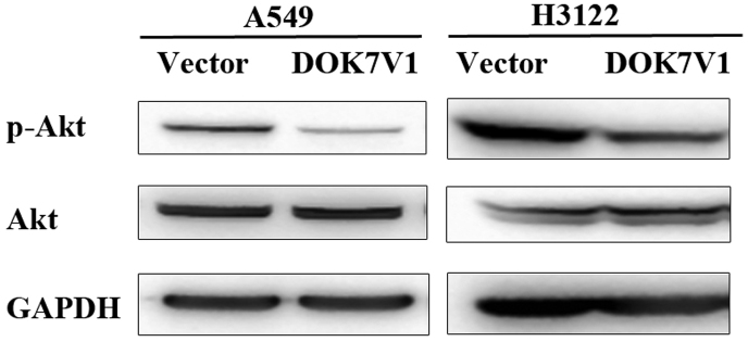
The downstream of tyrosine kinase 7 (DOK7) is an adaptor protein mediating signalling transduction between receptors and intracellular downstream molecules. Reduced expression of DOK7 has been observed in breast cancer. The present study aimed to investigate the role played by DOK7 in lung cancer. The expression of DOK7 at both mRNA and protein levels was evaluated in human lung cancer. A reduced expression of DOK7 transcripts was seen in lung cancers compared with normal lung tissues. Kaplan-Meier analyses showed that the reduced expression of DOK7 was associated with poorer overall survival and progression-free survival of patients with lung cancer. A further western blot analysis revealed a predominant expression of DOK7 isoform 1 (DOK7V1) in normal lung tissues, which was reduced in lung cancer. Forced overexpression of DOK7V1 in lung cancer cell lines, A549 and H3122 resulted in a decrease of in vitro cell proliferation and migration, while adhesion to extracellular matrix was enhanced following the expression. In conclusion, DOK7 was reduced in lung cancer and reduced DOK7 expression was associated with poorer survival. DOK7 isoform 1 plays an inhibitory role on the proliferation and migration of lung cancer cells in which Akt pathway may be involved.


Figures

Figure 1.
Figure 1.
Expression of DOK7 transcripts in human lung cancer. (A) Transcript levels of DOK7 were analysed in a cohort of lung cancers (515 cases) compared with normal lung tissues (59 cases) which were obtained from a collection of gene expression array data at TCGA (http://www.cbioportal.org/data_sets.jsp). (B) DOK7 expression in lung cancer compared with paired adjacent normal lung tissues (GSE19804) was analysed using paired t-test. ***p<0.001.
Figure 2.
Figure 2.
DOK7 expression and survival of patients with lung cancer. Association between DOK7 expression and overall survival (OS, A) and progression-free survival (PFS, B) was analysed using an online Kaplan-Meier survival analysis (KMplot, http://kmplot.com/analysis/). An auto-selected cut-off value of 115 was employed in the analysis.
Figure 3.
Figure 3.
DOK7 protein isoform-1 (DOK7V1) and −2 (DOK7V2) were determined in 12 lung cancer tumours together with paired adjacent normal lung tissues using western blotting. (A) Shown are representatives from the 12-paired lung cancer samples of western blotting. A semi-quantitative analysis was performed using ImageJ. Shown are values of band integrated density normalised against the corresponding loading control (GAPDH), DOK7V1 (B), and DOK7v2 (C), respectively. **p<0.01.
Figure 4.
Figure 4.
Overexpression of DOK7V1 in lung cancer cell lines and the effect on in vitro proliferation. (A) The forced overexpression of DOK7V1 was confirmed using western blotting. The influence of DOK7V1 was determined using an in vitro colorimetric growth assay for A549 (B) and H3122 (C) lung cancer cell lines in comparison with their corresponding control cell lines which were transfected with the empty plasmid vector. Six repeats were performed for each cell line in each experiments. Three independent experiments were carried out. Shown are representative data. Error bars represent standard deviations. *p<0.05; **p<0.01.
Figure 5.
Figure 5.
DOK7V1 overexpression and adhesiveness of lung cancer cell lines. The adhesion to extracellular matrix was determined using an adhesion assay for A549 (A) and H3122 (B) cell lines, respectively. Six repeats were included for each cell lines in every adhesion assay and three independent experiments were performed. Shown are representative data. **p<0.01.
Figure 6.
Figure 6.
Migration of DOK7V1 overexpression cell lines were determined using a wound assay for A549 (A and B) and H3122 (C and D). Three independent experiments were performed. Error bars are standard deviations. *p<0.05 and **p<0.01.
Figure 7.
Figure 7.
Phosphorylated Akt in DOK7 variant 1 overexpressing lung cancer cell lines. A reduced level of phosphorylated Akt was seen in both lung cancer cell lines with overexpression of the DOK7 variant 1 compared with the corresponding control using western blot analysis. GAPDH and Akt were used as loading controls.

Similar articles

Cited by

References

    1. Siegel RL, Miller KD, Jemal A. Cancer statistics, 2015. CA Cancer J Clin. 2015;65:5–29. doi: 10.3322/caac.21254. - DOI - PubMed
    1. Ferlay J, Soerjomataram I, Dikshit R, Eser S, Mathers C, Rebelo M, Parkin DM, Forman D, Bray F. Cancer incidence and mortality worldwide: Sources, methods and major patterns in GLOBOCAN 2012. Int J Cancer. 2015;136:E359–E386. doi: 10.1002/ijc.29210. - DOI - PubMed
    1. Yang M, Lewinska M, Fan X, Zhu J, Yuan ZM. PRR14 is a novel activator of the PI3K pathway promoting lung carcinogenesis. Oncogene. 2016;35:5527–5538. doi: 10.1038/onc.2016.93. - DOI - PMC - PubMed
    1. Lemmon MASJ, Schlessinger J. Cell signaling by receptor tyrosine kinases. Cell. 2010;141:1117–1134. doi: 10.1016/j.cell.2010.06.011. - DOI - PMC - PubMed
    1. Blume-Jensen P, Hunter T. Oncogenic kinase signalling. Nature. 2001;411:355–365. doi: 10.1038/35077225. - DOI - PubMed