. 2017 May;81(5):690-702.
doi: 10.1002/ana.24930. Epub 2017 May 9.

A mutation in the Tubb4a gene leads to microtubule accumulation with hypomyelination and demyelination

Affiliations

A mutation in the Tubb4a gene leads to microtubule accumulation with hypomyelination and demyelination

Ian D Duncan et al. Ann Neurol. 2017 May.

Abstract

Objective: Our goal was to define the genetic cause of the profound hypomyelination in the taiep rat model and determine its relevance to human white matter disease.

Methods: Based on previous localization of the taiep mutation to rat chromosome 9, we tested whether the mutation resided within the Tubb4a (β-tubulin 4A) gene, because mutations in the TUBB4A gene have been described in patients with central nervous system hypomyelination. To determine whether accumulation of microtubules led to progressive demyelination, we analyzed the spinal cord and optic nerves of 2-year-old rats by light and electron microscopy. Cerebral white matter from a patient with TUBB4A Asn414Lys mutation and magnetic resonance imaging evidence of severe hypomyelination were studied similarly.

Results: As the taiep rat ages, there is progressive loss of myelin in the brain and dorsal column of the spinal cord associated with increased oligodendrocyte numbers with accumulation of microtubules. This accumulation involved the entire cell body and distal processes of oligodendrocytes, but there was no accumulation of microtubules in axons. A single point mutation in Tubb4a (p.Ala302Thr) was found in homozygous taiep samples. A similar hypomyelination associated with increased oligodendrocyte numbers and arrays of microtubules in oligodendrocytes was demonstrated in the human patient sample.

Interpretation: The taiep rat is the first animal model of TUBB4 mutations in humans and a novel system in which to test the mechanism of microtubule accumulation. The finding of microtubule accumulation in a patient with a TUBB4A mutation and leukodystrophy confirms the usefulness of taiep as a model of the human disease. Ann Neurol 2017;81:690-702.


Conflict of interest statement

POTENTIAL CONFLICTS OF INTEREST:

None of the authors have any conflict of interest.

Figures

Fig. 1
Fig. 1. Myelin is initially present though reduced in the taiep brain and is lost with time
There is evidence of myelin in the brain of a 3 month old taiep rat, noticeably in the cerebellum, corpus callosum and fornix but much less than wild type. However by 9 months, demyelination has led to severe loss of myelin. In the cerebellum there is mild separation of the gyri suggesting atrophy that may have resulted from myelin loss. Scale bars: 0.5 mm (cerebellum), 1.0 mm (whole brain).
Fig. 2
Fig. 2. In the spinal cord, the dorsal column shows both hypomyelination followed by complete demyelination of certain tracts
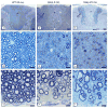
In the taiep rat spinal cord at early time points, all axons in the fasciculus gracilis (*, E) were myelinated but with thinner myelin sheaths than in controls (B). However, with time the dorsal column and particularly the fasciculus gracilis (*) and corticospinal tract (↑CT) show severe demyelination with only scattered large diameter axons being myelinated (G, H). In comparison, the fasciculus cuneatus was less severely affected (↑FC). There was an increase in glial cell nuclei in these tracts with time (H). In contrast, the ventral column from the same animals as in A-H showed that axons at 4 months were mainly thinly myelinated or non-myelinated in taiep and remained this way through 23 months (I). WT = wild type. Scale bars: 20 μm (B, C, E, F, H, I), 200 μm (A, D, G). (In color in online version)
Fig. 3
Fig. 3. Accumulation of microtubules, solely in oligodendrocytes, defines the defect
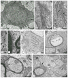
A) Oligodendrocytes contain densely packed microtubules and are often present in ER-associated arrays (arrows). On higher power, details of these arrays can be seen with microtubules linking adjacent (B) or separate (C) membranes where they appear discontinuous (C, arrows). In B) and C) there is also an increase in “free” microtubules. Microtubules also are aligned along RER and the outer nuclear membrane (D, E) (arrows). In D) the microtubules may have displaced ribosomes. F) Oligodendrocyte processes are frequently packed with microtubules that can also be aligned with ER (F). Numerous examples of excess microtubules circling myelinated axons were noted (G). High power from area ↑ shows more detail of the microtubules (G-inset). H) Collections of microtubules were frequently seen in duplicated inner loops (arrow). I) Axons of all diameters had normal density of microtubules in contrast to the adjacent oligodendrocyte ‘microprocesses’ (arrows). Scale bars: 50 nm (D), 100 nm (E), 200 nm (B, C, F, G-inset), 500 nm (A, H, I), 600 nm (G).
Fig. 4
Fig. 4. Identification of Tubb4a mutation in taiep rat
A) Previous work had localized the causative gene in the taiep mutant to rat chromosome 9q12. Analysis of candidate genes revealed Tubb4a, which is mutated in several patients with hypomyelination. Sequencing of taiep mutant rats and normal controls revealed an A to G mutation that changes Ala302 to Thr. The mutation also introduces a MscI restriction enzyme site. PCR fragments were generated from taiep rat and wild type littermate cDNA, and both fragments were digested with the MscI restriction enzyme, indicating a homozygous mutation in taiep. B) The crystal structure of α- and β-tubulins was used to indicate the position of the Ala302 residue that is mutated in the taiep rat and the Asn414 residue mutated in our patient. Additionally the position of the classical H-ABC mutation (Asp249) and two other patients with isolated hypomyelination (Glu410 and Gly96) are shown. The classical H-ABC mutation is located at the α-β interface where most H-ABC mutations are located, whereas the taiep mutation and the mutations observed in patients with isolated hypomyelination are located at the lateral side of β-tubulin.
Fig. 5
Fig. 5. Sox10 and Olig2 bind the Tubb4a gene promoter
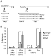
A) Schematic shown depicts location of genomic regions assayed by ChIP. Sox10 and Olig2 binding motifs are indicated and sequences are compared between species. Note that the motifs vary in relation to the consensus motifs for each: C(A/T)TTGT for Sox10 and CAGCTGC for Olig2. Also, the region at −8kb is not very conserved and the Sox10 motif near the gene promoter (Pro) is not conserved in humans. Gm11110 is a predicted long noncoding RNA annotated in the mouse genome build mm10. B) ChIP-qPCR analysis identifies Sox10 and Olig2 binding in P15 mouse thoracic spinal cord. Genomic regions assayed are listed on the x-axis with respect to the given gene translation start site and includes a negative control site within the Tekt3 gene and a positive control site −27kb upstream of Sox10. At each site, percent recoveries for Sox10 or Olig2 ChIP were compared with that of a control IP (goat IgG). Error bars represent the standard deviation of three independent experiments (*p<0.05).
Fig. 6
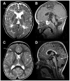
Fig. 6. MRI in the patient with TUBB4A-related severe hypomyelination and atrophy of the cerebellum
A, B) T2-weighted axial (A) and T1-weighted sagittal (B) images of the patient obtained at the age of 2½ years; C, D, same images of an age-matched neurologically normal child. In the patient the cerebral white matter is T2-hyperintense due to lack of myelin (arrow in A), while the white matter in the control is T2-hypointense (arrow in C). In the patient the cerebellum is mildly atrophic (arrow in B), while it is normal in the control (arrow in D).
Fig. 7
Fig. 7. Neuropathology of the patient with TUBB4A-related severe hypomyelination and atrophy of the cerebellum
A) Proteolipid protein (PLP) stain for myelin of the frontal lobe shows lack of white matter myelin with relative preservation of the U-fibers. B) Klüver stain for myelin shows profound lack of myelin in the deep white matter of the parietal lobe. C) At high magnification, the number of white matter oligodendrocytes (cells with small, compact, round nuclei and a clear perinuclear halo) appears increased on this H&E-stained section of the parietal lobe. D) CD68 stain for microglia/macrophages shows activation of microglia in the temporal white matter with clustering of macrophages around blood vessels. E) Bodian stain for axons shows axonal preservation in the temporal lobe white matter without axonal varicosities or spheroids. F) Stain against the astrocyte-specific glial fibrillary acidic protein (GFAP) shows moderate reactive astrogliosis in the frontal white matter. G) H&E stain of the cerebellum shows enlargement of the sulci and thinning of the cortex with loss of neurons in the molecular layer. H) Bodian stain of the cerebellar cortex shows mild drop out of Purkinje cells and presence of Purkinje cell dendritic swellings in the molecular layer. Scale bars: 20 μm (B–F), 40 μm (H), 500 μm (G), 1 mm (A).
Fig. 8
Fig. 8. Hypomyelination is associated with microtubule accumulation in the patient
Axons are either non-myelinated or have thin myelin sheaths (A). Close to a putative oligodendrocyte nucleus there are rows of microtubules (arrows in B). Magnification of one of these areas is shown in C). The most distal part of the oligodendrocyte process, the inner loop is enlarged and filled with microtubules (D, E). An oligodendrocyte process seen on longitudinal section contains prominent microtubules (F). Microtubules were also prominent in myelinated and non-myelinated axons (G). Scale bars: 100 nm (G), 200 nm (E, F), 400 nm (C, D), 500 nm (B), 2 μm (A).

Similar articles

Cited by

References

    1. van der Knaap MS, Naidu S, Pouwels PJ, et al. New syndrome characterized by hypomyelination with atrophy of the basal ganglia and cerebellum. AJNR Am J Neuroradiol. 2002 Oct;23(9):1466–74. - PMC - PubMed
    1. Hamilton EM, Polder E, Vanderver A, et al. Hypomyelination with atrophy of the basal ganglia and cerebellum: further delineation of the phenotype and genotype-phenotype correlation. Brain. 2014 Jul;137(Pt 7):1921–30. - PMC - PubMed
    1. van der Knaap MS, Linnankivi T, Paetau A, et al. Hypomyelination with atrophy of the basal ganglia and cerebellum: follow-up and pathology. Neurology. 2007 Jul 10;69(2):166–71. - PubMed
    1. Ferreira C, Poretti A, Cohen J, Hamosh A, Naidu S. Novel TUBB4A mutations and expansion of the neuroimaging phenotype of hypomyelination with atrophy of the basal ganglia and cerebellum (H-ABC) Am J Med Genet A. 2014 Jul;164A(7):1802–7. - PMC - PubMed
    1. Simons C, Wolf NI, McNeil N, et al. A de novo mutation in the beta-tubulin gene TUBB4A results in the leukoencephalopathy hypomyelination with atrophy of the basal ganglia and cerebellum. Am J Hum Genet. 2013 May 2;92(5):767–73. - PMC - PubMed