. 2017 Apr 10:8:15092.
doi: 10.1038/ncomms15092.

Cryo-EM structures of MERS-CoV and SARS-CoV spike glycoproteins reveal the dynamic receptor binding domains

Affiliations

Cryo-EM structures of MERS-CoV and SARS-CoV spike glycoproteins reveal the dynamic receptor binding domains

Yuan Yuan et al. Nat Commun. .

Abstract

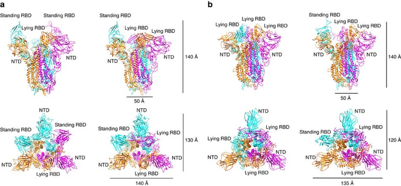
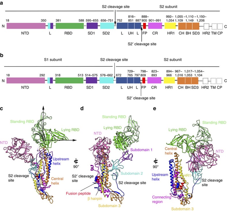
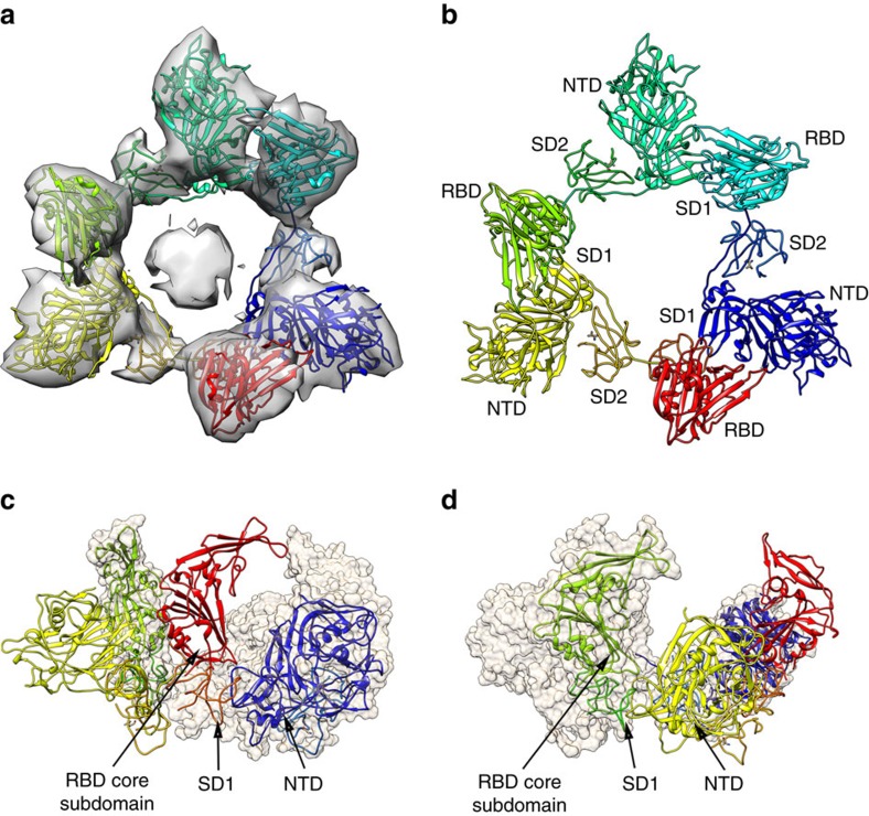
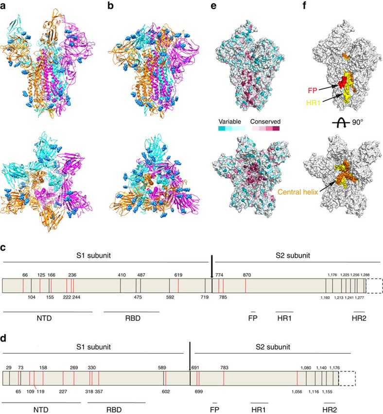
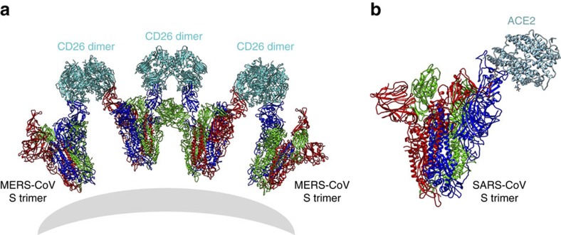
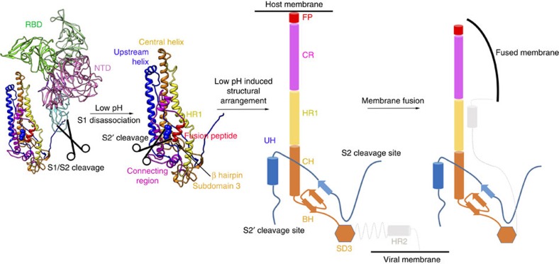
The envelope spike (S) proteins of MERS-CoV and SARS-CoV determine the virus host tropism and entry into host cells, and constitute a promising target for the development of prophylactics and therapeutics. Here, we present high-resolution structures of the trimeric MERS-CoV and SARS-CoV S proteins in its pre-fusion conformation by single particle cryo-electron microscopy. The overall structures resemble that from other coronaviruses including HKU1, MHV and NL63 reported recently, with the exception of the receptor binding domain (RBD). We captured two states of the RBD with receptor binding region either buried (lying state) or exposed (standing state), demonstrating an inherently flexible RBD readily recognized by the receptor. Further sequence conservation analysis of six human-infecting coronaviruses revealed that the fusion peptide, HR1 region and the central helix are potential targets for eliciting broadly neutralizing antibodies.


Conflict of interest statement

The authors declare no competing financial interests.

Figures

Figure 1
Figure 1. Overall structure of the MERS-CoV and SARS-CoV S ectodomain trimers.
(a) Two different conformations of the MERS-CoV S ectodomain trimer were determined without three-fold symmetry to resolutions of 4.1, 4.2 Å, respectively. The ribbon views of the structures are shown from both the side and the top. Two states (standing and lying) of the RBD were captured in the S ectodomain trimer structure. NTD domains are arranged in a triangular manner. (b) Two different conformations of SARS-CoV S ectodomain trimer were determined to resolutions of 3.2 and 3.7 Å, respectively. The ribbon views of the structures are shown from both the side and the top. Two states (standing and lying) of RBD were captured in the S ectodomain trimer structure. NTD domains are arranged in a triangular manner.
Figure 2
Figure 2. Architecture of the MERS-CoV and SARS-CoV S protomers.
(a) Schematic diagram of the MERS-CoV S glycoprotein organization. Black and grey dashed lines denote regions unresolved in the reconstruction and regions beyond the construct, respectively. NTD, N-terminal domain; L, linker region; RBD, receptor-binding domain; SD, subdomain; UH, upstream helix; FP, fusion peptide; CR, connecting region; HR, heptad repeat; CH, central helix; BH, β-hairpin; TM, transmembrane region/domain; CT, cytoplasmic tail. (b) Schematic diagram of the SARS-CoV S glycoprotein organization. The abbreviations of elements are the same as in a. (ce) Ribbon diagrams depicting three views of the S protomer coloured as in a. As the MERS-CoV and SARS-CoV S protomers have extremely similar structures, and thus only MERS-CoV S protomer was used to show the detailed architecture.
Figure 3
Figure 3. Low resolution cryo-EM structure of the disassociated S1 trimer.
(a) Cryo-EM electron density of the overall structure of the disassociated S1 trimer. (b) Ribbon view of the S1 trimer, including NTD, RBD, SD1 and SD2. (c,d) Interaction between S1 protomers. The RBD core subdomain and SD1 form quaternary interactions with the NTD to stabilize the disassociated S1 trimer.
Figure 4
Figure 4. N-linked glycosylation analysis of MERS-CoV and SARS-CoV S proteins and a potential strategy for antiviral intervention.
(a,b) Cartoon representation of MERS-CoV (a) and SARS-CoV (b) S trimers from the side and top views. The glycans are shown in spheres. (c,d) Schematic diagram of the N-linked glycosylation sites for MERS-CoV (c) and SARS-CoV (d) S proteins. The visible N-linked glycosylation sites are shown in red lines and the invisible N-linked glycosylation sites are shown in black lines. (e) Surface representation of the MERS-CoV S trimer from either the side or the top, coloured according the sequence conservation from the most conserved (magenta) to the most divergent (cyan), using the ConSurf server based on an alignment of S sequences from six human-infecting CoV in the NCBI database. (f) Surface representation of the MERS-CoV S trimer highlighting the highly conserved region for the design of broadly neutralizing antibodies, including exposed FP, HR1 region and central helix.
Figure 5
Figure 5. Models of MERS-CoV and SARS-CoV S trimers bound to their receptors.
The models were built by superimposition of the S trimer structures with the RBD-receptor complex structures through the RBD domains. The MERS-CoV S trimer can cross-link the dimeric CD26 receptor during the binding (a), whereas the SARS-CoV S trimer can bind one monomeric ACE2 receptor (b).
Figure 6
Figure 6. Proposed mechanism of membrane fusion promoted by MERS-CoV S protein.
After cleavage into S1/S2 subunits, the S1 subunit is easily disassociated from the S2 subunit. In the endosome, the S2′ cleavage site could be further cleaved by the host proteases, releasing its fusion peptide. Then, under low pH environment, the connecting region, HR1 helix and central helix undergo structural rearrangement to form a long helix to help the insertion of the fusion peptide into the host membrane. Finally, the HR1 and HR2 fold into an intra-hairpin helical structure that can trimerically assemble into a six-helix bundle, resulting in membrane fusion.

References

    1. Weinstein R. A. Planning for epidemics–the lessons of SARS. N. Engl. J. Med. 350, 2332–2334 (2004). - PubMed
    1. Hijawi B. et al.. Novel coronavirus infections in Jordan, April 2012: epidemiological findings from a retrospective investigation. East Mediterr. Health J. 19, (Suppl 1): S12–S18 (2013). - PubMed
    1. Zaki A. M., van Boheemen S., Bestebroer T. M., Osterhaus A. D. & Fouchier R. A. Isolation of a novel coronavirus from a man with pneumonia in Saudi Arabia. N. Engl. J. Med. 367, 1814–1820 (2012). - PubMed
    1. Wang Y. et al.. Origin and possible genetic recombination of the middle east respiratory syndrome coronavirus from the first imported case in china: phylogenetics and coalescence analysis. MBio 6, e01280–01215 (2015). - PMC - PubMed
    1. Su S. et al.. Epidemiology, genetic recombination, and pathogenesis of coronaviruses. Trends Microbiol. 24, 490–502 (2016). - PMC - PubMed

Publication types

MeSH terms