

Novel Wavelet Real Time Analysis of Neurovascular Coupling in Neonatal Encephalopathy
- PMID: 28393884
- PMCID: PMC5385531
- DOI: 10.1038/srep45958
Novel Wavelet Real Time Analysis of Neurovascular Coupling in Neonatal Encephalopathy
Abstract
Birth asphyxia constitutes a major global public health burden for millions of infants, despite hypothermia therapy. There is a critical need for real time surrogate markers of therapeutic success, to aid in patient selection and/or modification of interventions in neonatal encephalopathy (NE). This is a proof of concept study aiming to quantify neurovascular coupling (NVC) using wavelet analysis of the dynamic coherence between amplitude-integrated electroencephalography (aEEG) and near-infrared spectroscopy in NE. NVC coupling is assessed by a wavelet metric estimation of percent time of coherence between NIRS SctO2 and aEEG for 78 hours after birth. An abnormal outcome was predefined by a Bayley III score <85 by 18-24 m. We observed high coherence, intact NVC, between the oscillations of SctO2 and aEEG in the frequency range of 0.00025-0.001 Hz in the non-encephalopathic newborns. NVC coherence was significantly decreased in encephalopathic newborns who were cooled vs. non-encephalopathic controls (median IQR 3[2-9] vs.36 [33-39]; p < 0.01), and was significantly lower in those with abnormal 24 months outcomes relative to those with normal outcomes (median IQR 2[1-3] vs 28[19-26], p = 0.04). Wavelet coherence analysis of neurovascular coupling in NE may identify infants at risk for abnormal outcomes.
Conflict of interest statement
The authors declare no competing financial interests.
Figures
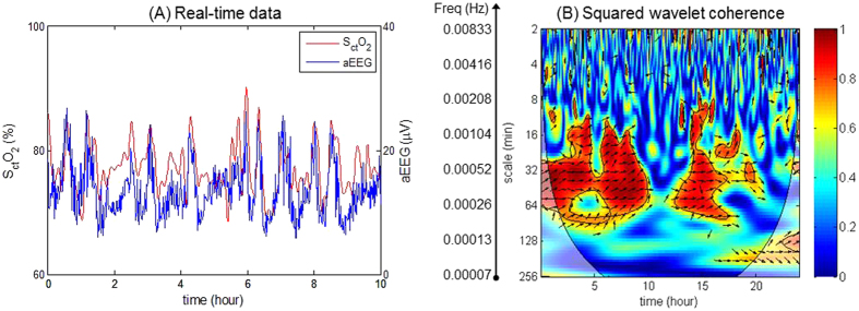
 , where the x-axis represents time, the y-axis represents scale in minute representing the range of frequencies, and the color scale represents the magnitude of R2. Significant coherence between the SctO2 and aEEG is seen in a very low-frequency (VLF) range of 0.00025–0.001 Hz.
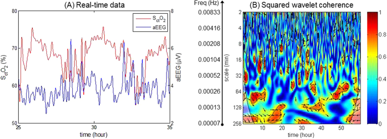
 , where the x-axis represents time, the y-axis represents scale of frequencies. No significant areas of coherence are seen through the range of time and frequencies studied.
References
Publication types
MeSH terms
Substances
Grants and funding
LinkOut - more resources
Full Text Sources
Other Literature Sources
Medical
