. 2017 Apr 10:7:46054.
doi: 10.1038/srep46054.

Involvement of a velvet protein ClVelB in the regulation of vegetative differentiation, oxidative stress response, secondary metabolism, and virulence in Curvularia lunata

Affiliations

Involvement of a velvet protein ClVelB in the regulation of vegetative differentiation, oxidative stress response, secondary metabolism, and virulence in Curvularia lunata

Jin-Xin Gao et al. Sci Rep. .

Abstract

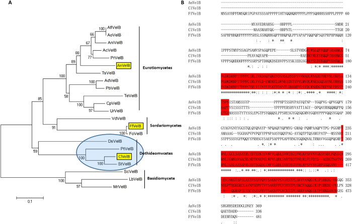
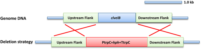
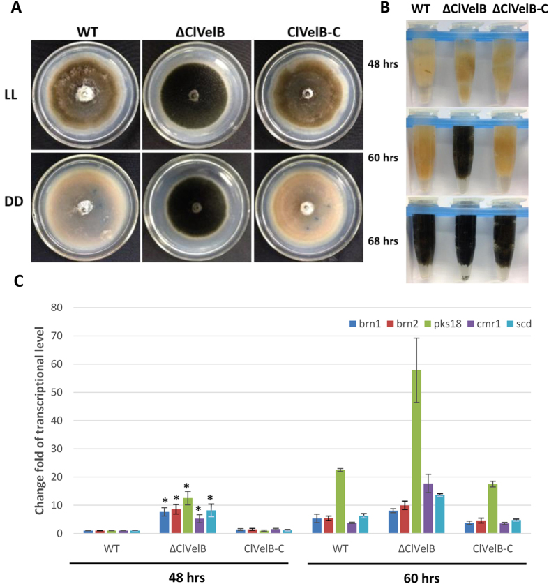
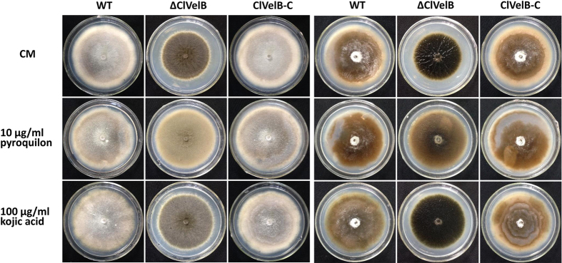
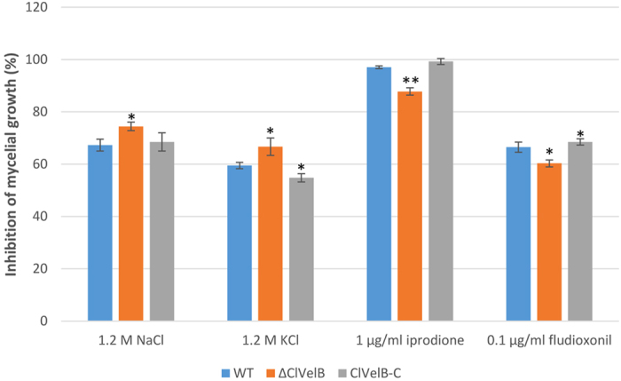
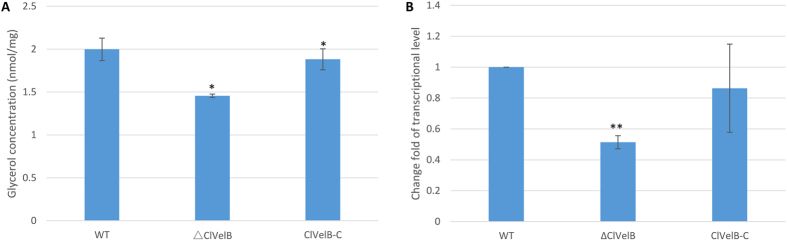
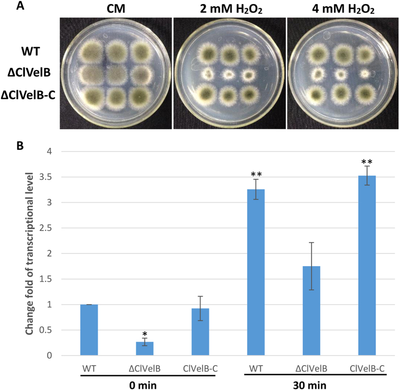
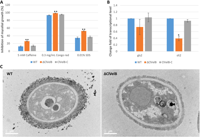
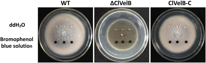
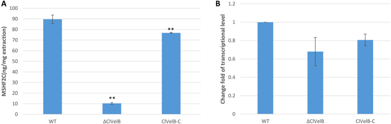
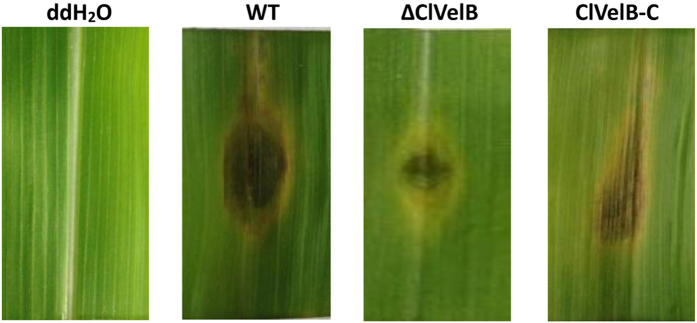
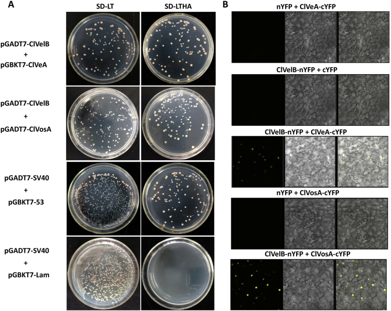
The ortholog of Aspergillus nidulans VelB, which is known as ClVelB, was studied to gain a broader insight into the functions of a velvet protein in Curvularia lunata. With the expected common and specific functions of ClVelB, the deletion of clvelB results in similar though not identical phenotypes. The pathogenicity assays revealed that ΔClVelB was impaired in colonizing the host tissue, which corresponds to the finding that ClVelB controls the production of conidia and the methyl 5-(hydroxymethyl) furan-2-carboxylate toxin in C. lunata. However, the deletion of clvelB led to the increase in aerial hyphae and melanin formation. In addition, ΔClVelB showed a decreased sensitivity to iprodione and fludioxonil fungicides and a decreased resistance to cell wall-damaging agents and osmotic stress and tolerance to H2O2. The ultrastructural analysis indicated that the cell wall of ΔClVelB became thinner, which agrees with the finding that the accumulated level of glycerol in ΔClVelB is lower than the wild-type. Furthermore, the interaction of ClVelB with ClVeA and ClVosA was identified in the present research through the yeast two-hybrid and bimolecular fluorescence complementation assays. Results indicate that ClVelB plays a vital role in the regulation of various cellular processes in C. lunata.


Conflict of interest statement

The authors declare no competing financial interests.

Figures

Figure 1
Figure 1. C. lunata VelB is an ortholog of A. nidulans VelB.
(A) Phylogenetic analysis. VelB protein sequences were obtained from GenBank using A. nidulans. AnVelB as a query. AnVelB, C. lunata ClVelB, and Fusarium fujikuroi FfVelB are marked in yellow highlights. A blue oval shadow marks the single candidate ortholog. (B) ClVelB, AnVelB, and FfVelB were aligned using ClustalW. Conserved velvet superfamily domains are highlighted in red, asterisks mark identical residues, colons mark conserved residues, and periods indicate semi-conserved residues.
Figure 2
Figure 2. ClvelB deletion strategy used by homologous recombination.
clvelB and hygromycin resistance (hph) genes are represented by blue and red boxes, respectively.
Figure 3
Figure 3. Effects of ClVelB on colony morphology and sporulation.
(A) Cultures grown on CM plates under constant light (LL) or dark (DD), and 12 h light/dark cycle (LD) conditions for 7 days at 28 °C. Note that in LL, WT and ClVelB-C are white and flat, while ΔClVelB is pigmented and fluffy, which reflects aerial hyphal growth. Alternating banding rhythm in the middle plate suggests that the conidiation of WT is responsive to light. This banding rhythm is greatly reduced in ΔClVelB. (B) Side view of the plates of WT, ΔClVelB, and ClVelB-C grown in LL or DD on CM. Note the aerial hyphae on the plates of ΔClVelB, especially from LL. By contrast, the surface of the WT and ClVelB-C only shows a few aerial hyphae. (C) Quantification of conidia from cultures grown under LL, LD, and DD conditions. Error bars are the standard deviation. A single asterisk indicates the p-value < 0.05 while double asterisks indicate the p-value < 0.001 in the T-test analysis. Sporulation of ΔClVelB is repressed in all circumstances, while this was not observed for WT and ClVelB-C. (D) Hyphae structures of WT and ΔClVelB were examined through scanning electron microscopy (Sirion 200, FEI).
Figure 4
Figure 4. ClVelB negatively regulates the mycelial melanization of C. lunata.
(A) Bottom of the CM plates of WT strain (CX-3), clvelB deletion mutant (ΔClVelB), and complemented strain (ClVelB-C) grown in constant light (LL) or dark (DD) for 7 days. Photos were taken after removing conidia. Note the heavy melanization of mycelia of ΔClVelB in both LL and DD compared to WT. (B) The mycelial pellet of WT, ΔClVelB, and ClVelB-C at different indicated time points. ΔClVelB is melanized by 60 h, which is ahead of WT and ClVelB-C. Pigmentation starts by 68 h in WT and ClVelB-C. (C) qRT-PCR analyses of pks18, cmr1, brn1, brn2, and scd. Expression was tested at 48 and 60 h. The expression level compared with the WT at 48 h is shown. Error bars are the standard deviation. A single asterisk indicates the p-value < 0.05 in a T-test analysis.
Figure 5
Figure 5. DHN type melanin produced by C. lunata.
(A) 10 μg/ml pyroquilon or 100 μg/ml kojic acid were added into the CM to confirm that the conidial and mycelial melanin of C. lunata is not the tyrosine-derived but DHN type. (B) Culture plates after removing conidia. The mycelial color had gone from black to light brown for all strains detected on the pyroquilon plates, but no change on the kojic acid medium.
Figure 6
Figure 6. Sensitivity of the WT strain (CX-3), clvelB deletion mutant (ΔClVelB), and complemented strain (ClVelB-C) to the osmotic stresses and fungicides.
1.2 M NaCl or KCl were added into the CM to study the osmotic stresses. Iprodione or fludioxonil was added into the CM at a final concentration of 1 μg/ml or 0.1 μg/ml, respectively, to test the tolerance of fungicides. Error bars are the standard deviation. (A) Single asterisk indicates the p-value < 0.05 while double asterisks indicate the p-value < 0.001 in the T-test analysis.
Figure 7
Figure 7. ClVelB regulates the glycerol accumulation.
(A) Determination of glycerol biosynthesis in WT strain (CX-3), clvelB deletion mutant (ΔClVelB), and complemented strain (ClVelB-C). After the mycelia of WT, ΔClVelB and ClVelB-C were treated with 1.2 M NaCl for 2 h, the intracellular glycerol concentrations (nmol/mg dried mycelia) were tested. Untreated mycelia were used as the controls. The bars indicate the standard errors of the three repeated trails. (B) qRT-PCR analysis of gpd1, which is responsible for glycerol biosynthesis. A single asterisk indicates the p-value < 0.05 while double asterisks indicate the p-value < 0.001 in the T-test analysis.
Figure 8
Figure 8. Sensitivity of the WT strain (CX-3), clvelB deletion mutant (ΔClVelB), and complemented strain (ClVelB-C) to oxidative stress.
(A) 4, 2, and 1 μl conidial suspensions prepared from WT, ΔClVelB, and ClVelB-C were dripped on a CM plate with the with the indicated concentrations of H2O2. ΔClVelB is more sensitive to H2O2 than WT and ClVelB-C. (B) qRT-PCR analysis of the catalase-encoding gene cat3. Error bars are the standard deviation. A single asterisk indicates the p-value < 0.05 while double asterisks indicate the p-value < 0.001 in a T-test analysis. The expression levels of cat3 were reduced in ΔClVelB (3.7-fold at time 0 and 3.3-fold 30 min after H2O2 addition).
Figure 9
Figure 9. ClVelB regulates cell wall integrity.
(A) Sensitivity of the WT strain (CX-3), clvelB deletion mutant (ΔClVelB), and complemented strain (ClVelB-C) to the cell wall damaging agents. The detection was made on a CM plate added with the corresponding cell wall damaging agent. (B) Expression changes of gls2 and slt2 in each strain. The relative expression levels of gls2 and slt2 in ΔClVelB are the relative cDNA amounts of the same gene in the WT strain. Line bars indicate the standard errors from the three trial replicates. A single asterisk indicates a p-value < 0.05 while double asterisks indicate a p-value < 0.001 in the T-test analysis. (C) Ultrastructural analyses of the cell of the clvelB deletion mutant. Cells of the WT and ΔClVelB were observed with a transmission electronic microscope (Tecnai G2 Spirit Biotwin, FEI). Mycelia were harvested and fixed in glutaraldehyde for 12 h at 4 °C.
Figure 10
Figure 10. Effects of the clvelB deletion on hyphal hydrophobicity.
20 μl of ddH2O or 2.5% bromophenol blue solution was dropped on the colony surfaces of the WT strain (CX-3), clvelB deletion mutant (ΔClVelB), and complemented strain (ClVelB-C), and photographed 10 min later. The droplet did not disperse on the colony of ΔClVelB, WT, and ClVelB-C.
Figure 11
Figure 11. ClVelB regulates the biosynthesis of M5HF2C toxin.
(A) Amount of M5HF2C (per mg extraction) produced by WT strain (CX-3), clvelB deletion mutant (ΔClVelB), and complemented strain (ClVelB-C) that were cultured in Fries 3 medium after 30 days. (B) qRT-PCR analysis of the M5HF2C biosynthesis related gene clt-1 in WT, ΔClVelB, and ClVelB-C. Error bars are the standard deviation. A single asterisk indicates the p-value < 0.05 while double asterisks indicate the p-value < 0.001 in a T-test analysis.
Figure 12
Figure 12. Virulence of the WT (CX-3), clvelB deletion mutant (ΔClVelB), and complemented strain (ClVelB-C) on maize leaves.
clvelB deletion mutants are impaired in the colonization of maize leaves. Detached leaves of HUANGZAO-4 were inoculated with conidial suspensions and incubated on two layers of filter papers moisturized with 10 mM 6-Benzyladenine (6-BA) in Petri dishes at 28 °C for 96 h.
Figure 13
Figure 13. Interaction of ClVelB with ClVeA and ClVosA in C. lunata.
(A) ClVelB interacts with ClVeA and ClVosA in a Y2H approach. For Y2H analysis, the clvelb cDNA was fused to the GAL4 activation domain, and the cDNAs of clveA and clvosA were respectively fused to the GAL4 binding domain. The cell suspensions of the Saccharomyces cerevisiae strain (AH109) containing the indicated vectors were dropped onto the selected media. SD-LT: synthetically defined (SD) medium without leucine and tryptophan was used to demonstrate the presence of both vectors. SD-LTHA: SD without leucine, tryptophan, histidine, and adenine supplemented with 11 mM 3-amino-1,2,4-triazole (SD_LWH+3-AT) was used to detect HIS3 reporter gene activity (displayed by colony growth). Plasmid pairs of pGADT7-SV40/pGBKT7-53 and pGADT7-SV40/pGBKT7-Lam served as the positive and negative controls, respectively. (B) Interaction analysis of ClVelB with ClVeA and ClVosA versions by BiFC assay in infiltrated Nicotiana benthamiana leaves. Co-expression of ClVelB with ClVeA and ClVosA versions fused to splitYFP. n/cYFP: n- or c-terminal part of splitYFP.

Similar articles

Cited by

References

    1. Liu T. et al. Clg2p interacts with Clf and ClUrase to regulate appressorium formation, pathogenicity and conidial morphology in Curvularia lunata. Sci. Rep. 6, 24047 (2016). - PMC - PubMed
    1. Liu T., Liu L. X., Jiang X., Huang X. L. & Chen J. A new furanoid toxin produced by Curvularia lunata, the causal agent of maize Curvularia leaf spot. Can. J. Plant Pathol. 31, 22–27 (2009).
    1. Gao J. X., Liu T. & Chen J. Insertional mutagenesis and cloning of the gene required for the biosynthesis of the non-host specific toxin in Cochliobolus lunatus that causes maize leaf spot. Phytopathology 104(4), 332–339 (2014). - PubMed
    1. Gao S. G. et al. Understanding resistant germplasm-induced virulence variation through analysis of proteomics and suppression subtractive hybridization in a maize pathogen Curvularia lunata. Proteomics 12, 1–12 (2012). - PubMed
    1. Merhej J., Richard-Forget F. & Barreau C. Regulation of trichothecene biosynthesis in Fusarium: recent advances and new insights. Appl. Microbiol. Biotechnol. 91, 519–528 (2011). - PubMed

Publication types

MeSH terms