Review
. 2017 Jun;17(6):363-375.
doi: 10.1038/nri.2017.21. Epub 2017 Apr 10.

Mitochondrial DNA in innate immune responses and inflammatory pathology

Affiliations
Review

Mitochondrial DNA in innate immune responses and inflammatory pathology

A Phillip West et al. Nat Rev Immunol. 2017 Jun.

Abstract

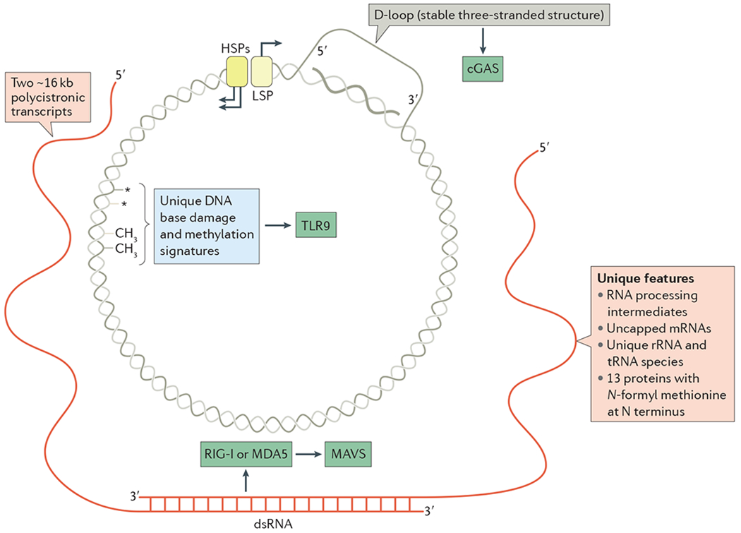
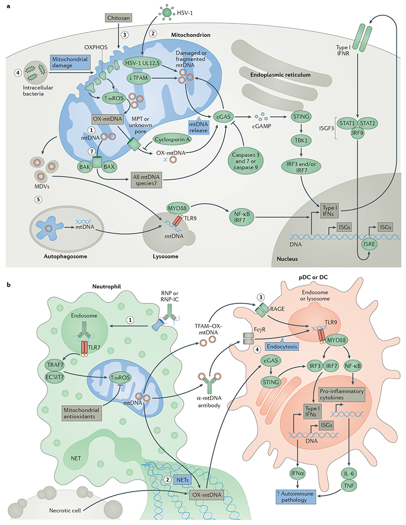
Mitochondrial DNA (mtDNA) - which is well known for its role in oxidative phosphorylation and maternally inherited mitochondrial diseases - is increasingly recognized as an agonist of the innate immune system that influences antimicrobial responses and inflammatory pathology. On entering the cytoplasm, extracellular space or circulation, mtDNA can engage multiple pattern-recognition receptors in cell-type- and context-dependent manners to trigger pro-inflammatory and type I interferon responses. Here, we review the expanding research field of mtDNA in innate immune responses to highlight new mechanistic insights and discuss the physiological and pathological relevance of this exciting area of mitochondrial biology.


Conflict of interest statement

Competing interests statement

The authors declare no competing interests.

Figures

Figure 1 |
Figure 1 |. Immunostimulatory features of mitochondrial DNA and related species.
The circular mitochondrial DNA (mtDNA) of mammals is depicted with nucleic acid species generated during transcription and replication (BOX 1). Transcription of mtDNA involves the divergent light-strand promoter (LSP) and heavy-strand promoters (HSPs), which are shown at the top with the direction of transcription indicated by arrows. The two polycistronic primary transcripts from each strand are shown in red. Because almost the entire genome is transcribed in both directions, there is ample opportunity for double-stranded RNA (dsRNA) to be formed, as depicted at the bottom, which could engage retinoic acid-inducible gene I protein (RIG-I; also known as DDX58) or melanoma differentiation-associated protein 5 (MDA5; also known as IFIH1) and trigger mitochondrial antiviral signalling protein (MAVS). Replication of mtDNA initiates downstream of the LSP, but is paused or terminated frequently, forming a stable ~1 kb nascent DNA strand that remains associated with the template and displaces the non-template strand. This forms the hallmark three-stranded D-loop structure of mammalian mtDNA, and may be the source of cytosolic DNA that activates cyclic GMP–AMP synthase (cGAS). Other potentially unique features of mtDNA and mtDNA-encoded proteins are indicated, as are the innate immune sensors that we postulate might sense these unique features. rRNA, ribosomal RNA; TLR9, Toll-like receptor 9.
Figure 2 |
Figure 2 |. Mitochondrial DNA in inflammasome activation and pro-inflammatory responses.
Event 1: tissue pathology and cellular damage trigger necrosis and/or mitochondrial stress, resulting in the release of mitochondrial DNA (mtDNA) or mtDNA-containing microparticles into the extracellular milieu. Event 2: mtDNA in the plasma engages intracellular Toll-like receptor 9 (TLR9)–myeloid differentiation primary response protein 88 (MYD88)–nuclear factor-κB (NF-κB) signalling on circulating leukocytes, resulting in increased production of pro-inflammatory mediators, such as tumour necrosis factor (TNF), interleukin-6 (IL-6) and adhesion molecules. This enhances leukocyte differentiation and extravasation into tissues and causes inflammasome priming (signal 1) in tissue-resident cells. The NOD, LRR and Pyrin domain-containing protein 3 (NLRP3) inflammasome (signal 2) in tissue-resident cells may also be activated by mtDNA, resulting in caspase 1-mediated cleavage of IL-1β and IL-18 to further amplify inflammatory responses. Event 3: mtDNA that enters the endocytic pathway by endocytosis or through mitochondria-derived vesicles (MDVs) can engage TLR9 on tissue-resident macrophages, resulting in increased NF-κB signalling for pro-inflammatory gene expression (signal 1). Event 4: exposure to cellular stress, inflammasome agonists or intracellular bacteria can trigger mitochondrial damage and enhance production of mitochondrial reactive oxygen species (mROS), resulting in the release of oxidized mtDNA (OX-mtDNA) into the cytosol to trigger NLRP3-, NLR family CARD domain-containing protein 4 (NLRC4)- or absent in melanoma 2 (AIM2)-dependent activation of caspase 1 (signal 2), which increases the processing and secretion of mature IL-1β and IL-18, further enhancing tissue inflammation and pathology (event 5). Event 6: increased expression of sequestosome 1 (SQSTM1; also known as p62) through NF-κB signalling increases mitophagy to clear damaged mitochondria and dampen inflammasome activation.
Figure 3 |
Figure 3 |. Mitochondrial DNA instability and release in type I interferon responses.
a | Event 1: initiation of apoptosis results in the BCL-2-like protein 4 (BAX)- and BCL-2 homologous antagonist/killer (BAK)-dependent release of mitochondrial DNA (mtDNA), which triggers cyclic GMP–AMP (cGAMP) synthase (cGAS) activation in the absence of apoptotic caspase 9 or both caspase 3 and caspase 7. Event 2: herpes simplex virus 1 (HSV-1) infection and mitochondrial expression of the HSV-1 protein UL12.5, or decreased expression of transcription factor A, mitochondrial (TFAM), results in mtDNA instability and release of fragmented mtDNA into the cytosol to activate cGAS. Events 3 and 4: exposure to the adjuvant chitosan, or infection with intracellular bacteria such as Mycobacterium tuberculosis, results in mitochondrial damage, increased levels of mitochondrial reactive oxygen species (mROS) and release of oxidized mtDNA (OX-mtDNA) into the cytosol to engage cGAS. Cyclosporin A, an inhibitor of the mitochondrial permeability transition (MPT) pore, can decrease cGAS activation by chitosan. On cGAS activation, cGAMP triggers conformational changes of the endoplasmic reticulum-resident protein stimulator of interferon genes (STING), which engages TANK-binding kinase 1 (TBK1) to activate interferon regulatory factor 3 (IRF3) and/or IRF7 to stimulate transcription of type I interferons (IFNs) and interferon-stimulated genes (ISGs). Type I IFNs can then activate the type I IFN receptor (IFNR) in an autocrine and/or paracrine manner to engage the interferon-stimulated gene factor 3 (ISGF3) complex, which consists of signal transducer and activator of transcription 1 (STAT1), STAT2 and IRF9. The ISGF3 complex further enhances ISG expression by binding to interferon-stimulated response elements (ISREs) in the promoters of these genes. Event 5: damaged mitochondria targeted to mitophagy or mtDNA in mitochondria-derived vesicles (MDVs) may also engage Toll-like receptor 9 (TLR9) in lysosomes if the mtDNA they contain is not completely degraded, resulting in engagement of the type I IFN response. b | Event 1: ribonucleoprotein-containing immune complexes (RNP-ICs) are internalized by neutrophils, where they stimulate a TLR7-dependent increase in mROS production, which enhances mtDNA oxidation and mitochondrial re-localization to the plasma membrane. Event 2: neutrophil extracellular traps (NETs) containing OX-mtDNA can be taken up by neighbouring conventional dendritic cells (DCs) or plasmacytoid dendritic cells (pDCs), resulting in engagement of the cGAS–STING axis to increase expression of type I IFNs, ISGs and pro-inflammatory cytokines. Event 3: in addition, TFAM–OX-mtDNA complexes can be endocytosed by DCs in a receptor for advanced glycosylation end products (RAGE)-dependent fashion to engage endosomal TLR9 and enhance type I IFN and inflammatory responses. Event 4: anti-mtDNA immune complexes can also engage Fcγ receptors (FcγRs) to stimulate endosomal TLR9 signalling. All outcomes enhance local and/or systemic type I IFN and inflammatory responses to promote pathology in systemic lupus erythematosus (SLE) or other autoimmune or autoinflammatory diseases. ECSIT, evolutionarily conserved signalling intermediate in Toll pathway, mitochondrial; IL-6, interleukin-6; MYD88, myeloid differentiation primary response protein 88; NF-κB, nuclear factor-κB; OXPHOS, oxidative phosphorylation; TNF, tumour necrosis factor; TRAF, TNF receptor-associated factor.

References

    1. Friedman JR & Nunnari J Mitochondrial form and function. Nature 505, 335–343 (2014). - PMC - PubMed
    1. Shadel GS & Clayton DA Mitochondrial DNA maintenance in vertebrates. Annu. Rev. Biochem 66, 409–435 (1997). - PubMed
    1. Shadel GS & Horvath TL Mitochondrial ROS signaling in organismal homeostasis. Cell 163, 560–569 (2015). - PMC - PubMed
    1. West AP, Shadel GS & Ghosh S Mitochondria in innate immune responses. Nat. Rev. Immunol 11, 389–402 (2011). - PMC - PubMed
    1. Weinberg SE, Sena LA & Chandel NS Mitochondria in the regulation of innate and adaptive immunity. Immunity 42, 406–417 (2015). - PMC - PubMed