Review
. 2017 Jul;17(7):421-436.
doi: 10.1038/nri.2017.24. Epub 2017 Apr 10.

Altered B cell signalling in autoimmunity

Affiliations
Review

Altered B cell signalling in autoimmunity

David J Rawlings et al. Nat Rev Immunol. 2017 Jul.

Abstract

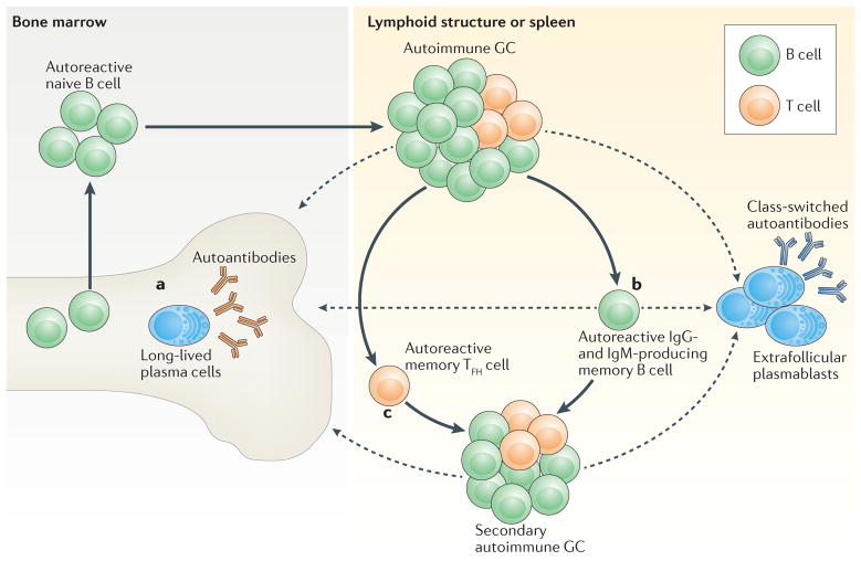
Recent work has provided new insights into how altered B cell-intrinsic signals - through the B cell receptor (BCR) and key co-receptors - function together to promote the pathogenesis of autoimmunity. These combined signals affect B cells at two distinct stages: first, in the selection of the naive repertoire; and second, during extrafollicular or germinal centre activation responses. Thus, dysregulated signalling can lead to both an altered naive BCR repertoire and the generation of autoantibody-producing B cells. Strikingly, high-affinity autoantibodies predate and predict disease in several autoimmune disorders, including type 1 diabetes and systemic lupus erythematosus. This Review summarizes how, rather than being a downstream consequence of autoreactive T cell activation, dysregulated B cell signalling can function as a primary driver of many human autoimmune diseases.


Conflict of interest statement

Competing interests statement

The authors declare no competing interests.

Figures

Figure 1
Figure 1. B cell receptor and co-receptor signalling govern B cell selection and maturation
Self-reactive B cells are subject to positive or negative selection throughout their development in the bone marrow and periphery (spleen). The selective fate of an individual B cell clone depends on multiple factors, including the location and form of self-antigen encounter, the strength of the B cell receptor (BCR) signal and synergy with co-receptor pathways. Negative selection mechanisms (such as deletion, receptor editing and anergy) are mediated primarily by BCR signalling, with potential input from specific Toll-like receptors (TLRs). By contrast, positive selection through survival and/or clonal expansion occurs primarily in transitional B cells in the periphery, and is driven by a complex interplay between BCR signalling and co-receptor signalling mediated by B cell-activating factor receptor (BAFFR), CD40 and TLRs. As the developing repertoire is fine-tuned, transitional B cells mature and populate the follicular mature (FM) compartment or the marginal zone (MZ) compartment. Although the majority of autoreactive BCR specificities are purged by negative selection, a proportion of mature naive B cells exhibit self-reactivity and/or polyreactivity, particularly within the MZ compartment. The dashed arrows indicate ongoing research regarding nonlinear routes for B cell development. T1, transitional type 1; T2, transitional type 2.
Figure 2
Figure 2. Altered B cell receptor and co receptor signalling promotes increased autoreactivity within the naive B cell repertoire
a | Under homeostatic conditions, self-reactive B cells are subjected to both positive and negative selection mechanisms as they transit into the naive B cell pool and establish the naive repertoire. Whereas tonic B cell receptor (BCR) signalling and BCR engagement with self-antigen primarily regulate these events, synergy between the BCR and co-receptors fine-tunes the tolerance programme within a given B cell. Among these co-receptors, B cell-activating factor receptor (BAFFR) signalling (1) synergizes with BCR signalling during late bone marrow and transitional development through a series of complex events, including proximal biochemical crosstalk and the downstream transcriptional regulation of both receptor and substrate expression. Dual BCR and Toll-like receptor (TLR) signalling (2) is mediated by internalization and delivery of self-antigens that contain TLR ligands to autophagosomes, which contain endosome-resident TLRs. CD40 signalling (3), which is triggered by interaction with CD40 ligand (CD40L) on T cells and possibly other cell types, also integrates with the BCR signalling pathway. Although BCR signalling can modulate CD40 expression, other biochemical or transcriptional events that affect this crosstalk are less well understood. b | In genetic (or environmental) settings that promote an increased risk of developing autoimmunity, the homeostatic signalling thresholds are modulated, and self-reactive B cells exhibit greater positive selection and/or reduced negative selection, leading to a naive repertoire that is skewed towards autoreactivity. For example, excess amounts of B cell-activating factor (BAFF; 1) in the Baff-transgenic mouse model rescue low-affinity self-reactive B cells from negative selection. A similar mechanism has been proposed to exist in individuals with systemic lupus erythematosus (SLE). Similarly, in mouse and human settings of Wiskott–Aldrich syndrome (WAS) deficiency (2), hyper-responsive dual BCR and TLR signalling promotes the positive selection of transitional B cells with BCRs that use a limited subset of genes that encode self-reactive heavy-chain variable (VH) domains. Healthy individuals with the autoimmunity-associated variant PTPN22R620W (3) exhibit altered BCR and CD40 signalling, and have an enrichment of self-reactive BCR specificities within the naive B cell compartment. Although it has not yet been definitively demonstrated, it is likely that enhanced positive selection, rather than relaxed negative selection, predominantly mediates this change. The thickness of the arrows indicates the strength of pathway activation. MAPK, mitogen-activated protein kinase; NF-κB, nuclear factor-κB; PI3K, phosphatidylinositol 3-kinase.
Figure 3
Figure 3. T cell-dependent and T cell-independent extrafollicular activation pathways in autoimmunity
Excess amounts of B cell-activating factor (BAFF) promote the survival of autoreactive B cells (part a), there by increasing the proportion of self-reactive B cells within the transitional and mature naive B cell compartments. Following engagement by self-antigen, autoreactive B cells undergo activation in response to dual B cell receptor (BCR)-dependent and Toll-like receptor (TLR)-dependent signalling. Subsequently, activated B cells differentiate into extrafollicular autoantibody-producing plasmablasts through either T cell-dependent (part b) or T cell-independent (part c) pathways. Activated B cells that engage cognate CD4+ T cells at the T cell–B cell border undergo class-switch recombination and differentiation into extrafollicular plasmablasts in response to co-stimulatory signals provided by T cells (part b), including CD40 ligand (CD40L; not shown), inducible T cell co-stimulator (ICOS; not shown) and interleukin-21 (IL-21). Following engagement by self-antigen, autoreactive B cells upregulate their surface expression of transmembrane activator and CAML interactor (TACI) in response to BCR-dependent signalling. TACI engagement by multimeric BAFF 60-mers (part c) promotes the expression of activation-induced cytidine deaminase (AID), resulting in somatic hypermutation, class-switch recombination and the subsequent generation of autoantibody-producing plasmablasts. Although additional B cell subsets may contribute to TACI-dependent autoantibody production in high BAFF settings, transitional B cells make a prominent contribution to the pool of IgG-producing plasmablasts in Baff-transgenic mice. BAFFR, BAFF receptor; TCR, T cell receptor.
Figure 4
Figure 4. Self reactive B cells initiate autoimmune germinal centre formation by facilitating breaks in T cell tolerance
a | After binding to self-antigen (either soluble or bound to antigen-presenting cells) derived from apoptotic particles or other disease-specific targets, autoreactive B cell receptors (BCRs) traffic nuclear antigens to the endosomal receptors Toll-like receptor 7 (TLR7) and TLR9, resulting in initial B cell activation in response to integrated BCR-dependent and TLR-dependent signalling. In parallel, endolysosomal enzymes also process internalized self-antigens (including a broad range of nucleic acid-associated proteins) into peptides for loading onto MHC class II. b | B cells function as antigen-presenting cells to present MHC class II-bound peptides to cognate self-reactive CD4+ T cells at the T cell–B cell border of lymphoid follicles. Together with co-stimulatory signals provided by CD80 and/or CD86 and inducible T cell co-stimulator ligand (ICOSL), self-reactive B cells initiate breaks in CD4+ T cell tolerance. Activated CD4+ T cells subsequently express CXC-chemokine receptor 5 (CXCR5) and B cell lymphoma 6 (BCL-6), resulting in their migration to the B cell follicle as early T follicular helper (TFH) cells (not shown). Activated B cells also produce interleukin-6 (IL-6), which may facilitate TFH cell differentiation by inducing BCL-6 expression, although this has not yet been directly tested. c | TFH cells promote germinal centre (GC) formation through the production of IL-21, which sustains B cell BCL-6 expression and promotes B cell activation, class-switch recombination and plasma cell differentiation. In autoimmune settings, interferon-γ (IFNγ; probably derived from activated CD4+ T cells) drives GC formation in a B cell-intrinsic, signal transducer and activator of transcription 1 (STAT1)-dependent manner, in part by enhancing BCL-6 expression. IFNγ also promotes B cell-intrinsic expression of the transcription factor T-bet (encoded by TBX21), which is required for class-switch recombination to pathogenic IgG2a and IgG2c isotypes, but is redundant for IFNγ-driven GC formation. Although not yet directly tested in autoimmune models, this dysregulated GC response is probably affected by additional cytokines, including B cell-activating factor (BAFF), which promotes the selection of high-affinity GC B cell clones, and IL-12, which facilitates T cell IFNγ production and TFH cell differentiation. d | Iterative interactions between GCB cells and cognate TFH cells within ongoing autoimmune GCs probably result in epitope spreading and the recruitment of additional autoreactive T cell and B cell clones. BAFFR, BAFF receptor; CD40L, CD40 ligand; ICOS, inducible T cell co-stimulator; IFNγR, IFNγ receptor; IL-6R, IL-6 receptor; TCR, T cell receptor.
Figure 5
Figure 5. A sequential model of the progression of systemic autoimmunity
After initial breaks in B cell and T cell tolerance, long-lived effector and memory populations are generated within spontaneous, autoimmune germinal centres (GCs) and contribute to the maintenance and progression of systemic autoimmunity. After iterative rounds of B cell proliferation and selection, B cells that express high-affinity B cell receptors (BCRs) exit the GC as either plasma cells or memory B cells. a | In response to CXC-chemokine receptor 4 (CXCR4)-dependent signalling, autoantibody-secreting plasma cells traffic to the bone marrow, where they exhibit long-term survival within bone marrow stromal niches. b | Alternatively, GC B cells can differentiate into autoreactive memory B cells. Long-lived memory B cells probably contribute to the progression of autoimmunity and to disease relapse following treatment owing to an increased propensity for either differentiation into short-lived, autoantibody-secreting, extrafollicular plasmablasts, or recruitment to and activation within secondary autoimmune GCs. c | In addition to memory B cells, emerging evidence suggests that a subset of T follicular helper (TFH) cells differentiate into memory TFH cells that can seed secondary GCs and promote the progression of autoimmunity. Importantly, after these initial breaks in T cell tolerance and the establishment of autoimmune T cell memory, additional naive B cell clones that exhibit self-reactivity may be recruited into established GCs in the absence of initial priming signals, thereby broadening the range of high-affinity autoantibodies that are generated. Dashed arrows indicate the movement of long-lived and other plasma cell populations to bone marrow or other lymphoid compartments, respectively.

Similar articles

Cited by

References

    1. Deng Y, Tsao BP. Advances in lupus genetics and epigenetics. Curr Opin Rheumatol. 2014;26:482–492. - PMC - PubMed
    1. Niewold TB. Advances in lupus genetics. Curr Opin Rheumatol. 2015;27:440–447. - PMC - PubMed
    1. Yarwood A, Huizinga TW, Worthington J. The genetics of rheumatoid arthritis: risk and protection in different stages of the evolution of RA. Rheumatology (Oxford) 2016;55:199–209. - PMC - PubMed
    1. Parkes M, Cortes A, van Heel DA, Brown MA. Genetic insights into common pathways and complex relationships among immune-mediated diseases. Nat Rev Genet. 2013;14:661–673. - PubMed
    1. Concannon P, Rich SS, Nepom GT. Genetics of type 1A diabetes. N Engl J Med. 2009;360:1646–1654. - PubMed

Substances