. 2017 Jul;215(1):309-322.
doi: 10.1111/nph.14540. Epub 2017 Apr 10.

Quantitative analysis of the tomato nuclear proteome during Phytophthora capsici infection unveils regulators of immunity

Affiliations

Quantitative analysis of the tomato nuclear proteome during Phytophthora capsici infection unveils regulators of immunity

Andrew J M Howden et al. New Phytol. 2017 Jul.

Abstract

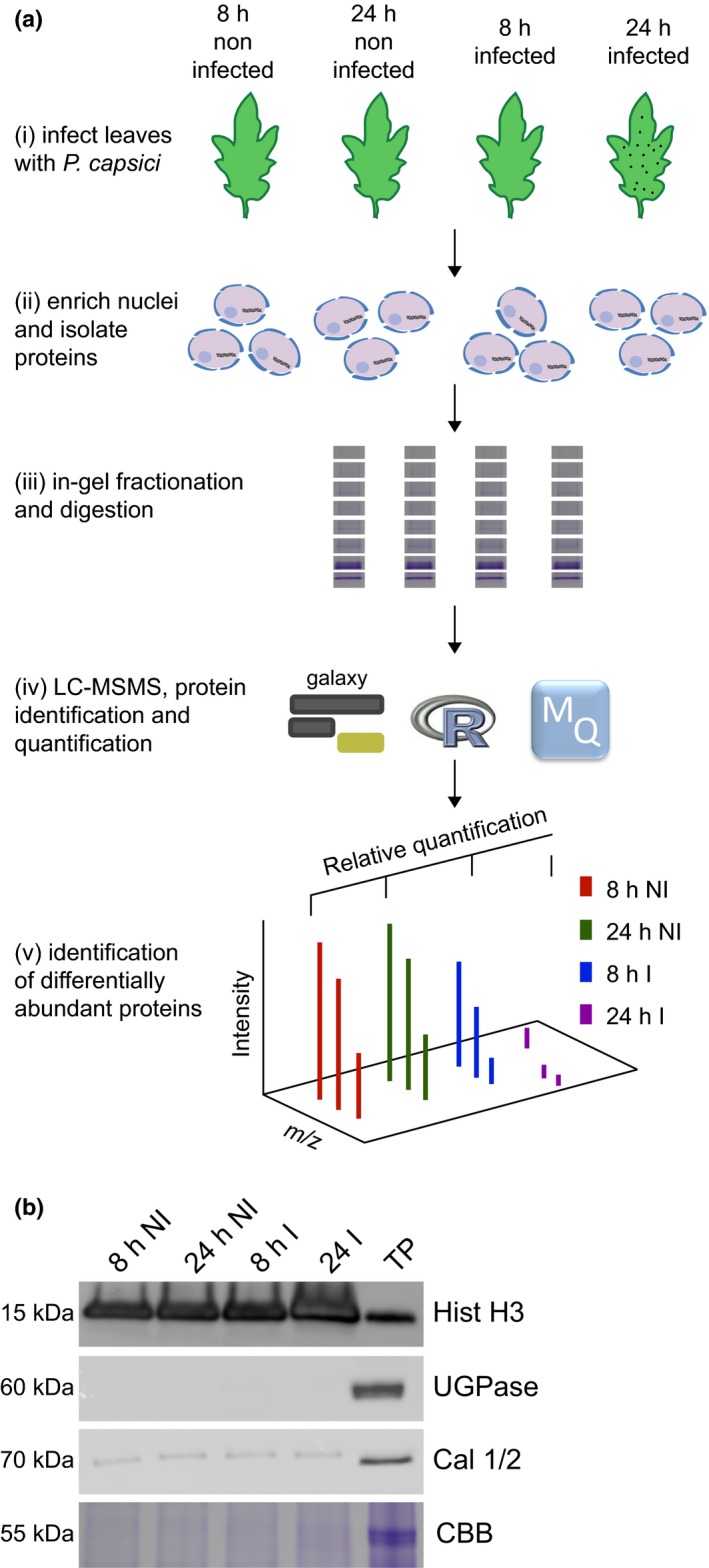
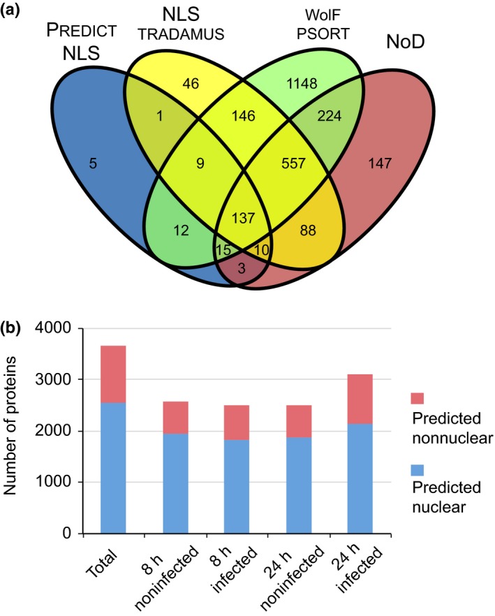
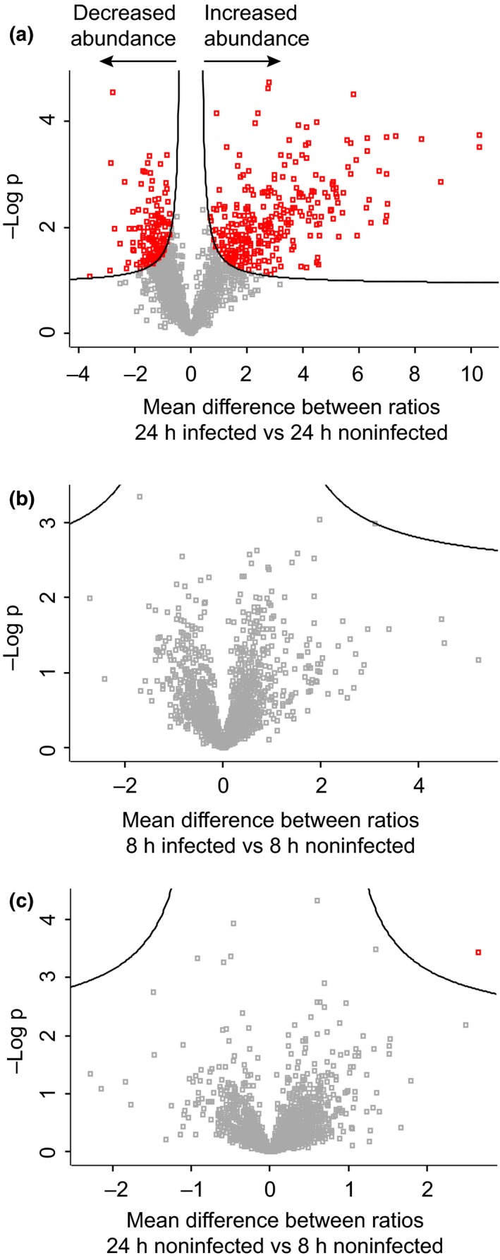
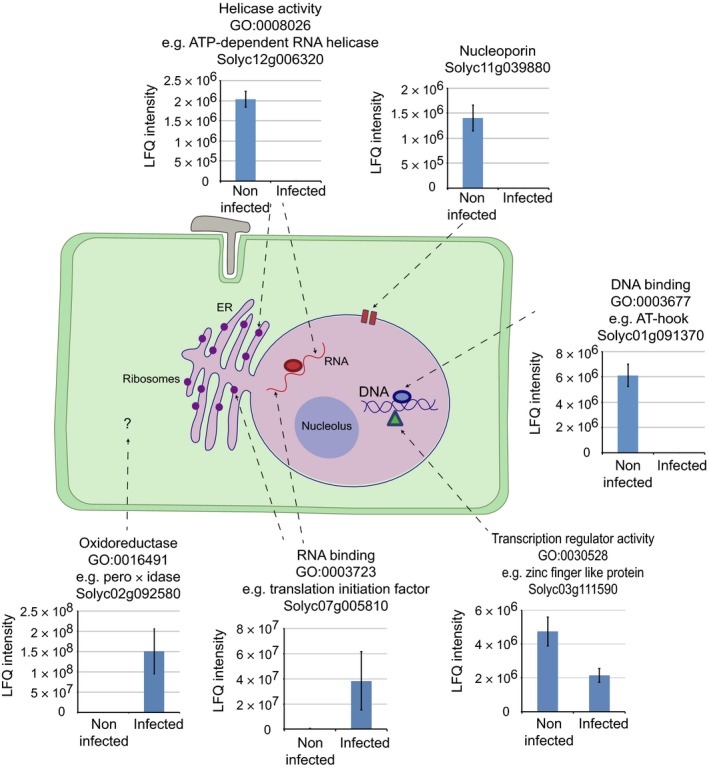
Plant-pathogen interactions are complex associations driven by the interplay of host and microbe-encoded factors. With secreted pathogen proteins (effectors) and immune signalling components found in the plant nucleus, this compartment is a battleground where susceptibility is specified. We hypothesized that, by defining changes in the nuclear proteome during infection, we can pinpoint vital components required for immunity or susceptibility. We tested this hypothesis by documenting dynamic changes in the tomato (Solanum lycopersicum) nuclear proteome during infection by the oomycete pathogen Phytophthora capsici. We enriched nuclei from infected and noninfected tissues and quantitatively assessed changes in the nuclear proteome. We then tested the role of candidate regulators in immunity through functional assays. We demonstrated that the host nuclear proteome dynamically changes during P. capsici infection. We observed that known nuclear immunity factors were differentially expressed and, based on this observation, selected a set of candidate regulators that we successfully implicated in immunity to P. capsici. Our work exemplifies a powerful strategy to gain rapid insight into important nuclear processes that underpin complex crop traits such as resistance. We have identified a large set of candidate nuclear factors that may underpin immunity to pathogens in crops.

Keywords: Phytophthora; immunity; nucleus; plant-microbe interactions; quantitative proteomics; tomato.


Figures

Figure 1
Figure 1
A simple workflow combining nuclear enrichment with quantitative mass spectrometry allows the study of host nuclear processes during infection. (a) Method overview. Detached leaves from 4‐wk‐old tomato plants were spray‐inoculated with Phytophthora capsici zoospores at a concentration of 500 000 spores ml−1, or water as a control (i). Leaves were harvested 8 and 24 h post‐infection and subject to nuclear enrichment (ii). Nuclear protein extracts were generated and fractionated in‐gel and digested with trypsin (iii). Peptide samples were subjected to liquid chromatography−tandem mass spectrometry (LC‐MSMS) analysis and peptide identification and label‐free quantification was carried out using the maxquant and perseus software packages, to identify proteins that were differentially expressed during infection. Nuclear predictions were performed using prediction software on our in‐house Galaxy server as described in the Methods. Subsequent data filtering was completed using the r software package (iv and v). Three independent biological replicates were generated. I, infected samples; NI, noninfected samples (v). (b) Successful enrichment of nuclear proteins demonstrated by western blotting with anti‐histone H3 antibody and subcellular markers. Protein extracts from enriched nuclear samples were compared to a total protein extract (TP) from tomato leaves. Protein concentrations were adjusted for equal loading. Nonnuclear contamination was assessed by probing with anti‐UDP‐glucose pyrophosphorylase (UGPase) antibody (cytoplasm) and calnexin homologue 1/2 antibody (endoplasmic reticulum). Samples were also run on gels and stained with Coomassie brilliant blue (CBB) to assess protein loading and Rubisco abundance. The figure shows the results from a single biological replicate. Blots for all three replicates are provided in Supporting Information Fig. S2.
Figure 2
Figure 2
Amino acid sequence analysis reveals that the majority of identified proteins have a predicted nuclear association. (a) Contaminant proteins and Phytophthora capsici proteins were removed from the data set and all remaining proteins were subjected to four nuclear prediction tools to establish how many proteins had a predicted nuclear association. In total, 2548 proteins were either found to have a nuclear localization signal (NLS) (according to predictnls and nlstradamus) or a nucleolar localization signal (using nod) or were predicted to localize to the nucleus according to wolf p‐sort. (b) Overview of protein numbers found within individual treatments and their predicted localization. Proteins that were not predicted to be nuclear in our analyses are classified as ‘predicted nonnuclear’. The full list of proteins found within the data set along with their quantification values is provided in Supporting Information Notes S1.
Figure 3
Figure 3
Identification of proteins that show a significant change in abundance during infection. Protein expression data were analysed within perseus using two‐sample t‐tests and a false discovery threshold of 0.05 to identify nuclear candidates significantly changing in abundance. A total of 285 proteins significantly increased in abundance 24 h post‐infection relative to the 24‐h noninfected sample, while 140 proteins significantly decreased in abundance upon infection (a). No proteins were found to be significantly altered in abundance 8 h post‐infection (b), while only one protein passed the significance threshold when comparing 8‐h and 24‐h noninfected samples (c).
Figure 4
Figure 4
Overview of host cellular processes changing during Phytophthora capsici infection. Quantitative proteomics revealed that diverse nuclear and nuclear‐associated processes may be modified during pathogen infection, including the activity of DNA‐ and RNA‐binding proteins, transcription factors, components of the nuclear pore complex and DNA and RNA helicases. Predicted nonnuclear proteins including peroxidases were also found to change in abundance. Graphs show the label‐free quantification data (LFQ) for selected proteins, with each bar representing the average LFQ intensity from three biological replicates ± SD.
Figure 5
Figure 5
Localization of AT‐Hook‐Like (AHL) proteins in planta. AHL1, AHL5, AHL9, AHL17a and AHL17b localized to the plant nucleus when transiently overexpressed in Nicotiana benthamiana using Agrobacterium tumefaciens‐mediated expression. Nicotiana benthamiana plants expressing histone‐RFP were used to visualize the nucleus. Leaves were infiltrated at an optical density (OD600) of 0.1 and imaged after 48 h. Bars, 5 μM. EGFP, enhanced green fluorescent protein.
Figure 6
Figure 6
Impact of five tomato AT‐Hook‐Like (AHL) proteins on immunity to Phytophthora capsici. Over‐expression of AHL1, AHL5 and AHL9 resulted in reduced P. capsici growth compared with an empty vector (EV) control. Virulence assays were performed in Nicotiana benthamiana plants over‐expressing enhanced green fluorescent protein (EGFP)‐AHLs. Plants were infiltrated with the Agrobacterium tumefaciens expression construct at OD 0.1. After 48 h, leaves were detached and 10‐μl drops of P. capsici spores (at a density of 50 000 ml−1) were placed on the leaf surface. Lesion diameters were measured 48 and 72 h post‐infection (hpi). Significant difference relative to the EV control according to Dunnett's multiple comparison testing: *, < 0.05. Error bars show ± SE.
Figure 7
Figure 7
Two AT‐Hook‐Like (AHL) proteins that enhance immunity to Phytophthora capsici also impact upon pattern triggered immunity (PTI) responses. PTI assays were performed in Nicotiana benthamiana plants over‐expressing enhanced green fluorescent protein (EGFP)‐AHLs or EGFP‐Avr3aKI (Engelhardt et al., 2012) on one half of the leaf and EGFPEV on the other. Leaves were then infiltrated with either Phytophthora capsici culture filtrate (CF) to trigger a PTI response, or pea broth (PB) as a negative control. Results show the levels of cell death observed 72 h after treatment with CF or PB. Cell death was scored from 0 to 6 (no cell death to high cell death) following the scoring method described by Stam et al. (2013). Significant difference relative to EV CF control according to Welch's t‐test: *P < 0.05. Error bars show ± SE. EV, empty vector.

Similar articles

Cited by

References

    1. Bos JIB, Armstrong MR, Gilroy EM, Boevink PC, Hein I, Taylor RM, Tian ZD, Engelhardt S, Vetukuri RR, Harrower B et al 2010. Phytophthora infestans effector AVR3a is essential for virulence and manipulates plant immunity by stabilizing host E3 ligase CMPG1. Proceedings of the National Academy of Sciences, USA 107: 9909–9914. - PMC - PubMed
    1. Canonne J, Marino D, Jauneau A, Pouzet C, Briere C, Roby D, Rivas S. 2011. The Xanthomonas Type III effector XopD targets the Arabidopsis transcription factor MYB30 to suppress plant defense. Plant Cell 23: 3498–3511. - PMC - PubMed
    1. Canonne J, Rivas S. 2012. Bacterial effectors target the plant cell nucleus to subvert host transcription. Plant Signaling & Behavior 7: 217–221. - PMC - PubMed
    1. Cock PJA, Gruning BA, Paszkiewicz K, Pritchard L. 2013. Galaxy tools and workflows for sequence analysis with applications in molecular plant pathology. PeerJ 1: e167. - PMC - PubMed
    1. Cokol M, Nair R, Rost B. 2000. Finding nuclear localization signals. Embo Reports 1: 411–415. - PMC - PubMed

LinkOut - more resources