. 2017 Jun;20(6):872-878.
doi: 10.1038/nn.4545. Epub 2017 Apr 10.

The neural correlates of dreaming

Affiliations

The neural correlates of dreaming

Francesca Siclari et al. Nat Neurosci. 2017 Jun.

Abstract

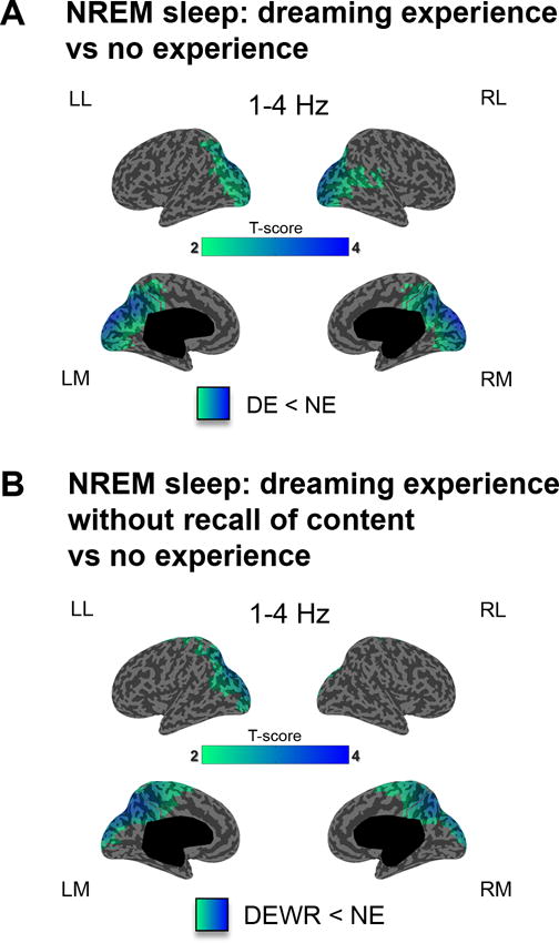
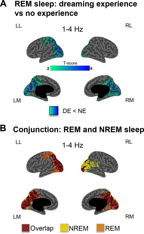
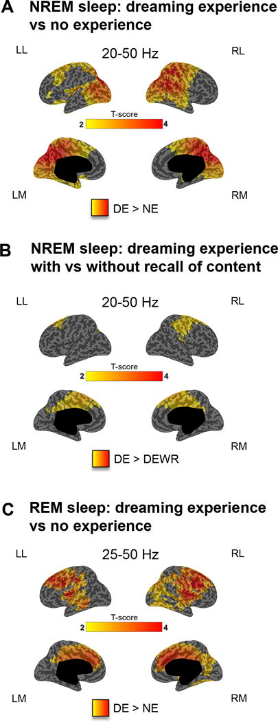
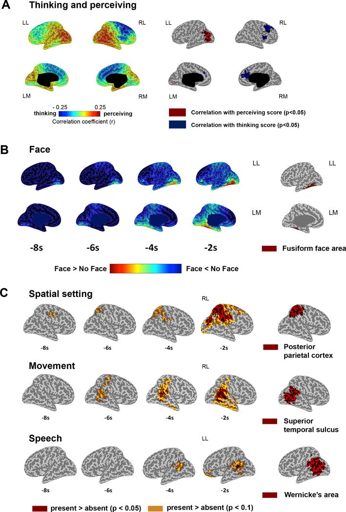
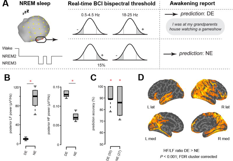
Consciousness never fades during waking. However, when awakened from sleep, we sometimes recall dreams and sometimes recall no experiences. Traditionally, dreaming has been identified with rapid eye-movement (REM) sleep, characterized by wake-like, globally 'activated', high-frequency electroencephalographic activity. However, dreaming also occurs in non-REM (NREM) sleep, characterized by prominent low-frequency activity. This challenges our understanding of the neural correlates of conscious experiences in sleep. Using high-density electroencephalography, we contrasted the presence and absence of dreaming in NREM and REM sleep. In both NREM and REM sleep, reports of dream experience were associated with local decreases in low-frequency activity in posterior cortical regions. High-frequency activity in these regions correlated with specific dream contents. Monitoring this posterior 'hot zone' in real time predicted whether an individual reported dreaming or the absence of dream experiences during NREM sleep, suggesting that it may constitute a core correlate of conscious experiences in sleep.


Conflict of interest statement

The other authors have indicated no financial conflicts of interest.

Figures

Figure 1
Figure 1. Dreaming experience vs. no experience in NREM sleep (low-frequency power)
A. Cortical distribution of t-values for the contrast between DEs and NEs at the source level for low-frequency power (1–4 Hz) in NREM sleep (20s before the awakening). p<0.05, after correction for multiple comparisons (two-tailed, paired t-tests, 32 subjects, t(31) > 2.04). B. Same as A for the contrast between NE and DEWR (two-tailed, paired t-tests, 20 subjects, t(19) > 2.09).
Figure 2
Figure 2. Dreaming experience vs. no experience in REM sleep (low-frequency power)
A. Cortical distribution of t-values for the contrast between DEs and NEs at the source level for low-frequency (1–4 Hz) power in REM sleep (20s before the awakening). p<0.05 after correction for multiple comparisons (two-tailed, paired t-tests, 10 subjects, t(9)>2.26). B. Conjunction maps: differences and overlap compared to the same contrast performed in NREM sleep.
Figure 3
Figure 3. Dreaming experience vs. no experience in NREM and REM sleep (high- frequency power)
A. Cortical distribution of t-values for the contrast between DEs and NEs at the source level for high-frequency power (20–50 Hz) in NREM sleep (20s before the awakening). p<0.05 after correction for multiple comparisons (two-tailed, paired t-tests, 32 subjects, t(31) > 2.04). B. Same as A for the contrast between DE and DEWR in NREM sleep (two-tailed, paired t-tests, 20 subjects, t(19) > 2.09) C. same as A for high-frequency power (25–50 Hz) in REM sleep (two-tailed, paired t-tests, 10 subjects, t(9) > 2.26).
Figure 4
Figure 4. The content of dream experiences in REM sleep
A. Correlation between the thinking/perceiving score and 25–50 Hz power (last 8s, 7 subjects). Left: mean Spearman rank correlation coefficients (7 subjects). Right: significant voxels p<0.05 (one-tailed permutation test, r>0.14). B. Left: 25–50 Hz power differences (DE with face minus DE without face). ROI contrast R FFA p=0.023; one-tailed paired t-test (7 subjects, t(6) = 2.52). Right: fusiform face area (red). C. Upper row: 25–50 Hz average power differences between DEs with and without a spatial setting (6 subjects, t(5) > 2.57). Right: right posterior parietal cortex. Middle row: movement vs. no movement (7 subjects, t(6) > 2.45). Right: superior temporal sulcus. Bottom row: speech vs. no speech (7 subjects, t(6) > 2.45). Right: Wernicke’s area. Two-tailed paired t-tests. p<0.05 (red) and p<0.01 (yellow).
Figure 5
Figure 5. Real-time prediction of dream experience
A. Awakenings were performed in NREM when neural activity surpassed a bispectral threshold in low- frequency (LF; 0.5–4.5 Hz) and high-frequency (HF; 18–25 Hz) power over the posterior hot zone. B. DE trials had significantly lower LF activity and significantly higher HF activity in DE compared to NE prediction trials. The bottom and top of the boxes show the 25th and 75th percentile (the lower and upper quartiles), the inner band shows the median, and the whiskers show the upper and lower quartiles +/− 1.5 * IQR (inter quartile range). Asterisks indicate significant differences (p=0.001; two-tailed t-test) between CE and NCE trials. C. Prediction accuracy for DE (55) and NE (27) trials. Asterisks indicate significant differences (p<0.001; one sample t-test two-tailed) between CE and NCE prediction accuracy and chance (50%). D. DE had a higher HF/LF power ratio compared to NE in cortical regions including bilateral occipital, medial and lateral parietal, medial temporal and inferior frontal cortex (p<0.001; two-tailed paired t-test, FDR cluster corrected).

Comment in

  • Unraveling the mystery of dreams.
    Lin X, Han Y, Lu L. Lin X, et al. J Thorac Dis. 2017 Sep;9(9):2732-2735. doi: 10.21037/jtd.2017.07.103. J Thorac Dis. 2017. PMID: 29221226 Free PMC article. No abstract available.

References

    1. Stickgold R, Malia A, Fosse R, Propper R, Hobson JA. Brain-mind states: I. Longitudinal field study of sleep/wake factors influencing mentation report length. Sleep. 2001;24:171–179. - PubMed
    1. Nir Y, Tononi G. Dreaming and the brain: from phenomenology to neurophysiology. Trends Cogn Sci. 2010;14:88–100. - PMC - PubMed
    1. Schwartz S, Maquet P. Sleep imaging and the neuro-psychological assessment of dreams. Trends Cogn Sci. 2002;6:23–30. - PubMed
    1. Aserinsky E, Kleitman N. Regularly occuring periods of eye motility, and concominant phenomena, during sleep. Science. 1953;118:273–274. - PubMed
    1. Moruzzi G, Magoun HW. Brain stem reticular formation and activation of the EEG. Electroencephalogr Clin Neurophysiol. 1949;1:455–473. - PubMed