. 2017 May;19(5):568-577.
doi: 10.1038/ncb3516. Epub 2017 Apr 10.

Long-term, hormone-responsive organoid cultures of human endometrium in a chemically defined medium

Affiliations

Long-term, hormone-responsive organoid cultures of human endometrium in a chemically defined medium

Margherita Y Turco et al. Nat Cell Biol. 2017 May.

Abstract

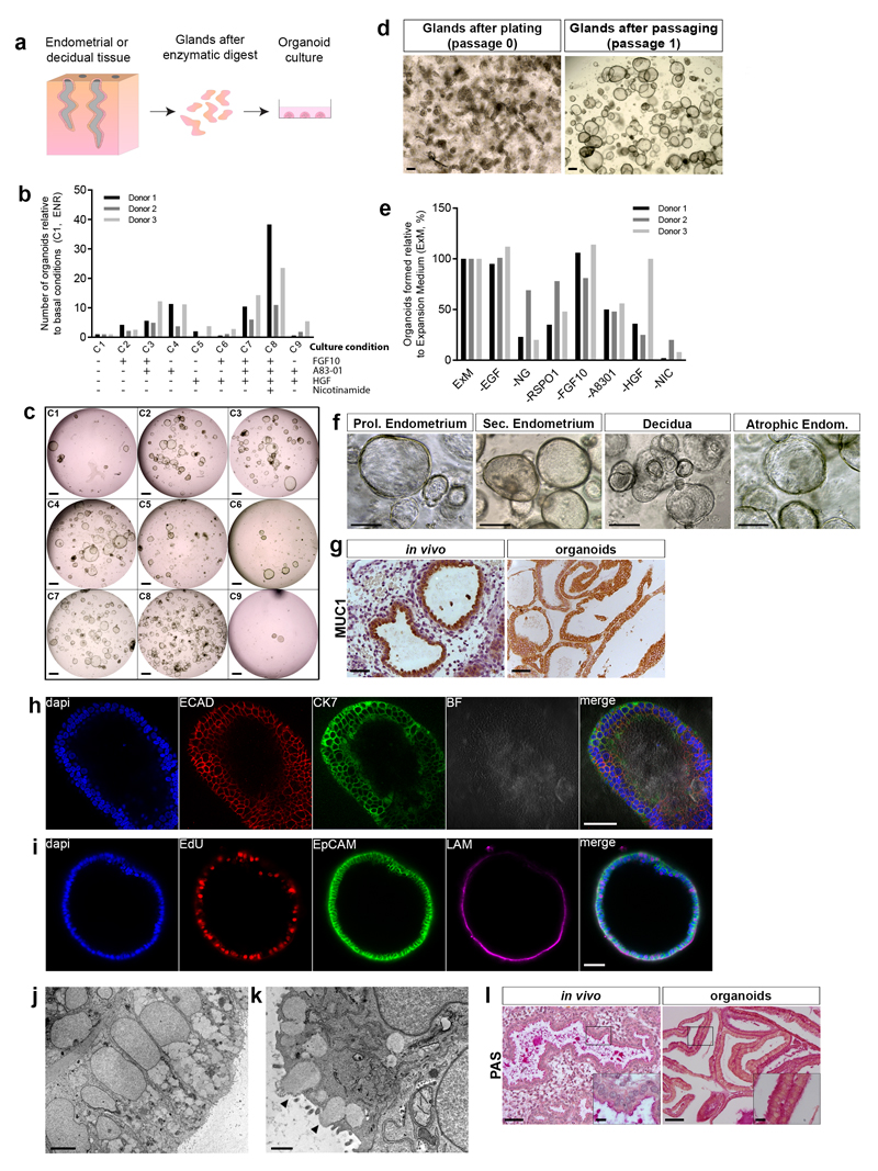
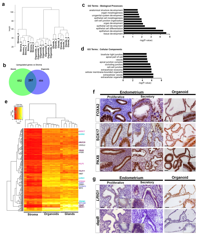
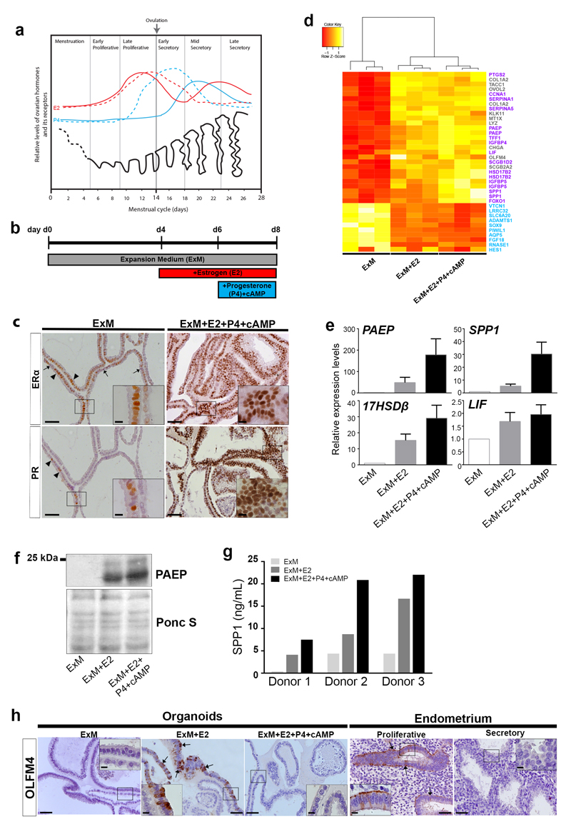
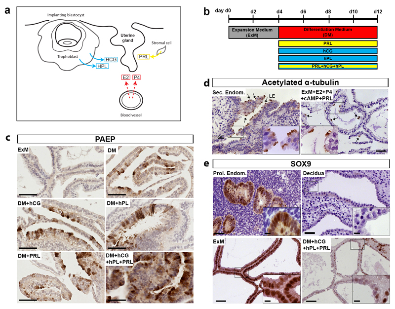
In humans, the endometrium, the uterine mucosal lining, undergoes dynamic changes throughout the menstrual cycle and pregnancy. Despite the importance of the endometrium as the site of implantation and nutritional support for the conceptus, there are no long-term culture systems that recapitulate endometrial function in vitro. We adapted conditions used to establish human adult stem-cell-derived organoid cultures to generate three-dimensional cultures of normal and decidualized human endometrium. These organoids expand long-term, are genetically stable and differentiate following treatment with reproductive hormones. Single cells from both endometrium and decidua can generate a fully functional organoid. Transcript analysis confirmed great similarity between organoids and the primary tissue of origin. On exposure to pregnancy signals, endometrial organoids develop characteristics of early pregnancy. We also derived organoids from malignant endometrium, and so provide a foundation to study common diseases, such as endometriosis and endometrial cancer, as well as the physiology of early gestation.


Figures

Figure 1
Figure 1. Long-term 3D organoid cultures can be established from human non-pregnant endometrium and decidua.
(a) Scheme for deriving organoids. (b) Screening conditions for generating organoids. FGF10, A83-01, HGF and Nicotinamide added in combinations to generic organoid medium (ENR). Number of organoids derived under each condition (C2 to C9) shown relative to basal conditions (C1). Decidual digests from 3 different patients. Source data in Supplementary Table 5. (c) Representative images for conditions C1-C9 in Fig. 1b. Scale bar, 500 μm. (d) Images of decidual gland isolates (passage 0) and organoids after one passage in Expansion Medium (ExM) (passage 1). Scale bar, 200 μm. Representative of all samples, summarized in Supplementary Table 1. (e) Effect of withdrawal of growth factors from ExM. Organoids grown in ExM and each factor withdrawn: EGF, Noggin (NG), Rspondin-1 (RSPO1), FGF10, A8301, HGF and Nicotinamide (NIC). Organoids formed shown relative to ExM (%). Shown are decidual cultures derived from 3 different patients. Source data in Supplementary Table 5. (f) Images of organoids established in ExM from proliferative (Prol.) endometrium (n=3), secretory (Sec.) endometrium (n=9), decidua (n=25) and post-menopausal (atrophic) endometrium (n=1). Scale bar, 100 μm. (g) IHC of decidua (in vivo) and organoids for Mucin 1 (MUC-1). Scale bar, 50 μm. Representative of 6 decidual and endometrial samples, and organoids derived from 2 endometrial and 2 decidual samples from different patients. (h) IF staining of organoid for E-CADHERIN (E-CAD) and CYTOKERATIN-7 (CK7). Scale bar, 50 μm. Experiment repeated twice (1 endometrial-derived and 1 decidua-derived organoids). (i) IF staining of organoid for cell proliferation (uptake of EdU), epithelial marker EPCAM and basement membrane marker laminin (LAM). Scale bar, 50 μm. Experiment repeated twice (1 endometrial-derived and 1 decidua-derived organoids). (j) Electron micrograph (EM) of organoid showing columnar epithelial cells with basally-located nuclei. Scale bar, 5 μm. Experiment repeated twice with different donors. (k) EM showing secretory activity (black arrowheads). Scale bar, 1 μm. Experiment repeated twice with different donors. (l) PAS staining for glycogen in endometrium and organoids. Scale bars, 50 μm (main image) and 10 μm (inset). Representative of 3 endometrial samples and 3 endometrial organoids.
Figure 2
Figure 2. Established human endometrial organoids recapitulate molecular signature of glands in vivo.
(a) Unsupervised hierarchical clustering analysis of global gene expression profiles by microarray of gland digests, stromal cells and corresponding established organoids from endometrium (n=7 independent donors). Analysis based on 15475 probes with sd/mean >0.1. Expression profiles of organoids cluster with glands while those of the stroma cluster in a separate tree. (b) Venn diagram showing overlap of 287 genes significantly upregulated in glands and organoids with a fold change ≥1.5 (p≤0.01) relative to stroma. (c) Gene ontology (GO) analysis of the 287 genes from (b) using HumanMine v2.2 database for GO Terms Biological processes and Benjamini Hochberg test correction with maximum p-value of 0.05. The top ten significantly enriched GO terms for each category are shown with the –log of their p-values and are enriched for terms describing epithelial tissue. (d) Gene ontology (GO) analysis of the 287 genes from (b) using same method as in (c). The top ten significantly enriched GO terms describe epithelial cells with secretory function. (e) Clustered heatmap of 287 genes commonly upregulated between organoids and glands compared to stroma from (b). Genes of interest are listed on the right. Epithelial markers (blue) (EPCAM, CLD10, CDH1), glandular products and markers of secretory cells (purple) (MUC20, PAX8, PAEP, MUC1), progenitor cell markers (cyan) (LRIG1, PROM1, AXIN2) and murine genes important for endometrial function (pink) (SOX17, KLF5, FOXA2). (f) IHC for genes selected from microarray, FOXA2, SOX17 and PAX8, in proliferative and secretory endometrium and organoids. Scale bars, 50 μm (main image) and 10 μm (insets). Representative of 3 proliferative and 7 secretory endometrial samples and endometrial organoids derived from 8 different patients. (g) ISH for LRIG1 on proliferative and secretory endometrium and organoids. Negative control probe is for the bacterial gene dapB. Scale bars, 50 μm (main image) and 10 μm (insets). Representative of 3 proliferative and 3 secretory endometrial samples and endometrial organoids derived from 4 different patients.
Figure 3
Figure 3. Human endometrial organoids respond to sex hormones.
(a) Ovarian hormones, Estrogen (E2)(red), Progesterone (P4)(blue), and the cycling endometrium. Expression of Estrogen Receptor (ERα)(dashed red) and Progesterone Receptor (PR)(dashed blue) are specific for glands of the functional layer. Adapted from Reference. (b) Hormonal stimulation. Organoids grown in ExM, day 0 (d0), are primed with E2 for 48 h on day 4 (d4) followed by stimulation with P4 and cyclic AMP (cAMP) for 48 h. (c) IHC for ERα and PR on organoids after hormonal stimulation. In ExM expression of ERα is weak, but some cells are either ERαhigh (arrowheads) or ERαnegative (arrows). Few cells are positive for PR (arrowheads). After E2 and P4 treatment, levels of ERα and PR are higher. Scale bars, 50 μm (main image), 10 μm (insets). Representative of endometrial organoids from 6 patients and decidual organoids from 9 patients. (d) Clustered heatmap of selected genes from organoids grown in ExM, ExM+E2 or ExM+E2+P4+cAMP (n=3 donors). Shown are genes known to reflect differentiation in response to hormones (purple), uncharacterized genes (grey) and downregulated genes (cyan). (e) QRT-PCR analysis for differentiation markers (PAEP, SPP1, 17HSDβ2 and LIF) of organoids grown in ExM, ExM+E2 or ExM+E2+P4+cAMP. Shown is the mean±SEM levels of expression relative to housekeeping genes and ExM conditions (δδCt). Data from endometrial organoids from n=6 different patients. Source data in Supplementary Table 5. (f) Western blot for PAEP in organoids after hormonal stimulation. Levels of glycosylated and non-glycosylated PAEP increase upon exposure to E2 and E2+P4+cAMP. Ponceau S staining (Ponc S) for loading control. Experiment repeated twice using endometrial organoids from 2 patients. Unprocessed blots in Supplementary Figure 7. (g) ELISA for SPP1 production by endometrial organoids upon exposure to hormones. Three independent experiments (Donors 1-3). SPP1 secretion increases following exposure to E2 and further after E2+P4+cAMP. Source data in Supplementary Table 5. (h) IHC for OLFM4 on organoids under ExM, ExM+E2 and ExM+E2+P4+cAMP, and proliferative and secretory endometrium. Scale bars, 50 μm (main image) and 10 μm (insets). Representative of 2 proliferative and 2 secretory endometrial tissues and organoids derived from 3 different patients.
Figure 4
Figure 4. Signals from decidualised stroma and the placenta can further stimulate differentiation of human endometrial gland organoids.
(a) Hormonal environment of endometrium during the first trimester of pregnancy. Estrogen (E2) and Progesterone (P4) are ovarian products, human chorionic gonadotropin (hCG) and human placental lactogen (hPL) are secreted by trophoblast and prolactin (PRL) by decidualized stromal cells. (b) Protocol for stimulation of endometrial organoids. Organoids are passaged and plated on day 0 (d0) in ExM. On d4, ExM is changed to Differentiation Medium (DM; ExM with E2+P4+cAMP). hCG, hPL and/or PRL were added for 8 d. (c) IHC for PAEP on endometrial organoids under the following conditions: ExM, DM, DM with hCG/hPL or PRL or all three combined. Maximal production of PAEP and differentiated morphology of cells is seen upon exposure to DM with hCG, hPL and PRL. Scale bar, 50 μm. Representative of endometrial organoids derived from 3 different patients. (d) IHC for acetylated α-tubulin to visualize cilia in secretory endometrium (Sec. Endom.) and endometrial organoids following stimulation with PRL. Ciliated cells (arrows) are present in the luminal epithelium (LE) and within organoids. GE, glandular epithelium. Scale bars, 50 μm (main image) and 10 μm (insets). Representative of 4 secretory endometrial samples and endometrial organoids derived from 4 different patients. (e) Immunohistochemistry for SOX9 on endometrial glands (in vivo) and organoids. Organoids in ExM express high levels of SOX9 similar to proliferative endometrium (Prol. Endom.). After hormonal stimulation, SOX9 is downregulated in organoids (ExM+HCG+HPL+PRL) similar to glands in decidua. Scale bars, 50 μm (main image) and 10 μm (insets). Representative of 4 proliferative endometrial samples, 7 decidual samples and endometrial organoids derived from 4 different patients.
Figure 5
Figure 5. Human endometrial organoids have clonogenic ability and are bipotent.
(a) Phase-contrast images of (from top to bottom row): an organoid forming from a single cell; a single cell forming a spheroid with no further growth, and a single cell showing no growth. Images were taken every two days. Scale bar, 50 μm. Experiment was performed with 3 clonal lines derived from 2 endometrial and 1 decidual organoid cultures. (b) Representative image showing expansion of a clonal culture at passage 1 (p1) from a single organoid (at passage 0, p0) in a 96-well. Scale bar, 500 μm. 12 clonal cultures were established from organoids from 5 different patient samples (4 endometrial-derived and 1 decidual-derived). (c) IF on clonally-derived endometrial organoid cultures subjected to the full cocktail of hormonal stimuli to visualize two main endometrial epithelial cell types: ciliated cells (acetylated α-tubulin) (cyan) and secretory cells (PAEP) (red). Scale bars from left to right: 100 μm, 20 μm and 5 μm. Representative of 4 clonal lines derived from 2 different endometrial organoid cultures. (d) EM on clonally-derived endometrial organoid cultures subjected to the full cocktail of hormonal stimuli showing basal bodies of fully formed cilia. Scale bars: 10 μm and 1 μm. Experiment performed twice using 1 clonal endometrial organoid culture.
Figure 6
Figure 6. Organoids can be derived from endometrial cancer.
Derivation of organoids from endometrial carcinomas. From left to right: H&E stained sections of normal atrophic endometrium showing gland surrounded by dense stroma and a FIGO Grade I endometrioid carcinoma with dense glandular structures from the same patient, scale bar, 100 μm; images of organoids derived from matched normal and malignant endometrium cultured in ExM (passage 1), scale bar, 100 μm; H&E stained sections showing marked differences in morphology between organoids derived from normal endometrium and those from tumours which show nuclear pleomorphism, a disorganized epithelium with irregular basement membrane and isolated cells present in surrounding Matrigel (arrows), scale bar, 20 μm; IHC for MUC-1 and SOX17 on tumour and normal organoids confirm their glandular origin, scale bar, 20 μm. Representative of organoids derived from 3 different endometrial carcinomas and 1 matching normal tissue.

References

    1. Burton GJ, Watson AL, Hempstock J, Skepper JN, Jauniaux E. Uterine glands provide histiotrophic nutrition for the human fetus during the first trimester of pregnancy. The Journal of clinical endocrinology and metabolism. 2002;87:2954–2959. - PubMed
    1. Hempstock J, Cindrova-Davies T, Jauniaux E, Burton GJ. Endometrial glands as a source of nutrients, growth factors and cytokines during the first trimester of human pregnancy: a morphological and immunohistochemical study. Reproductive biology and endocrinology : RB&E. 2004;2:58. - PMC - PubMed
    1. Gray CA, Burghardt RC, Johnson GA, Bazer FW, Spencer TE. Evidence that absence of endometrial gland secretions in uterine gland knockout ewes compromises conceptus survival and elongation. Reproduction. 2002;124:289–300. - PubMed
    1. Filant J, Spencer TE. Endometrial glands are essential for blastocyst implantation and decidualization in the mouse uterus. Biology of reproduction. 2013;88:93. - PubMed
    1. Zhang S, et al. Physiological and molecular determinants of embryo implantation. Molecular aspects of medicine. 2013;34:939–980. - PMC - PubMed

MeSH terms