. 2017 Apr 10;13(4):e1006327.
doi: 10.1371/journal.ppat.1006327. eCollection 2017 Apr.

Analysis of host microRNA function uncovers a role for miR-29b-2-5p in Shigella capture by filopodia

Affiliations

Analysis of host microRNA function uncovers a role for miR-29b-2-5p in Shigella capture by filopodia

Ushasree Sunkavalli et al. PLoS Pathog. .

Abstract

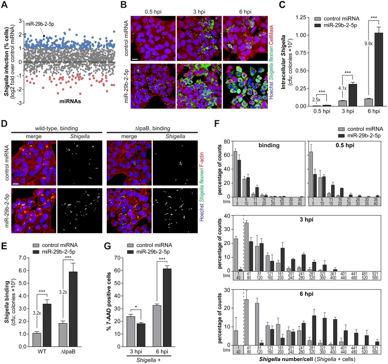
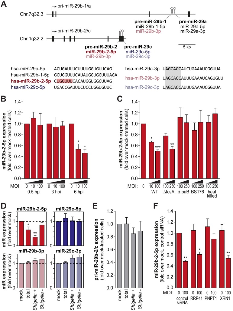
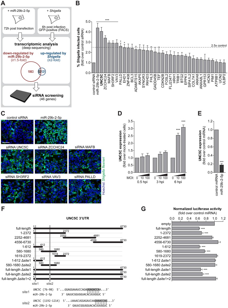
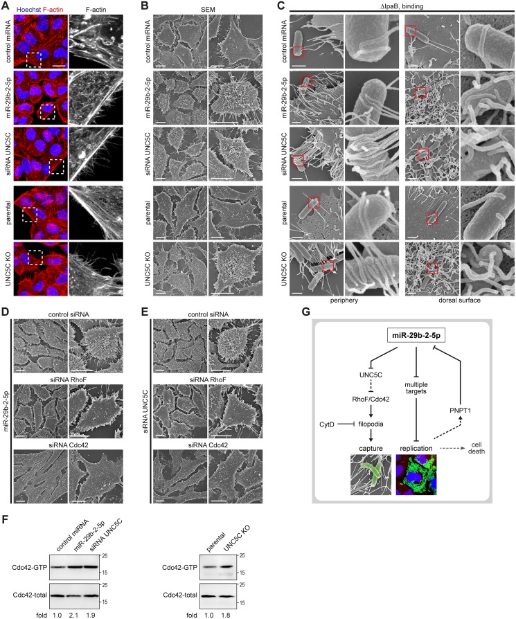
MicroRNAs play an important role in the interplay between bacterial pathogens and host cells, participating as host defense mechanisms, as well as exploited by bacteria to subvert host cellular functions. Here, we show that microRNAs modulate infection by Shigella flexneri, a major causative agent of bacillary dysentery in humans. Specifically, we characterize the dual regulatory role of miR-29b-2-5p during infection, showing that this microRNA strongly favors Shigella infection by promoting both bacterial binding to host cells and intracellular replication. Using a combination of transcriptome analysis and targeted high-content RNAi screening, we identify UNC5C as a direct target of miR-29b-2-5p and show its pivotal role in the modulation of Shigella binding to host cells. MiR-29b-2-5p, through repression of UNC5C, strongly enhances filopodia formation thus increasing Shigella capture and promoting bacterial invasion. The increase of filopodia formation mediated by miR-29b-2-5p is dependent on RhoF and Cdc42 Rho-GTPases. Interestingly, the levels of miR-29b-2-5p, but not of other mature microRNAs from the same precursor, are decreased upon Shigella replication at late times post-infection, through degradation of the mature microRNA by the exonuclease PNPT1. While the relatively high basal levels of miR-29b-2-5p at the start of infection ensure efficient Shigella capture by host cell filopodia, dampening of miR-29b-2-5p levels later during infection may constitute a bacterial strategy to favor a balanced intracellular replication to avoid premature cell death and favor dissemination to neighboring cells, or alternatively, part of the host response to counteract Shigella infection. Overall, these findings reveal a previously unappreciated role of microRNAs, and in particular miR-29b-2-5p, in the interaction of Shigella with host cells.


Conflict of interest statement

The authors have declared that no competing interests exist.

Figures

Fig 1
Fig 1. MiR-29b-2-5p favors Shigella binding and intracellular replication.
A. Effect of human miRNA mimics (genome-wide library of miRNA mimics corresponding to miRBase 19) on the percentage of Shigella infected cells (expressed as log2 fold change compared to control miRNA). MiRNAs highlighted in blue and red significantly increase or decrease infection by at least 2-fold, respectively (P<0.05). B and C. Representative images (B) and cfu quantification (C) of intracellular bacteria in HeLa cells infected with Shigella WT, upon treatment with miR-29b-2-5p or control miRNA mimics, and analyzed at three times post-infection (0.5, 3 and 6 hpi). Scale bar, 20 μm. D and E. Representative images (D) and cfu quantification (E) of bacteria bound to HeLa cells transfected with miR-29b-2-5p or control miRNA mimics and incubated with Shigella WT or ΔIpaB mutant strain for 10 min. Scale bar, 20 μm. F. Distribution of the number of Shigella per infected cell at different times post-infection (binding, 0.5, 3 and 6 hpi), in HeLa cells transfected with miR-29b-2-5p or control miRNA mimics. Results are shown for at least 50 infected cells per condition and independent experiment. Values in the X-axis correspond to the extremities of the defined bins. G. Percentage of 7-AAD positive cells following treatment with miR-29b-2-5p or control miRNA mimics for the cell population with internalized Shigella (Shigella +), analyzed at 3 and 6 hpi. Shigella infection was performed at MOI 10, except for the binding experiments (panels D and E) in which MOI 50 was used. Results are shown as mean ± s.e.m. from 4 (panel F), 5 (panels C and E) or 15 (panel G) independent experiments; *P<0.05, ***P<0.001.
Fig 2
Fig 2. MiR-29b-2-5p is decreased upon Shigella infection.
A. Schematic representation of the pri-miR-29b-1/a and pri-miR-29b-2/c genomic loci, located respectively on Chr.7q32.3 and Chr.1q32.2. Precursor miRNAs are represented as hairpins in the diagram and exons as black boxes. The sequences of the mature miRNAs derived from the pri-miR-29b-1/a and pri-miR-29b-2/c precursors are shown below the diagram. The unique seed sequence of hsa-miR-29b-2-5p is highlighted in red and the seed sequence common to hsa-miR-29a/b/c-3p is highlighted in gray. B. Levels of mature miR-29b-2-5p in HeLa cells infected with Shigella WT (MOI 10 and 100), determined at 0.5, 3 and 6 hpi. C. Mature miR-29b-2-5p levels in HeLa cells infected with Shigella WT, ΔIcsA (defective in intercellular spreading), ΔIpaB (able to bind, but invasion deficient) and BS176 (unable to bind and invade) strains, or incubated with heat-killed Shigella. Infection was performed at MOI 10 and 100 for Shigella WT and MOI 100 and 250 for all other strains, and analyzed at 6 hpi. D. Levels of mature miR-29b-2-5p, miR-29b-3p, miR-29c-5p and miR-29c-3p in the total cell population, Shigella + and Shigella - fractions, at 6 hpi. HeLa cells were infected with Shigella WT expressing GFP at MOI 10 and subjected to cell sorting to separate the population of cells with internalized bacteria (Shigella +) and bystander cells (Shigella -). E. Expression levels of pri-miR-29b-2/c in the total cell population, Shigella + and Shigella - fractions, at 6 hpi. F. Levels of miR-29b-2-5p in HeLa cells infected with Shigella WT (MOI 100), determined at 6 hpi, in cells transfected with control siRNA or with siRNAs targeting RRP41, PNPT1 or XRN1, which have been involved in mature miRNA degradation. Expression levels of pri-miR-29b-2/c and mature miRNAs was determined by qRT-PCR. Results are normalized to mock-treated cells and shown as mean ± s.e.m. from 3 (panels B, D and E) or 5 (panel C and F) independent experiments; *P<0.05, **P<0.01, ***P<0.001.
Fig 3
Fig 3. Increase of Shigella infection induced by miR-29b-2-5p is dependent on multiple targets.
A. Schematic of the workflow for identification of miR-29b-2-5p targets relevant during Shigella infection. Transcriptomic analysis revealed fifty-two genes down-regulated by miR-29b-2-5p overexpression (≥1.5-fold) and up-regulated upon Shigella infection (≥2-fold, Shigella + cell population; 6 hpi). A RNAi screening targeting 46 of these genes was performed to identify siRNAs able to recapitulate the phenotype of miR-29b-2-5p. B. Percentage of HeLa cells infected with Shigella WT upon treatment with control siRNA or siRNAs targeting 33 genes, selected as described in Fig 3A. SiRNAs that decreased cell viability to less than 65% of control were excluded. MiR-29b-2-5p is shown for comparison. Results are shown normalized to control siRNA. C. Representative images of HeLa cells infected with Shigella WT, upon treatment with the 6 siRNAs that increase percentage of infected cells by at least 2.5-fold, compared with control siRNA. Cells treated with control siRNA and miR-29b-2-5p mimic are shown for comparison. Scale bar, 100 μm. D. UNC5C expression in HeLa cells infected with Shigella WT (MOI 10 and 100), at 0.5, 3 and 6 hpi. Results are shown normalized to mock-treated cells. E. UNC5C expression in HeLa cells treated with miR-29b-2-5p mimic. Results are shown normalized to cells transfected with control miRNA mimic. F. Schematic representation of the UNC5C 3'UTR constructs used for the miRNA binding site reporter assays and of the two identified regions of miR-29b-2-5p complementarity to the UNC5C 3’UTR. Shaded regions denote the UNC5C 3’UTR regions deleted in the reporter constructs. G. Results of the luciferase reporter assays. Luciferase activity in cells treated with miR-29b-2-5 is shown compared to control miRNA mimic. For panels B and C, Shigella infection was performed at MOI 10 and analyzed at 6 hpi. Results are shown as mean ± s.e.m. from 3 (panel B and E), 4 (panel D), >5 (panel G) independent experiments; *P<0.05, ***P<0.001.
Fig 4
Fig 4. UNC5C is a negative regulator of Shigella binding to host cells.
A and B. Cfu quantification (A) and representative images (B) of intracellular bacteria in HeLa cells transfected with UNC5C siRNA or control siRNA, as well as UNC5C knockout (UNC5C KO) and parental cells, infected with Shigella WT and analyzed at 0.5 and 6 hpi. Scale bar, 20 μm. C and D. Cfu quantification (C) and representative images (D) of bacteria bound to HeLa cells transfected with UNC5C or control siRNA, as well as UNC5C KO and parental cells, incubated with Shigella WT or ΔIpaB mutant strain for 10 min. Scale bar, 20 μm. E. UNC5C expression in HeLa cells treated with UNC5C siRNA (left panel) and in parental and UNC5C KO cells (right panel). Results are shown normalized to cells transfected with control siRNA or parental cells, respectively. F. Percentage of 7-AAD positive cells following transfection with UNC5C siRNA or control siRNA for the cell population with internalized Shigella (Shigella +), analyzed at 3 and 6 hpi. Y-axis was left unchanged to facilitate comparison with Fig 1G. G. Heat-map representing the gene expression analysis of a panel of pro-inflammatory cytokines and downstream genes in mock-treated and Shigella infected cells, transfected with control or miR-29b-2-5p miRNA mimics or UNC5C siRNA, analyzed at 0.5, 3 and 6 hpi. Results are shown normalized to mock-treated cells, transfected with control miRNA. H-K. Representative images (H, I) and cfu quantification (J, K) of Shigella bound to HCT-8 colon cancer cells (H, J) or CCD 841 CoN normal colon cells (I, K) transfected with miR-29b-2-5p, UNC5C siRNA or control miRNA mimics, and infected with Shigella WT. Scale bar, 20 μm. L. UNC5C expression in HeLa, HCT-8 and CCD 841 CoN cells. Results are shown normalized to UNC5C expression levels in HeLa cells. Shigella infection was performed at MOI 50 (HeLa and CCD 841 CoN cells) or MOI 10 (HCT-8 cells) for the binding experiments and MOI 10 for intracellular bacterial load (0.5, 3 and 6 hpi). Results are shown as mean ± s.e.m. from 3 (panel E), 4 (panels L and G), 5 (panels A, C, J and K), or 11 (panel F) independent experiments; *P<0.05, **P<0.01, ***P<0.001.
Fig 5
Fig 5. MiR-29b-2-5p, through repression of UNC5C expression, increases filopodia formation and Shigella capture by host cells.
A. Representative images of HeLa cells treated with UNC5C siRNA, miR-29b-2-5p or control miRNA mimics, as well as UNC5C KO and parental cells, stained for F-actin with fluorescently labeled phalloidin. F-actin staining corresponding to the dashed boxes are enlarged in the rightmost panels. Scale bar, 20 μm. B. Scanning electron microscopy (SEM) of HeLa cells transfected with UNC5C siRNA, miR-29b-2-5p or control miRNA mimics, as well as UNC5C KO and parental cells. Images obtained at lower (left) and higher (right) magnifications are shown for each treatment. Scale bar, 10 μm. C. Scanning electron microscopy of HeLa cells treated with UNC5C siRNA, miR-29b-2-5p or control miRNA mimics, as well as UNC5C KO and parental cells, infected with Shigella ΔIpaB mutant strain (MOI 50) for 10 min. Images representative of filopodia at cell periphery (left) or at the dorsal cell surface (right) are shown; red boxes are enlarged in the rightmost panels. Scale bar, 1 μm. D and E. Scanning electron microscopy of HeLa cells co-transfected with miR-29b-2-5p mimic (D) or UNC5C siRNA (E) and control, RhoF or Cdc42 siRNAs. Images obtained at lower (left) and higher (right) magnifications are shown for each treatment. Scale bar, 10 μm. Representative images of the scanning electron microscopy were selected from 3 independent experiments. F. Cdc42 activation in miR-29b-2-5p treated cells, UNC5C knockdown (left panel) and UNC5C knockout cells (right panel), when compared to control. G. Model of the dual role of host miRNA miR-29b-2-5p during Shigella infection. MiR-29b-2-5p induces formation of filopodia, through repression of UNC5C expression, thus increasing Shigella capture by host cells; in addition, miR-29b-2-5p increases Shigella intracellular replication, presumably through repression of several target mRNAs. Internalization and replication of Shigella within host cells lead to decreased levels of miR-29b-2-5p, which contributes to a balanced intracellular replication, avoiding premature cell death and favoring the efficient dissemination of Shigella to neighboring cells.

Similar articles

Cited by

References

    1. Kotloff KL, Nataro JP, Blackwelder WC, Nasrin D, Farag TH, Panchalingam S, et al. Burden and aetiology of diarrhoeal disease in infants and young children in developing countries (the Global Enteric Multicenter Study, GEMS): a prospective, case-control study. Lancet. 2013;382(9888):209–22. 10.1016/S0140-6736(13)60844-2 - DOI - PubMed
    1. Kotloff KL, Winickoff JP, Ivanoff B, Clemens JD, Swerdlow DL, Sansonetti PJ, et al. Global burden of Shigella infections: implications for vaccine development and implementation of control strategies. Bulletin of the World Health Organization. 1999;77(8):651–66. - PMC - PubMed
    1. Romero S, Grompone G, Carayol N, Mounier J, Guadagnini S, Prevost MC, et al. ATP-mediated Erk1/2 activation stimulates bacterial capture by filopodia, which precedes Shigella invasion of epithelial cells. Cell host & microbe. 2011;9(6):508–19. - PMC - PubMed
    1. Brotcke Zumsteg A, Goosmann C, Brinkmann V, Morona R, Zychlinsky A. IcsA is a Shigella flexneri adhesin regulated by the type III secretion system and required for pathogenesis. Cell host & microbe. 2014;15(4):435–45. - PubMed
    1. Clerc P, Sansonetti PJ. Entry of Shigella flexneri into HeLa cells: evidence for directed phagocytosis involving actin polymerization and myosin accumulation. Infection and immunity. 1987;55(11):2681–8. - PMC - PubMed