Review
. 2017 Mar 15;113(4):411-421.
doi: 10.1093/cvr/cvx017.

Metabolism in cardiomyopathy: every substrate matters

Review

Metabolism in cardiomyopathy: every substrate matters

Julia Ritterhoff et al. Cardiovasc Res. .

Abstract

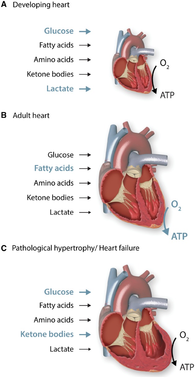
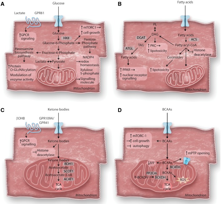
Cardiac metabolism is highly adaptive to changes in fuel availability and the energy demand of the heart. This metabolic flexibility is key for the heart to maintain its output during the development and in response to stress. Alterations in substrate preference have been observed in multiple disease states; a clear understanding of their impact on cardiac function in the long term is critical for the development of metabolic therapies. In addition, the contribution of cellular metabolism to growth, survival, and other signalling pathways through the generation of metabolic intermediates has been increasingly noted, adding another layer of complexity to the impact of metabolism on cardiac function. In a quest to understand the complexity of the cardiac metabolic network, genetic tools have been engaged to manipulate cardiac metabolism in a variety of mouse models. The ability to engineer cardiac metabolism in vivo has provided tremendous insights and brought about conceptual innovations. In this review, we will provide an overview of the cardiac metabolic network and highlight alterations observed during cardiac development and pathological hypertrophy. We will focus on consequences of altered substrate preference on cardiac response to chronic stresses through energy providing and non-energy providing pathways.

Keywords: Cardiac metabolism; Energy metabolism; Metabolic flexibility; Metabolic signalling; Pathological hypertrophy.


Figures

Figure 1
Figure 1
Contribution of different substrates to ATP production during cardiac development and disease progression. The heart is capable of using all classes of substrates, including carbohydrates, lipids, amino acids and ketone bodies to meet its energetic demands. Its substrate preference changes throughout the life cycle as well as under physiological and pathological conditions, which allows the heart to adapt to environmental changes. (A) The developing heart is highly dependent on aerobic glycolysis and lactate oxidation, while oxidation of fatty acids plays only a minor role. This metabolic phenotype is well suited for the biosynthesis of cellular building blocks, which are necessary for cardiomyocyte proliferation and cellular growth. (B) The adult heart is exposed to an increased haemodynamic load and oxygen tension. It mainly uses oxidative metabolism for ATP production, which is reflected by a substantial increase in mitochondrial volume mass. Fatty acids become the predominant fuel for the adult hearts. (C) Hearts with pathological hypertrophy revert to a foetal metabolic profile, demonstrating increased reliance on glucose and reduced oxidative capacity. In advanced heart failure, increased use of ketone bodies as fuel has been suggested.
Figure 2
Figure 2
Consequences of altered cardiac metabolism on non-energy providing functions. The primary role of cardiac metabolism is to provide energy for cardiac contraction. However, altered substrate utilization and metabolic remodelling in the hypertrophied heart can affect various cellular functions beyond energy supply. (A) Glucose metabolism and cardiac growth: After entering the cell, glucose is phosphorylated to glucose-6-phosphate (G6P) before feeding into glycolysis, the hexosamine biosynthesis pathway (HBP) or the pentose phosphate pathway (PPP). In cardiac hypertrophy, increased glucose reliance results in increased flux through all three pathways. Accumulation of G6P has been linked to the activation of the mechanistic target of rapamycin complex 1 (mTORC1) and cell growth. Enhanced flux through the HBP pathway results in increased protein O-GlcNAcylation and modulation of enzyme activity. Intermediates of the PPP have been recognized to act as signalling molecules (Xylulose-5-phosphate) or modulate redox homeostasis (NADPH). Recently, identification of cardiac lactate receptor (GPR81) expression proposes a role of lactate as a signalling molecule. (B) Fatty acid metabolism and cardiotoxicity: Cytosolic free fatty acids are activated by acyl CoA synthetase (ACS) to form fatty acyl-CoAs. Besides entering mitochondria for oxidation, acyl-CoAs can also form ceramides, diacylglycerol (DAG) and triacylglycerol (TAG). Formation of DAG can also occur through phospholipid breakdown which will result in protein kinase C (PKC) activation. DAG as well as ceramide species are increased in the hypertrophied heart and have been associated with lipotoxicity, while redirecting these toxic lipid intermediates into the TAG pool prevents lipotoxicity. TAG can be hydrolyzed by adipose tissue triglyceride lipase (ATGL) and can either enter the fatty acid oxidation pathway or serve as ligand for nuclear receptor activation. Acyl-CoAs have recently been identified to contribute to epigenetic regulation by acting as endogenous histone deacetylase inhibitors. (C) Ketone body metabolism and cellular signalling: Ketone bodies are oxidized in the heart for ATP generation. After entering mitochondria, they rapidly from acetyl-CoA via βOHB dehydrogenase (BDH1), succinyl-CoA:3-oxoacid-CoA transferase (SCOT) and mitochondrial acetyl-CoA acetyltransferase 1 (ACAT1). βOHB can further act as endogenous histone deacetylase inhibitor or as signalling molecule through its G protein coupled receptor (GPR109A/GPR41). (D) Branched-chain amino acids catabolism in cardiomyocyte growth and survival: Catabolism of the branched-chain amino acids (BCAA) feeds into the TCA cycle although their contribution to energy provision is rather minor under normal conditions. The first steps in BCAA catabolism yields branched-chain keto acids (BCKA) through mitochondrial branched-chain aminotransferase (BCATm) reaction. BCKA is processed by a dehydrogenase complex (BCKDH) to form CoA compounds, before further breakdown for TCA cycle. BCKDH is the rate-limiting enzyme in BCAA catabolism, it is activated by dephosphorylation through a mitochondrial-localized 2C-type protein phosphatase (PP2Cm). Accumulation of BCAA results in mitochondrial dysfunction, characterized by loss of mitochondrial membrane potential (ΔΨ) and mitochondrial permeability transition pore (mPTP) opening. BCKA accumulation stimulates ROS formation. Additionally, leucine can directly activate mTORC1 and simulate cell growth.

References

    1. Ingwall JS. ATP and the Heart. Boston: Kluwer Academic Publishers; 2002.
    1. Doenst T, Nguyen TD, Abel ED.. Cardiac metabolism in heart failure: implications beyond ATP production. Circul Research 2013;113:709–724. - PMC - PubMed
    1. Kolwicz SC Jr., Purohit S, Tian R.. Cardiac metabolism and its interactions with contraction, growth, and survival of cardiomyocytes. Circul Res 2013;113:603–616. - PMC - PubMed
    1. Neubauer S. The failing heart – an engine out of fuel. New Eng J Med 2007;356:1140–1151. - PubMed
    1. Abdulla R, Blew GA, Holterman MJ.. Cardiovascular embryology. Pediatr Cardiol 2004;25:191–200. - PubMed