. 2017 Apr 1;58(4):2095-2105.
doi: 10.1167/iovs.16-20641.

Inhibition of Stat3 by a Small Molecule Inhibitor Slows Vision Loss in a Rat Model of Diabetic Retinopathy

Affiliations

Inhibition of Stat3 by a Small Molecule Inhibitor Slows Vision Loss in a Rat Model of Diabetic Retinopathy

Phillip A Vanlandingham et al. Invest Ophthalmol Vis Sci. .

Abstract

Purpose: Diabetic retinopathy is a leading cause of vision loss. Previous studies have shown signaling pathways mediated by Stat3 (signal transducer and activator of transcription 3) play a primary role in diabetic retinopathy progression. This study tested CLT-005, a small molecule inhibitor of Stat3, for its dose-dependent therapeutic effects on vision loss in a rat model of diabetic retinopathy.

Methods: Brown Norway rats were administered streptozotocin (STZ) to induce diabetes. CLT-005 was administered daily by oral gavage for 16 weeks at concentrations of 125, 250, or 500 mg/kg, respectively, beginning 4 days post streptozotocin administration. Systemic and ocular drug concentration was quantified with mass spectrometry. Visual function was monitored at 2-week intervals from 6 to 16 weeks using optokinetic tracking to measure visual acuity and contrast sensitivity. The presence and severity of cataracts was visually monitored and correlated to visual acuity. The transcription and translation of multiple angiogenic factors and inflammatory cytokines were measured by real-time polymerase chain reaction and Multiplex immunoassay.

Results: Streptozotocin-diabetic rats sustain progressive vision loss over 16 weeks, and this loss in visual function is rescued in a dose-dependent manner by CLT-005. This positive therapeutic effect correlates to the positive effects of CLT-005 on vascular leakage and the presence of inflammatory cytokines in the retina.

Conclusions: The present study indicates that Stat3 inhibition has strong therapeutic potential for the treatment of vision loss in diabetic retinopathy.


Figures
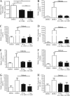
Figure 1
Figure 1
Intravitreal administration of CLT-005 downregulates vascular leakage and angiogenic factor expression in STZ-diabetic rats. (AH) Two weeks following induction of diabetes with STZ, the rats received an intravitreal administration of dimethyl sulfoxide (DMSO) vehicle in one eye, and CLT-005 at the indicated dose in the contralateral eye. (A) At 7 days post intravitreal administration, Evans blue-albumin leakage from blood vessels into the retina was quantified as an indication of retinal vascular permeability. Treatment with either 1 μg or 5 μg of CLT-005 significantly decreases retinal vascular leakage relative to vehicle alone. (BH) At 24 hours post intravitreal administration, the retinas were collected and qRT-PCR was used to quantify the mRNA levels of the indicated molecules. Nondiabetic BN rats were also treated with DMSO as an additional control. STZ-diabetic rats show a dramatic increase in gene expression for each of the examined molecules, and treatment with CLT-005 brings transcript levels near nondiabetic levels (n = 4; mean ± SEM; *P < 0.05; **P < 0.01; Mann-Whitney nonparametric t-test).
Figure 2
Figure 2
CLT-005 is effectively delivered systemically via oral gavage and is well tolerated by BN rats. (A) The baseline (4 days post STZ) and endpoint (16 weeks post STZ) weights were averaged for each group. Daily dosing of CLT-005 did not adversely affect weight gain at week 16 for any of the given doses when compared with the vehicle treatment. (B) Following 16 weeks of daily doses, whole blood was collected 3 hours after the terminal dose, and CLT-005 concentration in the plasma was measured by liquid chromatography/mass spectrometry (LC/MS). CLT-005 concentration in the plasma was dose dependent (mean ± SEM; *P < 0.05; **P < 0.01; Mann-Whitney nonparametric t-test). (C) Glucose levels were measured once a week for 16 weeks post STZ. All measurements within each group during the duration of the study were averaged and plotted (mean ± SEM). The groups receiving the two highest doses of CLT-005 had significantly lower average blood glucose levels during the course of the study when compared with the animals receiving either vehicle or 125 mg/kg CLT-005 (***P > 0.001; Mann-Whitney nonparametric t-test). (D) The baseline (4 days post STZ) and endpoint (16 weeks post STZ) nonfasting glucose levels were averaged for each group and plotted. The differences between baseline and endpoint glucose levels were greatest for the groups administered the two highest doses of CLT-005. (E) Retinas were dissected from animals within the indicated treatment group, and equal concentrations of protein were processed for SDS-PAGE, transferred to polyvinylidene fluoride (PVDF), and immunoblotted with the indicated antibodies. Stat3 activity is higher in the retinas of STZ-diabetic rats treated with vehicle or 125 mg/kg CLT-005 compared to the two highest doses of CLT-005.
Figure 3
Figure 3
CLT-005 rescues vision loss in STZ-diabetic rats in a dose-dependent manner. (A, B) OKT was used to measure the SFT values in BN rats at 2-week intervals post STZ. At 6 and 8 weeks post STZ, the SFT values are comparable in all STZ-diabetic groups. However, by week 10, the SFT values significantly diverge between the vehicle and 125 mg/kg CLT-005 groups and the 250 and 500 mg/kg groups. For the 250 mg/kg CLT-005 group, P < 0.01 at 10, 14, and 16 weeks when compared with the vehicle group. For the 500 mg/kg CLT-005 group, P < 0.01 at 10 weeks, and P < 0.001 at 14 and 16 weeks when compared with the vehicle group (1-way ANOVA with Tukey's posttest). (B) At 16 weeks post STZ, both the 250 and 500 mg/kg CLT-005 groups have significantly higher SFT values when compared with the vehicle treated group and are not significantly different than the insulin-rescued group (mean ± SEM; *P < 0.05; ***P < 0.0001; 1-way ANOVA with Tukey's posttest). (C) OKT was used to measure the contrast thresholds in BN rats at 2-week intervals post STZ. STZ-diabetic animals receiving 500 mg/kg CLT-005 retained their ability to distinguish contrast during the course of 16 weeks at levels comparable to insulin-rescued STZ-diabetic rats. For the 250 mg/kg CLT-005 group, P < 0.05 at 10 and 12 weeks when compared with the vehicle group. For the 500 mg/kg CLT-005 group, P < 0.05 at 12 weeks, P < 0.001 at 10, 14, and 16 weeks when compared with the vehicle group (1-way ANOVA with Tukey's posttest). (D) The average contrast threshold values within each group at week 16 were plotted and the values were compared with the vehicle treated group (mean ± SEM; *P < 0.05; 1-way ANOVA with Tukey's posttest). The 500 mg/kg CLT-005 group had significantly higher contrast threshold values and is not significantly different than the insulin-rescued group. (E) Contrast sensitivity was evaluated as a measure of contrast threshold values at the indicated spatial frequencies at 16 weeks post STZ. STZ-diabetic rats receiving 500 mg/kg CLT-005 have greater contrast sensitivity across spatial frequencies when compared with the other treated STZ-diabetic groups. (A, C, E) red line, naive; gray line, vehicle; blue line, 500 mg/kg CLT-005; orange line, 250 mg/kg CLT-005; green line, 125 mg/kg CLT-005; black line, insulin rescue.
Figure 4
Figure 4
CLT-005 reduces cataract severity and incidence in STZ-diabetic rats. (A) Representative images of cataracts from naïve and STZ-diabetic groups. (B) Cataracts were scored every 2 weeks beginning at 6 weeks post STZ treatment. The development of severe cataracts (score > 2) was delayed in rats receiving the two highest doses of CLT-005, with the average score remaining lower than the vehicle-treated animals through 16 weeks post STZ. (C) The average SFT values from eyes with a cataract score of 2, which represents the upper level of severity at this time point, were separated from the average SFT values of all other eyes within each group and plotted side by side. The data show the SFT values are comparable for different cataract scores within each group. (D) Scatterplot representation of each eye with a cataract score of 2 in either the vehicle or 500 mg/kg CLT-005 groups. The data show that even in animals with the same cataract scores across treatment groups, the group receiving 500 mg/kg CLT-005 maintain higher overall SFT values (mean ± SEM; *P < 0.05; two-tailed paired t-test).
Figure 5
Figure 5
Orally administered CLT-005 is delivered to the eye. LC/MS quantification of CLT-005 in (A) retina and (B) PECS after daily gavage for 16 weeks at 125, 250, and 500 mg/kg of CLT-005. Eyes were enucleated within 3 hours of final gavage treatment and immediately processed for analysis (n = 5, mean ± SEM).
Figure 6
Figure 6
CLT-005 downregulates inflammatory mediators in STZ-diabetic rat eyes. Equal concentrations of protein from individual eyes were analyzed using a Bio Rad Bio-plex Pro Rat Cytokine group I 23-Plex Assay according to the manufacturer's protocol (see Materials and Methods; n = 4; mean ± SEM; *P < 0.05; 1-way ANOVA with Tukey's posttest). The data presented in this figure is selected from more than 20 examined cytokines (see Table).

Similar articles

Cited by

References

    1. Vaz JA,, Patnaik A. Diabetes mellitus: exploring the challenges in the drug development process. Perspect Clin Res. 2012; 3109–3112. - PMC - PubMed
    1. Klein R,, Knudtson MD,, Lee KE,, Gangnon R,, Klein BE. The Wisconsin Epidemiologic Study of Diabetic Retinopathy: XXII the twenty-five-year progression of retinopathy in persons with type 1 diabetes. Ophthalmology. 2008; 115: 1859–1868. - PMC - PubMed
    1. Ko F,, Vitale S,, Chou CF,, Cotch MF,, Saaddine J,, Friedman DS. Prevalence of nonrefractive visual impairment in US adults and associated risk factors, 1999-2002 and 2005-2008. JAMA. 2012; 308: 2361–2368. - PMC - PubMed
    1. Aiello LM. Preserving human vision: eliminating blindness from diabetes mellitus. J Am Optom Assoc. 1998; 69: 690–692. - PubMed
    1. Fong DS,, Aiello L,, Gardner TW,, et al. Diabetic retinopathy. Diabetes Care. 2003; 26: 226–229. - PubMed

Publication types

MeSH terms

Substances