. 2017 Feb 1;147(2):129-152.
doi: 10.1093/ajcp/aqw214.

EBV-Positive B-Cell Proliferations of Varied Malignant Potential: 2015 SH/EAHP Workshop Report-Part 1

Affiliations

EBV-Positive B-Cell Proliferations of Varied Malignant Potential: 2015 SH/EAHP Workshop Report-Part 1

Yasodha Natkunam et al. Am J Clin Pathol. .

Abstract

Objectives: The 2015 Workshop of the Society for Hematopathology/European Association for Haematopathology aimed to review B-cell proliferations of varied malignant potential associated with immunodeficiency.

Methods: The Workshop Panel reviewed all cases of B-cell hyperplasias, polymorphic B-lymphoproliferative disorders, Epstein-Barr virus (EBV)-positive mucocutaneous ulcer, and large B-cell proliferations associated with chronic inflammation and rendered consensus diagnoses. Disease definitions, boundaries with more aggressive B-cell proliferations, and association with EBV were explored.

Results: B-cell proliferations of varied malignant potential occurred in all immunodeficiency backgrounds. Presentation early in the course of immunodeficiency and in younger age groups and regression with reduction of immunosuppression were characteristic features. EBV positivity was essential for diagnosis in some hyperplasias where other specific defining features were absent.

Conclusions: This spectrum of B-cell proliferations show similarities across immunodeficiency backgrounds. Localized forms of immunodeficiency disorders arise in immunocompetent patients most likely due to chronic immune stimulation and, despite aggressive histologic features, often show indolent clinical behavior.

Keywords: Autoimmune; EBV; Early lesion; HIV; Iatrogenic; Nondestructive lesion; Polymorphic lymphoproliferative disorder; Posttransplant lymphoproliferative disorder.


Figures

Image 1
Image 1
Distribution of Epstein-Barr virus-encoded small RNA (EBER)–positive cells in follicular hyperplasias. Four case examples of follicular hyperplasia are illustrated. The distribution and number of EBER-positive cells were variable and often localized to one or a few follicles with scant interfollicular EBER-positive cells (case SH2015-123, A and B [x10], and case SH2015-145, C and D [x20]).
Image 1
Image 1
Distribution of Epstein-Barr virus-encoded small RNA (EBER)–positive cells in follicular hyperplasias. Four case examples of follicular hyperplasia are illustrated. The distribution and number of EBER-positive cells were variable and often localized to one or a few follicles with scant interfollicular EBER-positive cells (case SH2015-123, A and B [x10], and case SH2015-145, C and D [x20]).
Image 2
Image 2
Polymorphic B-lymphoproliferative disorder in a patient with human immunodeficiency virus (HIV) (case SH2015-83). Initially diagnosed as primary tuberculosis, this polymorphic B-lymphoproliferative disorder presented as bilateral pulmonary nodules in a 40-year-old woman with HIV (A, x4). A polymorphous infiltrate with increased immunoblasts is seen in the lung biopsy specimen (B, x40), which shows expression of CD20 (C, x40) and CD30 (D, x40),
Image 2
Image 2
Polymorphic B-lymphoproliferative disorder in a patient with human immunodeficiency virus (HIV) (case SH2015-83). Initially diagnosed as primary tuberculosis, this polymorphic B-lymphoproliferative disorder presented as bilateral pulmonary nodules in a 40-year-old woman with HIV (A, x4). A polymorphous infiltrate with increased immunoblasts is seen in the lung biopsy specimen (B, x40), which shows expression of CD20 (C, x40) and CD30 (D, x40),
Image 3
Image 3
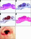
Mucocutaneous ulcer (MCU), Epstein-Barr virus positive (EBV+). Low-power view of MCU, EBV+ shows a sharply circumscribed ulcerating lesion in the jejunum (A, x2), which was resected following small bowel perforation in a 57-year-old kidney transplant recipient (case SH2015-169). The lesion shows CD20-positive cells (B, x2) in a background rich in small T cells (C, x2) and Epstein-Barr virus-encoded small RNA (EBER)–positive cells (D, x2). A second case example of MCU, EBV+ shows an ulcerated nasal lesion (E) in a 74-year-old woman with no known immunodeficiency (case SH2015-344).
Image 3
Image 3
Mucocutaneous ulcer (MCU), Epstein-Barr virus positive (EBV+). Low-power view of MCU, EBV+ shows a sharply circumscribed ulcerating lesion in the jejunum (A, x2), which was resected following small bowel perforation in a 57-year-old kidney transplant recipient (case SH2015-169). The lesion shows CD20-positive cells (B, x2) in a background rich in small T cells (C, x2) and Epstein-Barr virus-encoded small RNA (EBER)–positive cells (D, x2). A second case example of MCU, EBV+ shows an ulcerated nasal lesion (E) in a 74-year-old woman with no known immunodeficiency (case SH2015-344).
Image 4
Image 4
Large B-cell proliferations associated with chronic inflammation. Three case examples are illustrated, all of which occurred in immunocompetent patients. A left atrial myxoma in a 50-year-old otherwise healthy man (case SH2015-68) shows a gelatinous mass with a rim of highly pleomorphic large lymphoid cells (arrow) (A, x4; B, x40), expressing CD20 (C, x40), and Epstein-Barr virus–encoded small RNA in a subset of cells (D, x40).
Image 4
Image 4
Large B-cell proliferations associated with chronic inflammation. Three case examples are illustrated, all of which occurred in immunocompetent patients. A left atrial myxoma in a 50-year-old otherwise healthy man (case SH2015-68) shows a gelatinous mass with a rim of highly pleomorphic large lymphoid cells (arrow) (A, x4; B, x40), expressing CD20 (C, x40), and Epstein-Barr virus–encoded small RNA in a subset of cells (D, x40).
Figure 1
Figure 1
Histologic features of B-cell hyperplasias in immunodeficiency settings. The cartoon illustrates the relationship among the three types of hyperplasias. There is retention of hyperplastic follicles in all types. Interfollicular proliferation ranges from scant in follicular hyperplasia, to a mixed inflammatory infiltrate with increased immunoblasts in infectious mononucleosis-like hyperplasia, to one that is rich in polytypic plasma cells in plasmacytic hyperplasia.

References

    1. Swerdlow SH, Weber SA, Chadburn A, et al. Post-transplant lymphoproliferative disorders. In: Swerdlow SH, Campo E, Harris NL, et al., eds. WHO Classification of Tumours of Haematopoietic and Lymphoid Tissues. 4th ed Lyon, France: IARC; 2008:343-349.
    1. Swerdlow SH, Campo E, Pileri SA, et al. The 2016 revision of the World Health Organization classification of lymphoid neoplasms. Blood. 2016;127:2375-2390. - PMC - PubMed
    1. Meru N, Davison S, Whitehead L, et al. Epstein-Barr virus infection in paediatric liver transplant recipients: detection of the virus in post-transplant tonsillectomy specimens. Mol Pathol. 2001;54:264-269. - PMC - PubMed
    1. Harris NL, Swerdlow SH, Frizzera G, et al. Post-Transplant Lymphoproliferative Disorders. Jaffe ES, Harris NL, Stein H, Vardiman JW, eds. Lyon, France: IARC; 2001:264-269.
    1. Williamson RA, Huang RY, Shapiro NL. Adenotonsillar histopathology after organ transplantation. Otolaryngology Head Neck Surg. 2001;125:231-240. - PubMed