

Beta 2-adrenergic receptor agonists are novel regulators of macrophage activation in diabetic renal and cardiovascular complications
- PMID: 28396116
- PMCID: PMC5483383
- DOI: 10.1016/j.kint.2017.02.013
Beta 2-adrenergic receptor agonists are novel regulators of macrophage activation in diabetic renal and cardiovascular complications
Abstract
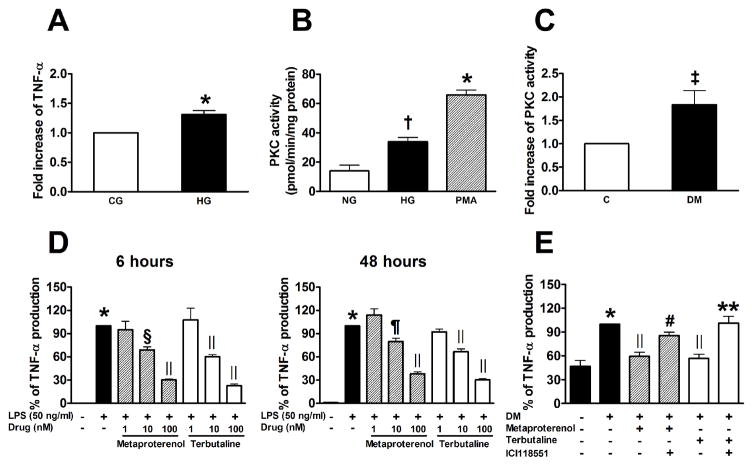
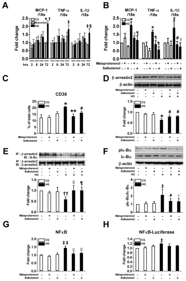
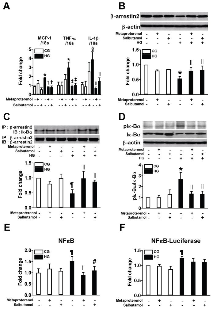
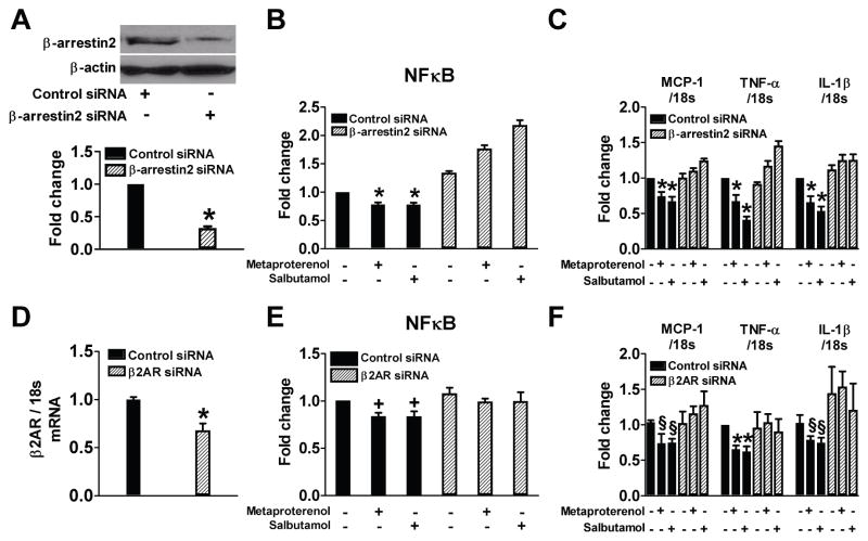
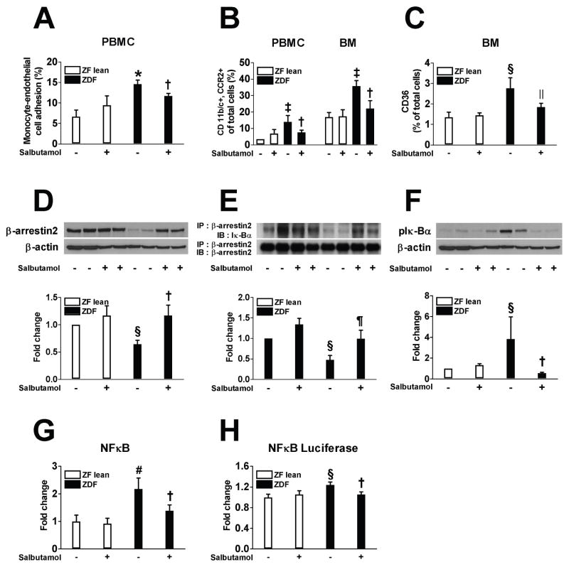
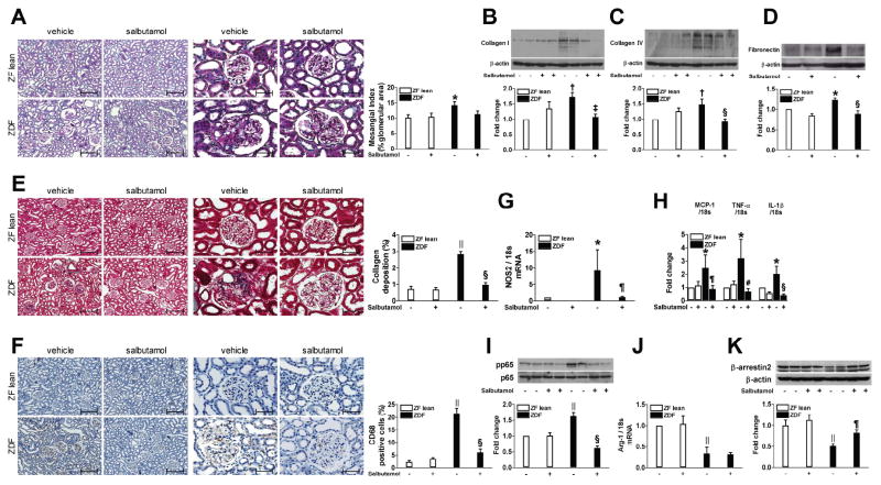
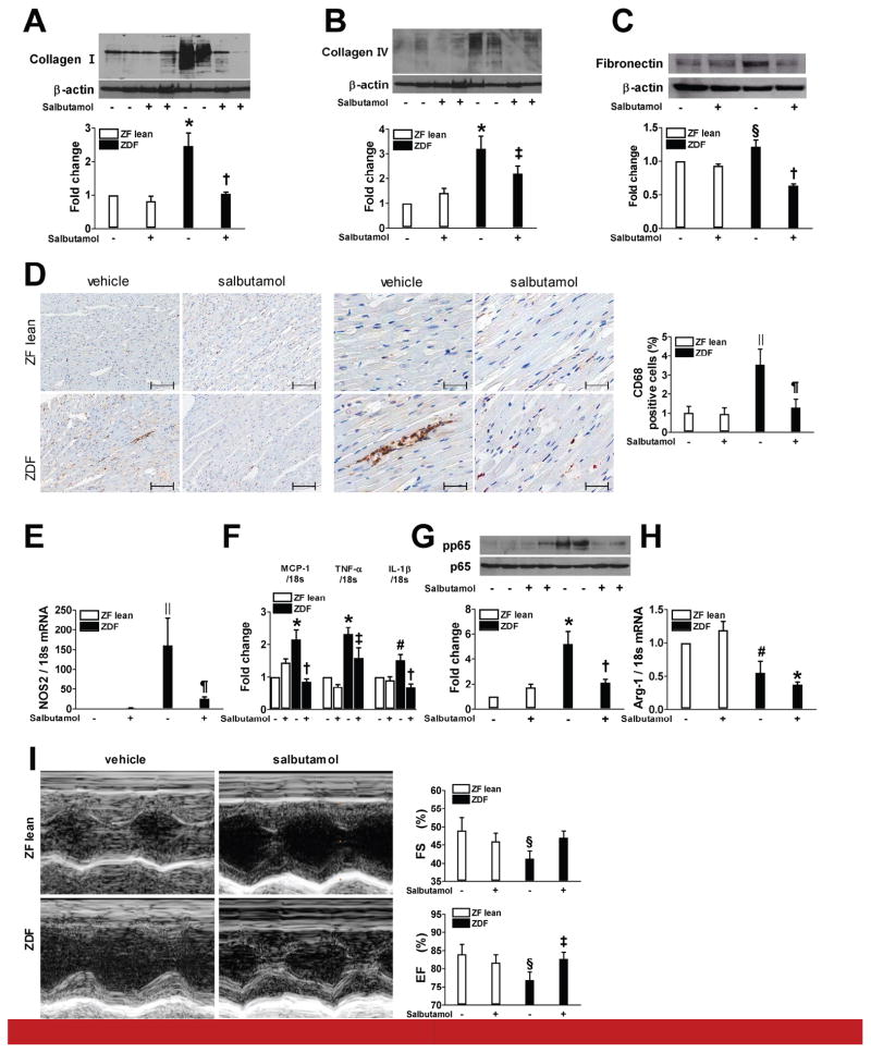
Macrophage activation is increased in diabetes and correlated with the onset and progression of vascular complications. To identify drugs that could inhibit macrophage activation, we developed a cell-based assay and screened a 1,040 compound library for anti-inflammatory effects. Beta2-adrenergic receptor (β2AR) agonists were identified as the most potent inhibitors of phorbol myristate acetate-induced tumor necrosis factor-α production in rat bone marrow macrophages. In peripheral blood mononuclear cells isolated from streptozotocin-induced diabetic rats, β2AR agonists inhibited diabetes-induced tumor necrosis factor-α production, which was prevented by co-treatment with a selective β2AR blocker. To clarify the underlying mechanisms, THP-1 cells and bone marrow macrophages were exposed to high glucose. High glucose reduced β-arrestin2, a negative regulator of NF-κB activation, and its interaction with IκBα. This subsequently enhanced phosphorylation of IκBα and activation of NF-κB. The β2AR agonists enhanced β-arrestin2 and its interaction with IκBα, leading to downregulation of NF-κB. A siRNA specific for β-arrestin2 reversed β2AR agonist-mediated inhibition of NF-κB activation and inflammatory cytokine production. Treatment of Zucker diabetic fatty rats with a β2AR agonist for 12 weeks attenuated monocyte activation as well as pro-inflammatory and pro-fibrotic responses in the kidneys and heart. Thus, β2AR agonists might have protective effects against diabetic renal and cardiovascular complications.
Keywords: diabetes; fibrosis; inflammation; macrophages.
Copyright © 2017 International Society of Nephrology. Published by Elsevier Inc. All rights reserved.
Figures
Comment in
-
β2-adrenergic receptors in inflammation and vascular complications of diabetes.Kidney Int. 2017 Jul;92(1):14-16. doi: 10.1016/j.kint.2017.03.024. Kidney Int. 2017. PMID: 28646990
Similar articles
-
Anti-inflammatory activities of fenoterol through β-arrestin-2 and inhibition of AMPK and NF-κB activation in AICAR-induced THP-1 cells.Biomed Pharmacother. 2016 Dec;84:185-190. doi: 10.1016/j.biopha.2016.09.044. Epub 2016 Sep 19. Biomed Pharmacother. 2016. PMID: 27657826
-
Attenuation of inflammatory response by a novel chalcone protects kidney and heart from hyperglycemia-induced injuries in type 1 diabetic mice.Toxicol Appl Pharmacol. 2015 Oct 15;288(2):179-91. doi: 10.1016/j.taap.2015.07.009. Epub 2015 Jul 20. Toxicol Appl Pharmacol. 2015. PMID: 26206226
-
Renal-protective effect of thalidomide in streptozotocin-induced diabetic rats through anti-inflammatory pathway.Drug Des Devel Ther. 2018 Jan 9;12:89-98. doi: 10.2147/DDDT.S149298. eCollection 2018. Drug Des Devel Ther. 2018. PMID: 29386886 Free PMC article.
-
NF-κB pathway as a molecular target for curcumin in diabetes mellitus treatment: Focusing on oxidative stress and inflammation.Cell Biochem Funct. 2024 Jun;42(4):e4030. doi: 10.1002/cbf.4030. Cell Biochem Funct. 2024. PMID: 38720663 Review.
-
Beta2-adrenergic receptor in kidney biology: A current prospective.Nephrology (Carlton). 2019 May;24(5):497-503. doi: 10.1111/nep.13584. Nephrology (Carlton). 2019. PMID: 30848004 Review.
Cited by
-
Adipose and serum zinc alpha-2-glycoprotein (ZAG) expressions predict longitudinal change of adiposity, wasting and predict survival in dialysis patients.Sci Rep. 2022 May 31;12(1):9087. doi: 10.1038/s41598-022-13149-6. Sci Rep. 2022. PMID: 35641588 Free PMC article.
-
Mitochondrial biogenesis induced by the β2-adrenergic receptor agonist formoterol accelerates podocyte recovery from glomerular injury.Kidney Int. 2019 Sep;96(3):656-673. doi: 10.1016/j.kint.2019.03.023. Epub 2019 May 6. Kidney Int. 2019. PMID: 31262488 Free PMC article.
-
The emerging role of leptin in obesity-associated cardiac fibrosis: evidence and mechanism.Mol Cell Biochem. 2023 May;478(5):991-1011. doi: 10.1007/s11010-022-04562-6. Epub 2022 Oct 10. Mol Cell Biochem. 2023. PMID: 36214893 Review.
-
Obesity: a neuroimmunometabolic perspective.Nat Rev Endocrinol. 2020 Jan;16(1):30-43. doi: 10.1038/s41574-019-0283-6. Epub 2019 Nov 27. Nat Rev Endocrinol. 2020. PMID: 31776456 Review.
-
Cardiovascular diseases or type 2 diabetes mellitus and chronic airway diseases: mutual pharmacological interferences.Ther Adv Chronic Dis. 2023 May 31;14:20406223231171556. doi: 10.1177/20406223231171556. eCollection 2023. Ther Adv Chronic Dis. 2023. PMID: 37284143 Free PMC article. Review.
References
-
- Sassy-Prigent C, Heudes D, Mandet C, et al. Early glomerular macrophage recruitment in streptozotocin-induced diabetic rats. Diabetes. 2000;49:466–475. - PubMed
-
- Tsao PS, Niebauer J, Buitrago R, et al. Interaction of diabetes and hypertension on determinants of endothelial adhesiveness. Arterioscler Thromb Vasc Biol. 1998;18:947–953. - PubMed
-
- Li AC, Glass CK. The macrophage foam cell as a target for therapeutic intervention. Nat Med. 2002;8:1235–1242. - PubMed
-
- Cipolletta C, Ryan KE, Hanna EV, et al. Activation of peripheral blood CD14+ monocytes occurs in diabetes. Diabetes. 2005;54:2779–2786. - PubMed
Publication types
MeSH terms
Substances
Grants and funding
LinkOut - more resources
Full Text Sources
Other Literature Sources
Medical
