

Hypoxia Response Element-Regulated MMP-9 Promotes Neurological Recovery via Glial Scar Degradation and Angiogenesis in Delayed Stroke
- PMID: 28396199
- PMCID: PMC5474960
- DOI: 10.1016/j.ymthe.2017.03.020
Hypoxia Response Element-Regulated MMP-9 Promotes Neurological Recovery via Glial Scar Degradation and Angiogenesis in Delayed Stroke
Abstract
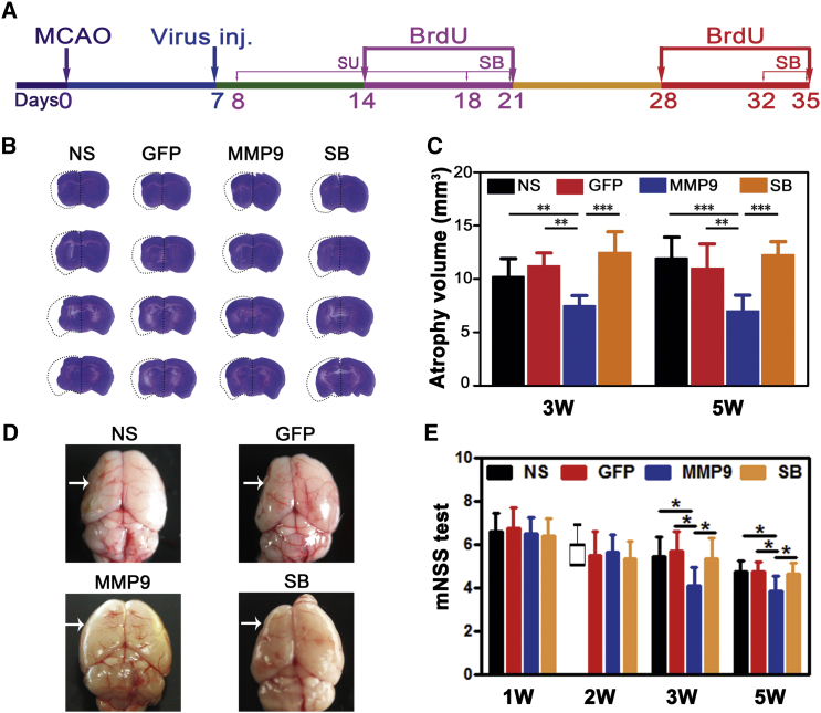
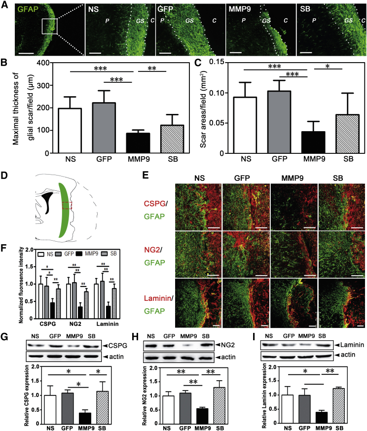
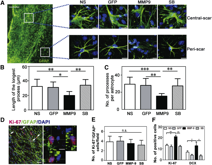
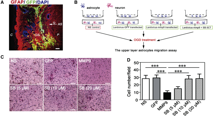
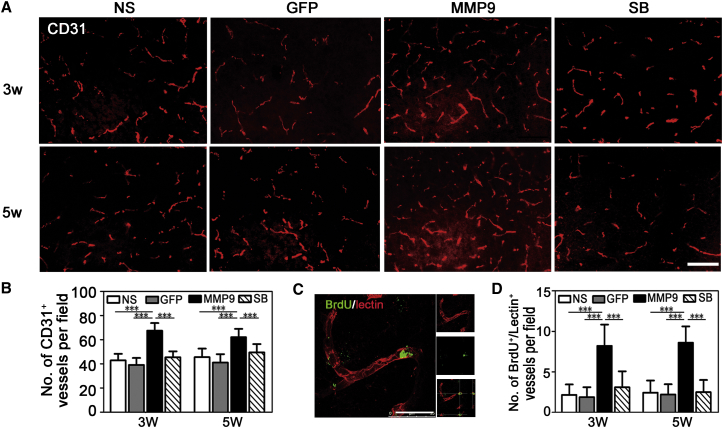
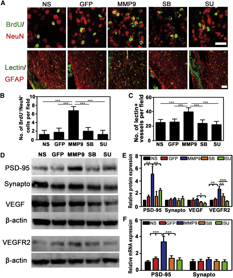
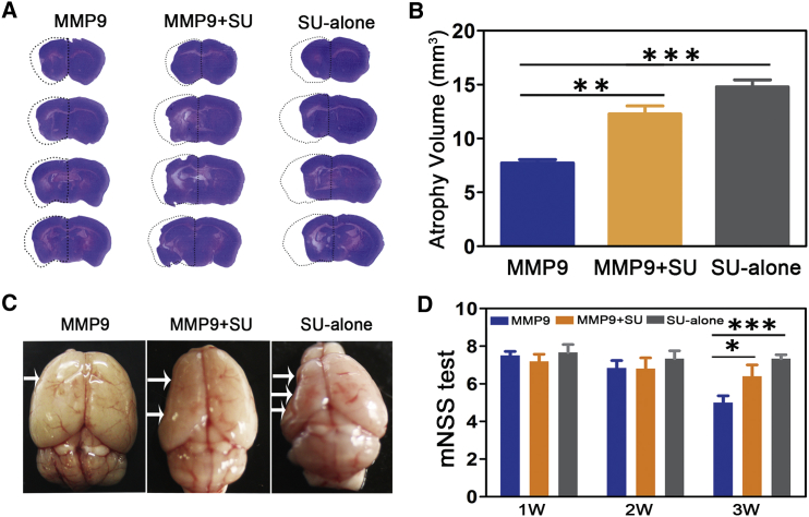
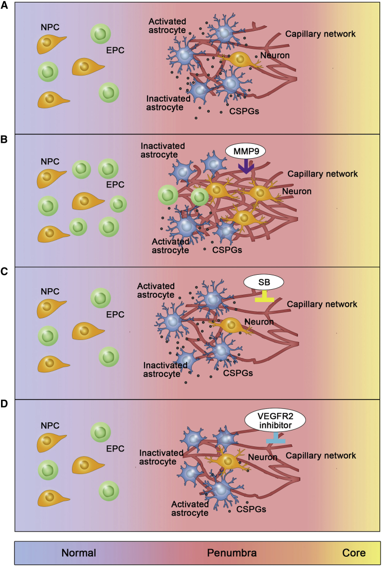
Matrix metalloproteinase 9 (MMP-9) plays a beneficial role in the delayed phase of middle cerebral artery occlusion (MCAO). However, the mechanism is obscure. Here, we constructed hypoxia response element (HRE)-regulated MMP-9 to explore its effect on glial scars and neurogenesis in delayed ischemic stroke. Adult male Institute of Cancer Research (ICR) mice underwent MCAO and received a stereotactic injection of lentivirus carrying HRE-MMP-9 or normal saline (NS)/lentivirus-GFP 7 days after ischemia. We found that HRE-MMP-9 improved neurological outcomes, reduced ischemia-induced brain atrophy, and degraded glial scars (p < 0.05). Furthermore, HRE-MMP-9 increased the number of microvessels in the peri-infarct area (p < 0.001), which may have been due to the accumulation of endogenous endothelial progenitor cells (EPCs) in the peri-infarct area after glial scar degradation. Finally, HRE-MMP-9 increased the number of bromodeoxyuridine-positive (BrdU+)/NeuN+ cells and the expression of PSD-95 in the peri-infarct area (p < 0.01). These changes could be blocked by vascular endothelial growth factor receptor 2 (VEGFR2) inhibitor SU5416 and MMP-9 inhibitor 2-[[(4-phenoxyphenyl)sulfonyl]methyl]-thiirane (SB-3CT). Our results provided a novel mechanism by which glial scar degradation and vascular endothelial growth factor (VEGF)/VEGFR2-dependent angiogenesis may be key procedures for neurological recovery in delayed ischemic stroke after HRE-MMP-9 treatment. Therefore, HRE-MMP-9 overexpression in the delayed ischemic brain is a promising approach for neurological recovery.
Keywords: HRE-MMP-9; angiogenesis; glial scar; neurogenesis; stroke.
Copyright © 2017 The American Society of Gene and Cell Therapy. Published by Elsevier Inc. All rights reserved.
Figures
References
-
- Ring H., Rosenthal N. Controlled study of neuroprosthetic functional electrical stimulation in sub-acute post-stroke rehabilitation. J. Rehabil. Med. 2005;37:32–36. - PubMed
-
- Villa P., van Beek J., Larsen A.K., Gerwien J., Christensen S., Cerami A., Brines M., Leist M., Ghezzi P., Torup L. Reduced functional deficits, neuroinflammation, and secondary tissue damage after treatment of stroke by nonerythropoietic erythropoietin derivatives. J. Cereb. Blood Flow Metab. 2007;27:552–563. - PubMed
-
- Yenari M.A., Fink S.L., Sun G.H., Chang L.K., Patel M.K., Kunis D.M., Onley D., Ho D.Y., Sapolsky R.M., Steinberg G.K. Gene therapy with HSP72 is neuroprotective in rat models of stroke and epilepsy. Ann. Neurol. 1998;44:584–591. - PubMed
-
- Montaner J., Alvarez-Sabín J., Molina C., Anglés A., Abilleira S., Arenillas J., González M.A., Monasterio J. Matrix metalloproteinase expression after human cardioembolic stroke: temporal profile and relation to neurological impairment. Stroke. 2001;32:1759–1766. - PubMed
Publication types
MeSH terms
Substances
LinkOut - more resources
Full Text Sources
Other Literature Sources
Medical
Miscellaneous
