. 2017 May 23;114(21):5431-5436.
doi: 10.1073/pnas.1617035114. Epub 2017 Apr 10.

Structural hierarchy controlling dimerization and target DNA recognition in the AHR transcriptional complex

Affiliations

Structural hierarchy controlling dimerization and target DNA recognition in the AHR transcriptional complex

Seung-Hyeon Seok et al. Proc Natl Acad Sci U S A. .

Abstract

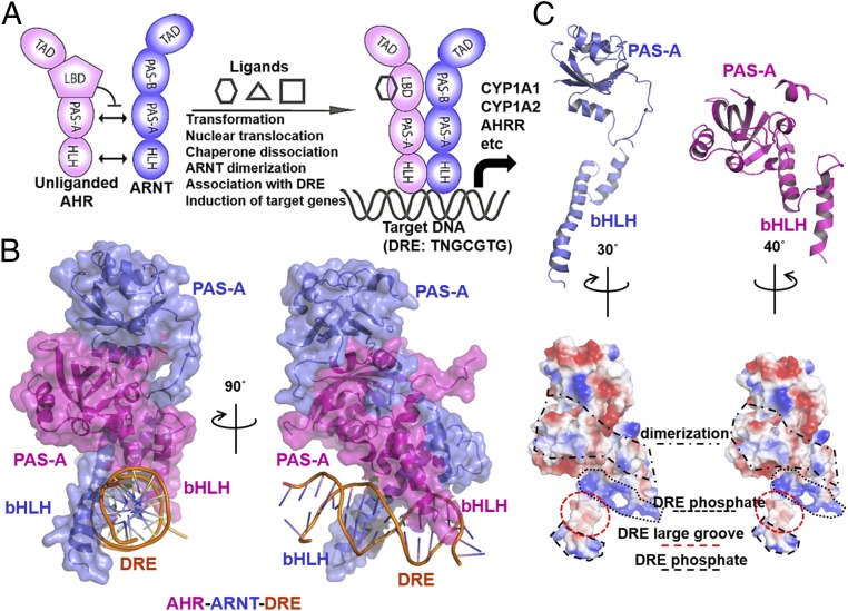
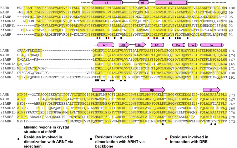
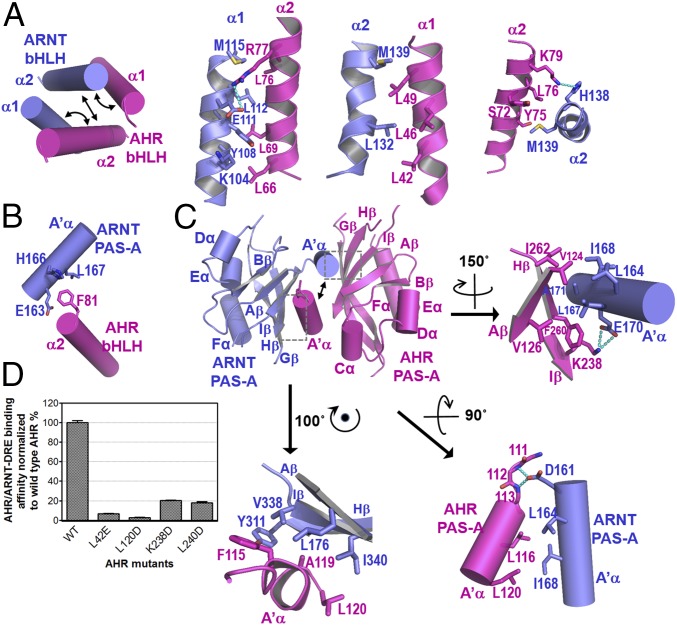
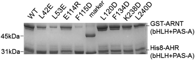
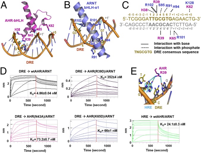
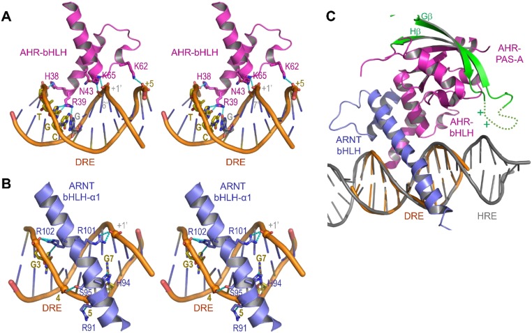
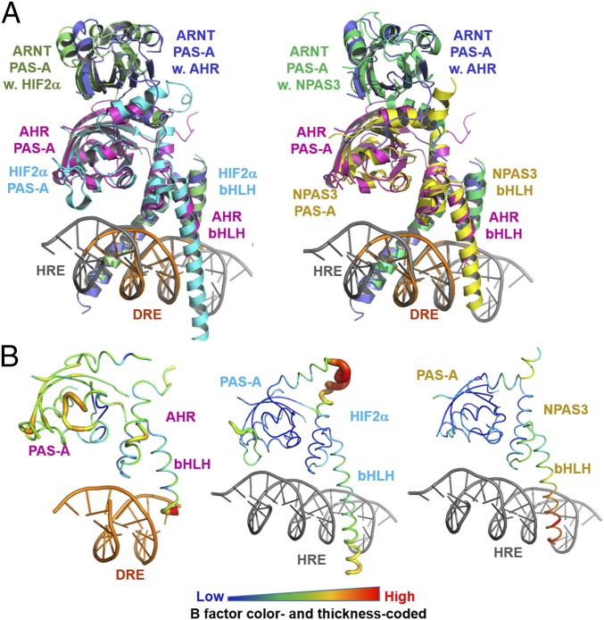
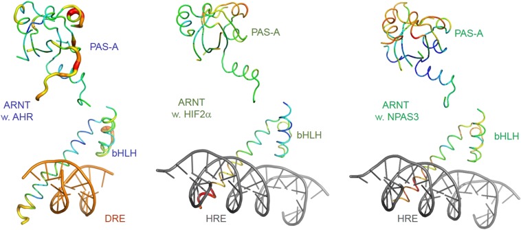
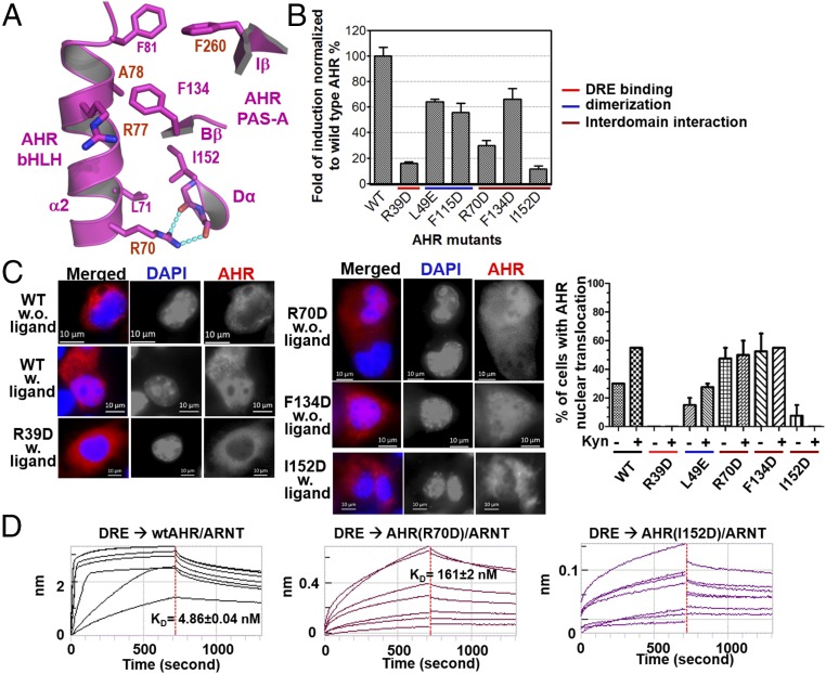
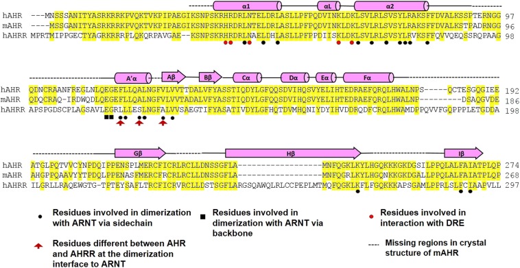
The aryl hydrocarbon receptor (AHR) belongs to the PAS (PER-ARNT-SIM) family transcription factors and mediates broad responses to numerous environmental pollutants and cellular metabolites, modulating diverse biological processes from adaptive metabolism, acute toxicity, to normal physiology of vascular and immune systems. The AHR forms a transcriptionally active heterodimer with ARNT (AHR nuclear translocator), which recognizes the dioxin response element (DRE) in the promoter of downstream genes. We determined the crystal structure of the mammalian AHR-ARNT heterodimer in complex with the DRE, in which ARNT curls around AHR into a highly intertwined asymmetric architecture, with extensive heterodimerization interfaces and AHR interdomain interactions. Specific recognition of the DRE is determined locally by the DNA-binding residues, which discriminates it from the closely related hypoxia response element (HRE), and is globally affected by the dimerization interfaces and interdomain interactions. Changes at the interdomain interactions caused either AHR constitutive nuclear localization or failure to translocate to nucleus, underlying an allosteric structural pathway for mediating ligand-induced exposure of nuclear localization signal. These observations, together with the global higher flexibility of the AHR PAS-A and its loosely packed structural elements, suggest a dynamic structural hierarchy for complex scenarios of AHR activation induced by its diverse ligands.

Keywords: AHR; ARNT; DNA recognition; dimerization; transcriptional complex.


Conflict of interest statement

The authors declare no conflict of interest.

Figures

Fig. 1.
Fig. 1.
Overall architecture of the AHR–ARNT heterodimer in complex with DRE. (A) Schematic illustration of domain arrangements of AHR (magenta) and ARNT (blue), and ligand-induced AHR activation and assembly of the transcriptional complex. (B) Overall structure of the AHR–ARNT–DRE complex in two perpendicular views. (C) Individual structures and electrostatic contours of ARNT (blue) and AHR (magenta), illustrating surface features for DRE recognition and heterodimerization.
Fig. S1.
Fig. S1.
Sequence alignment of the AHR from human (h), mouse (m), Xenopus laevis (xl), zebra fish (zf), and C. elegans (ce). Secondary structural elements are indicated above the sequences. Conserved residues are highlighted in yellow. Residues that participate in dimerization with ARNT are indicated by black circles (via sidechain) and black squares (via backbone). Residues that participates in DRE binding are indicated by red circles.
Fig. 2.
Fig. 2.
Heterodimerization interfaces of the AHR–ARNT complex. (A) Overall arrangements of α1 and α2 helices of AHR and ARNT bHLHs highlighting three subinterfaces for heterodimerization (Left), and close-up views of detailed interactions (right three images). (B) Cross-domain interactions involved in heterodimerization between the A′α helix of the ARNT PAS-A domain and the α2 helix of the AHR bHLH domain. (C) Overall pseudoasymmetric structure of the AHR–ARNT PAS-A domains highlighting three subinterfaces at the dimerization interface (Upper Left). A close-up view of the interactions between A′α of ARNT and Aβ, Iβ, and Hβ strands of AHR is shown at Upper Right, that between A′α of AHR and Aβ, Iβ, and Hβ strands of ARNT at Lower Left, and that between A′α helices of AHR and ARNT at Lower Right. For both A and C, H-bonds interactions are shown in cyan dash. (D) Effects of the AHR mutations at the dimerization interfaces to ARNT on the binding affinities of the AHR–ARNT heterodimer to an optimized DRE detailed in Fig. 3C.
Fig. S2.
Fig. S2.
Coexpression of GST-ARNT and His8-tagged AHR wild type and mutants in BL21DE3 followed by copurification over GS4B resin. The eluents were visualized on SDS/PAGE by Coomassie blue staining. AHR residues L53 and L115 were not located at the dimerization interfaces. Their mutations, L53E and F115D, likely affected the protein folding and interdomain interactions of AHR, and were used as control.
Fig. 3.
Fig. 3.
DRE recognition by the DRE-reading head formed by the bHLH domains of AHR and ARNT. (A) A close-up view of H bonds and salt bridge interactions of the AHR bHLH domain to the base moieties and phosphate groups of DRE. (B) A close-up view of H bonds and salt bridge interactions of ARNT bHLH to bases and phosphate groups of DRE. (C) Summary of interactions between AHR and ARNT residues and bases and phosphate groups of an optimized DRE. (D) Determination of binding affinities between the optimized DRE (as in C) and the AHR–ARNT deterodimers containing wild-type AHR or AHR bearing mutations to residues interacting with bases (R39) and phosphate groups (N43 and K65), respectively. The BLI signals for association at titrated concentrations and dissociation, and the calculated KD were shown. (E) A close-up view of H-bond interactions of R39 to the GC/CG base pairs of DRE overlaid with the adenine base of HRE in the HIF-2α–ARNT–HRE complex (Upper). The BLI association and dissociation signals of AHR–ARNT heterodimer to an HRE, with single G→A replacement to the optimized DRE (as in C) (Lower). The calculated KD was shown. The binding to the DRE control was the same as in D.
Fig. S3.
Fig. S3.
DRE recognition by the DRE-reading head formed by the bHLH domains of AHR and ARNT and predicted interaction with the AHR PAS-A domain. (A) A close-up stereoview of H bonds and salt bridge interactions of the AHR bHLH domain to the base moieties and phosphate groups of DRE. (B) A close-up stereoview of H bonds and salt bridge interactions of ARNT bHLH to bases and phosphate groups of DRE. (C) The invisible Gβ-Hβ loop (dashed curve) bearing positively charged residues is expected to be near the DNA (gray) outside the DRE consensus core (orange).
Fig. 4.
Fig. 4.
Comparison of structures of AHR–ARNT–DRE, HIF–2α-ARNT–HRE, and NPAS3–ARNT–HRE complexes. (A) Overall view of the overlay of the HIF-2α–ARNT–HRE and AHR–ARNT–DRE complexes (Left) and NPAS3–ARNT–HRE and AHR–ARNT–DRE complexes (Right). AHR is colored magenta; HIF-2α, cyan; NPAS3, yellow; DRE, orange; and HRE, gray. ARNT in complex AHR, HIF-2α, and NPAS3 are colored slate, lime, and green, respectively. (B) Structures of AHR, HIF-2α, and NPAS3 in worm with the color and thickness reflecting the scale of the B factors.
Fig. S4.
Fig. S4.
Structures of ARNT in the AHR–ARNT–DRE (Left) HIF-2α–ARNT–HRE (Center), and NPAS3–ARNT–HRE (Right) in worm with the color and thickness reflecting the scale of the B factors as in Fig. 4B.
Fig. 5.
Fig. 5.
AHR interdomain interactions and hierarchical control of AHR induction, AHR localization and DRE recognition. (A) A close-up view of interdomain interactions between the α2 helix of the AHR bHLH domain and the Iβ/Bβ strands and Dα helix of the AHR PAS-A domain. H-bonds interactions are shown in cyan dash. (B) The effects of AHR mutations on the induction of AHR activity by 2 nM FICZ, normalized to wild-type AHR–ARNT heterodimer (Upper). The mutations at the interdomain interfaces were compared with those at the DRE reading head and the dimerization interfaces to ARNT. (C) Immunofluorescence staining to detect nuclear translocation of AHR WT and mutants. Cells with ligand were treated with 10 nM FICZ. AHR was stained red, and DNA was stained with DAPI (blue). The percentages of cells with AHR nuclear translocation with and without ligand induction were calculated by counting the transfected cells (Right). (D) The effects of mutations at the interdomain interfaces on DRE binding, measured as in Fig. 3D.
Fig. S5.
Fig. S5.
Sequence alignment of the human and mouse AHR with human AHRR. Similar to Fig. S1, secondary structural elements are indicated above the sequences; conserved residues are highlighted in yellow. Residues that participate in dimerization with ARNT are indicated by black circles (via sidechain) and black squares (via backbone). Residues that participates in DRE binding are indicated by red circles. Residues that are different between AHR and AHRR at the dimerization interface with ARNT are indicated by red arrowhead.

Comment in

  • Assembly and function of bHLH-PAS complexes.
    Fribourgh JL, Partch CL. Fribourgh JL, et al. Proc Natl Acad Sci U S A. 2017 May 23;114(21):5330-5332. doi: 10.1073/pnas.1705408114. Epub 2017 May 15. Proc Natl Acad Sci U S A. 2017. PMID: 28507151 Free PMC article. No abstract available.

References

    1. Schmidt JV, Bradfield CA. Ah receptor signaling pathways. Annu Rev Cell Dev Biol. 1996;12:55–89. - PubMed
    1. Nguyen LP, Bradfield CA. The search for endogenous activators of the aryl hydrocarbon receptor. Chem Res Toxicol. 2008;21:102–116. - PMC - PubMed
    1. Thomas PE, Hutton JJ. Genetics of aryl hydrocarbon hydroxylase induction in mice: Additive inheritance in crosses between C3H-HeJ and DBA-2J. Biochem Genet. 1973;8:249–257. - PubMed
    1. Thomas PE, Kouri RE, Hutton JJ. The genetics of aryl hydrocarbon hydroxylase induction in mice: A single gene difference between C57BL-6J and DBA-2J. Biochem Genet. 1972;6:157–168. - PubMed
    1. Poland AP, Glover E, Robinson JR, Nebert DW. Genetic expression of aryl hydrocarbon hydroxylase activity. Induction of monooxygenase activities and cytochrome P1-450 formation by 2,3,7,8-tetrachlorodibenzo-p-dioxin in mice genetically “nonresponsive” to other aromatic hydrocarbons. J Biol Chem. 1974;249:5599–5606. - PubMed

Publication types

Substances

Associated data