. 2017 Apr 25;114(17):4507-4512.
doi: 10.1073/pnas.1620432114. Epub 2017 Apr 10.

Enterococcus faecalis bacteriocin EntV inhibits hyphal morphogenesis, biofilm formation, and virulence of Candida albicans

Affiliations

Enterococcus faecalis bacteriocin EntV inhibits hyphal morphogenesis, biofilm formation, and virulence of Candida albicans

Carrie E Graham et al. Proc Natl Acad Sci U S A. .

Abstract

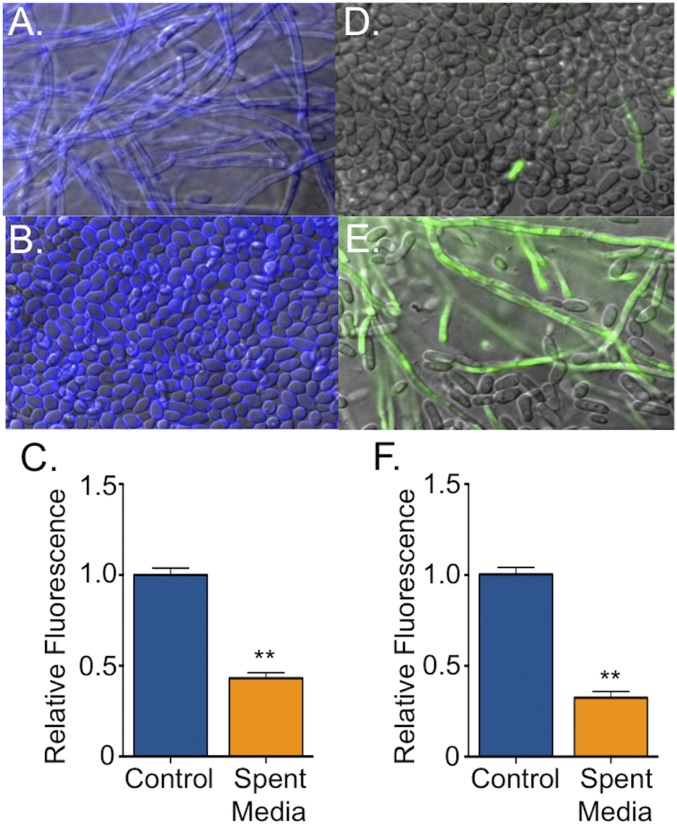
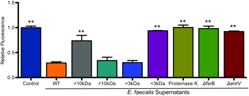
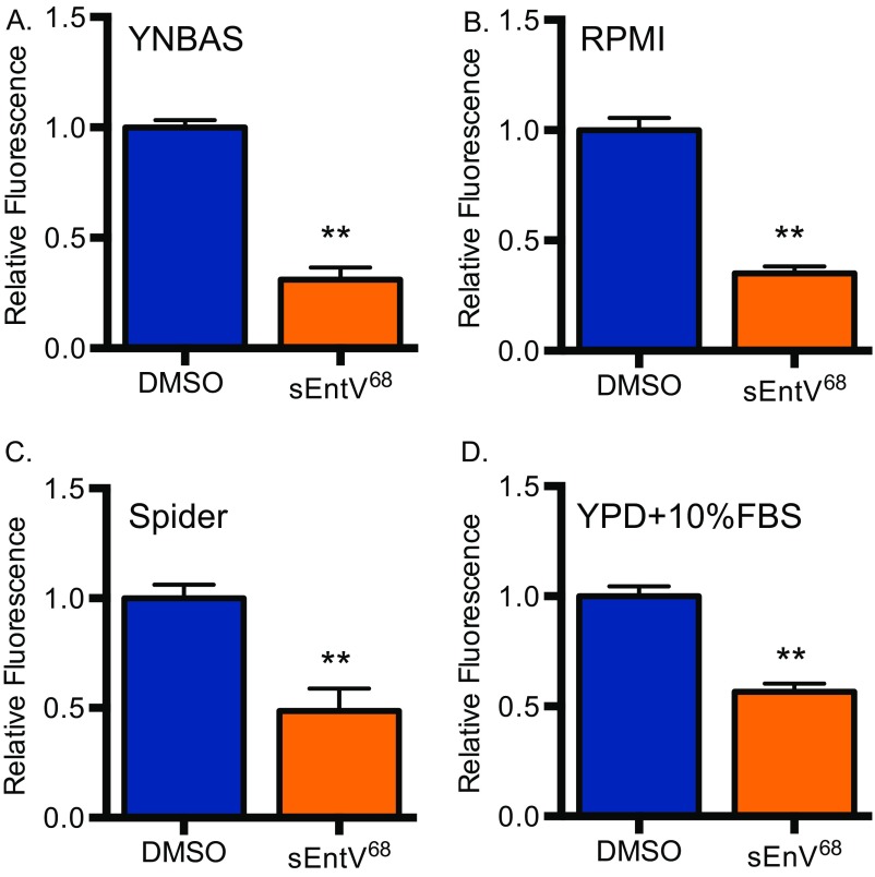
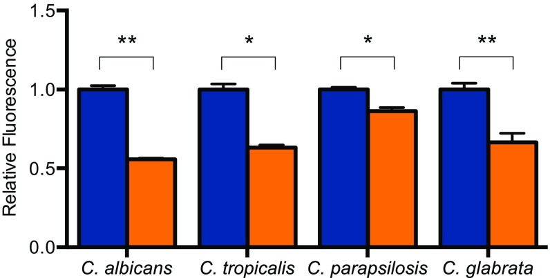
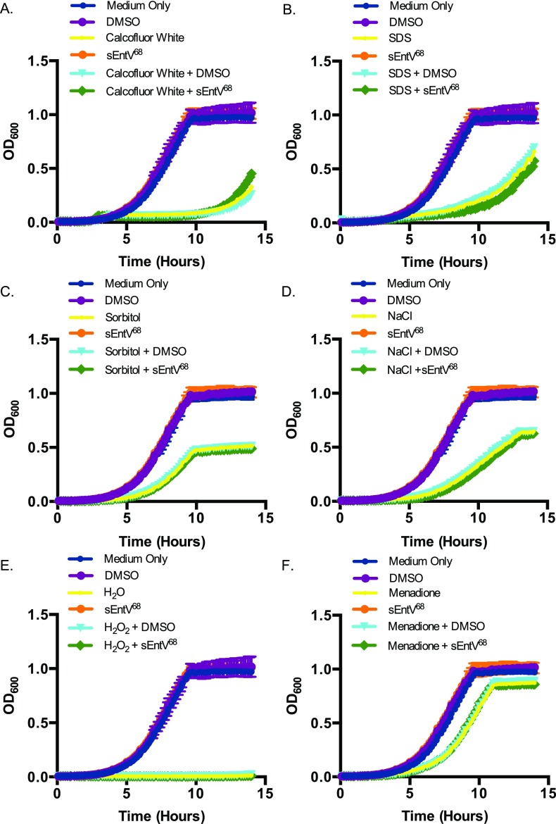
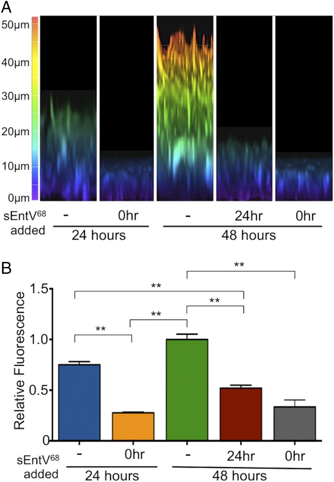
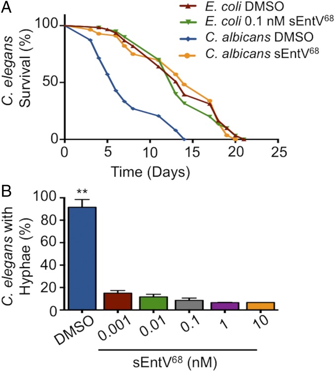
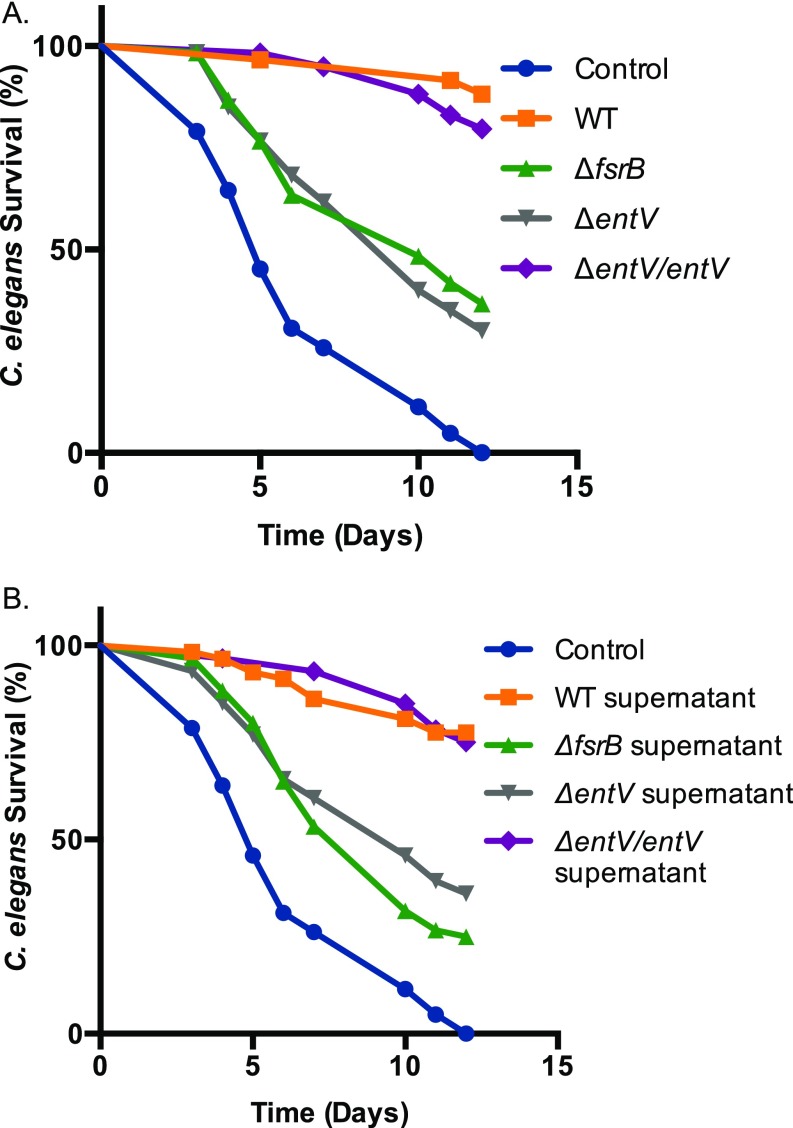
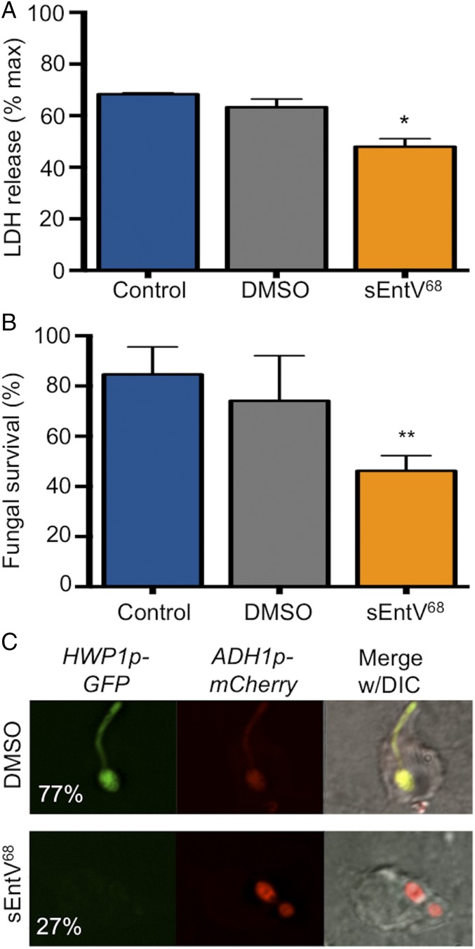
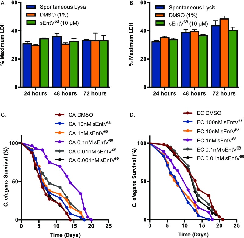
Enterococcus faecalis, a Gram-positive bacterium, and Candida albicans, a fungus, occupy overlapping niches as ubiquitous constituents of the gastrointestinal and oral microbiome. Both species also are among the most important and problematic, opportunistic nosocomial pathogens. Surprisingly, these two species antagonize each other's virulence in both nematode infection and in vitro biofilm models. We report here the identification of the E. faecalis bacteriocin, EntV, produced from the entV (ef1097) locus, as both necessary and sufficient for the reduction of C. albicans virulence and biofilm formation through the inhibition of hyphal formation, a critical virulence trait. A synthetic version of the mature 68-aa peptide potently blocks biofilm development on solid substrates in multiple media conditions and disrupts preformed biofilms, which are resistant to current antifungal agents. EntV68 is protective in three fungal infection models at nanomolar or lower concentrations. First, nematodes treated with the peptide at 0.1 nM are completely resistant to killing by C. albicans The peptide also protects macrophages and augments their antifungal activity. Finally, EntV68 reduces epithelial invasion, inflammation, and fungal burden in a murine model of oropharyngeal candidiasis. In all three models, the peptide greatly reduces the number of fungal cells present in the hyphal form. Despite these profound effects, EntV68 has no effect on C. albicans viability, even in the presence of significant host-mimicking stresses. These findings demonstrate that EntV has potential as an antifungal agent that targets virulence rather than viability.

Keywords: Candida albicans; Enterococcus faecalis; bacteriocins; biofilms.


Conflict of interest statement

The authors declare no conflict of interest.

Figures

Fig. 1.
Fig. 1.
E. faecalis supernatant inhibits C. albicans hyphal morphogenesis and biofilm formation. C. albicans biofilms grown for 24 h in YNBAS. Representative images of C. albicans (strain SC5314) biofilms in the absence (A) and presence (B) of E. faecalis supernatant and stained with calcofluor white. (C) Biofilm density was quantified by measuring resazurin fluorescence. Representative images of C. albicans hyphal reporter strain (HWP1p::GFP) in the absence (D) or presence (E) of E. faecalis supernatant. (F) Hyphal morphogenesis was quantified by measuring GFP fluorescence as a ratio to the OD600. Experiments were performed three times and analyzed by using Student’s t test (**P < 0.01).
Fig. S1.
Fig. S1.
Characterization of inhibitory activity from E. faecalis supernatants. C. albicans (HWP1p::GFP) biofilms grown at 37 °C for 24 h in YNBAS. Hyphal morphogenesis was quantified by measuring GFP fluorescence as a ratio to the OD600. Inhibitory activity from E. faecalis supernatant was retained between 3–10 kDa after size exclusion centrifugation. A reduction of inhibitory activity was observed in supernatants treated with proteinase K and from deletion mutants of fsrB or entV. Experiments were repeated at least three times and statistically significant differences relative to treatment with the WT supernatant were calculated by using a one-way ANOVA, and are indicated by asterisks (**P < 0.01).
Fig. 2.
Fig. 2.
EntV inhibition of hyphal morphogenesis and biofilm formation in C. albicans. (A) C. albicans SC5314 biofilms grown for 24 h in YNBAS with sterile media (M9HY) or spent supernatant from wild-type and mutant E. faecalis strains. Hyphal morphogenesis was quantified by measuring GFP fluorescence as a ratio to the OD600. The experiment was performed three times and analyzed by using one-way ANOVA (**P < 0.01). (B) Biofilms were grown with increasing concentrations of rEntV136 or sEntV68 and the IC50 calculated at 1,000 nM and 0.3 nM, respectively. (C) C. albicans was grown at 30 °C in YPD for 15 h in a 96-well plate with increasing concentrations of sEntV68, and OD600 was measured every 2 min in a microplate reader.
Fig. S2.
Fig. S2.
sEntV68 inhibitory activity in different media types. C. albicans (HWP1p::GFP) biofilms grown for 24 h at 37 °C in YNBAS (A), RPMI (B), Spider (C), or YPD (D) with 10% FBS with 0.01% DMSO (blue) or 100 nM sEntV (orange). Hyphal morphogenesis was quantified by measuring GFP fluorescence as a ratio to the OD600 for three separate experiments and statically significant differences were calculated by using a Student’s t test (**P < 0.01).
Fig. S3.
Fig. S3.
sEntV68 inhibitory activity against different Candida species. C. albicans, C. tropicalis, C. parapsilosis, and C. glabrata biofilms grown for 24 h in RPMI (with l-glutamine and Hepes) at 37 °C for 24 h with 0.01% DMSO (blue) or 100 nM sEntV (orange). Biofilm density was quantified by measuring resazurin fluorescence for three separate experiments and statically significant differences were calculated by using a Student’s t test (*P < 0.05; **P < 0.01).
Fig. S4.
Fig. S4.
Impact of host-relevant stresses on sEntV68 activity. C. albicans (SC5314) was grown for 15 h in YPD at 30 °C with sEntV68 (100 nM) in different stress conditions: cell wall stress calcofluor white (100 μg/mL) (A) and SDS (10%) (B); cell membrane stress sorbitol (1 M) (C) and sodium chloride (1 M) (D); and oxidative stress hydrogen peroxide (5 mM) (E) and menadione (100 μM) (F) in a 96-well plate. OD600 was measured every 2 min in a microplate reader.
Fig. 3.
Fig. 3.
Characterization of sEntV68 inhibitory activity. C. albicans SC5314 biofilms were grown with 0.01% DMSO or 100 nM sEntV68 added at different time points during biofilm formation (-, not added; 0hr, added at the beginning of the experiment. (A) Representative image of 24-h and 48-h biofilms observed by confocal microscopy. (B) Hyphal morphogenesis was quantified by measuring GFP fluorescence as a ratio to the OD600 for three separate experiments (**P < 0.01).
Fig. 4.
Fig. 4.
sEntV68 protects C. elegans from killing by C. albicans. C. elegans was exposed to C. albicans SC5314 on BHI agar or E. coli OP50 on NB agar for 2 h, then washed with sterile PBS and transferred to six-well plates containing increasing concentrations of sEntV68 or DMSO at ∼30 nematodes per well. (A) Representative data of three independent experiments where nematode viability was scored daily. (B) Nematodes were assayed for evidence of visible C. albicans filaments penetrating the cuticle after 7 d of infection (**P > 0.01).
Fig. S5.
Fig. S5.
EntV inhibition of virulence and hyphal morphogenesis of C. albicans in C. elegans. (A) C. elegans was exposed to E. faecalis on BHI agar or E. coli OP50 on NG agar for 2 h, followed by exposure to C. albicans on BHI agar for 2 h then washed with sterile PBS and transferred to six-well plates at ∼30 nematodes per well. (B) C. elegans was exposed to C. albicans SC5314 on BHI agar or E. coli OP50 on NB agar for 2 h, then washed with sterile PBS and transferred to six-well plates containing 20% E. faecalis supernatants or sterile BHI media at ∼30 nematodes per well. Both graphs are representative data of three independent experiments where nematode viability was scored daily.
Fig. 5.
Fig. 5.
Protection of murine macrophages by sEntV68 during C. albicans infection. (A) RAW264.7 murine macrophages were incubated with C. albicans SC5314 for 4 h with or without 100 nM sEntV68. Macrophage killing was evaluated by using the LDH cell toxicity assay, and the percentage of killed macrophages was calculated. (B) C. albicans survival in murine macrophages was assessed by using the XTT cell viability assay. (*P < 0.05; **P < 0.01). (C) RAW264.7 cells were infected with C. albicans (HWP1p::GFP, ADH1p::mCherry) for 1 h ± peptide followed by fixation and visualized by fluorescence microscopy. The percentage of hyphal cells was scored after 2 h of coculture and is given in the left image for both conditions. At least 200 cells were counted per replicate, and the experiment was repeated three times.
Fig. S6.
Fig. S6.
Activity of sEntV68 in mammalian cells and C. elegans. LDH was measured from murine macrophages (RAW 264.7) (A) and HeLa (B) exposed to 10 μM sEntV68 or 1% DMSO for up to 72 h. C. elegans was exposed to C. albicans SC5314 on BHI agar (C) or E. coli OP50 (D) on NB agar for 2 h, then washed with sterile PBS and transferred to six-well plates containing increasing concentrations of sEntV68 or DMSO at ∼30 nematodes per well. Representative data of three independent experiments where nematode viability was scored daily.
Fig. 6.
Fig. 6.
sEntV68 is protective in a murine OPC model. Immunosuppressed mice were inoculated sublingually with C. albicans SC5314 with 0.01% DMSO or 100 nM sEntV68. Water containing sEntV68 (100 nM) or vehicle (DMSO) alone was provided ad libitum. After 3 or 5 d, mice were euthanized and the tongues were excised for histological examination of DMSO control (A) or sEntV68-treated (B) mice. Red arrowheads indicate hyphal cells, and black arrowheads indicate yeast cells. (C) The percentage of the epithelial surface showing evidence of fungal invasion was calculated for control and treated tongues. (D) DNA was extracted from the tongues and the fungal burden was estimated from qPCR amplification of the 5.8S ITS2 region. Statistically significant differences were calculated by using one-way ANOVA (**P < 0.01).

References

    1. Agudelo Higuita NI, Huycke MM. Enterococcal disease, epidemiology, and implications for treatment. In: Gilmore MS, Clewell DB, Ike Y, Shankar N, editors. Enterococci: From Commensals to Leading Causes of Drug Resistant Infection. Mass Eye Ear Infirmary; Boston: 2014.
    1. Lebreton F, Willems RJL, Gilmore MS. Enterococcus diversity, origins in nature, and gut colonization. In: Gilmore MS, Clewell DB, Ike Y, Shankar N, editors. Enterococci: From Commensals to Leading Causes of Drug Resistant Infection. Mass Eye Ear Infirmary; Boston: 2014. - PubMed
    1. Moran G, Coleman D, Sullivan D. 2012. An introduction to the medically relevant Candida species. Candida and Candidiasis, eds Calderone RA, Clancy CJ (Am Soc Microbiol, Washington), pp 11–26. - PubMed
    1. Oever JT, Netea MG. The bacteriome-mycobiome interaction and antifungal host defense. Eur J Immunol. 2014;44:3182–3191. - PubMed
    1. Mayr FB, Yende S, Angus DC. Epidemiology of severe sepsis. Virulence. 2014;5:4–11. - PMC - PubMed

Publication types