. 2017 Jul;23(7):995-1011.
doi: 10.1261/rna.061234.117. Epub 2017 Apr 10.

Riboswitch diversity and distribution

Affiliations

Riboswitch diversity and distribution

Phillip J McCown et al. RNA. 2017 Jul.

Abstract

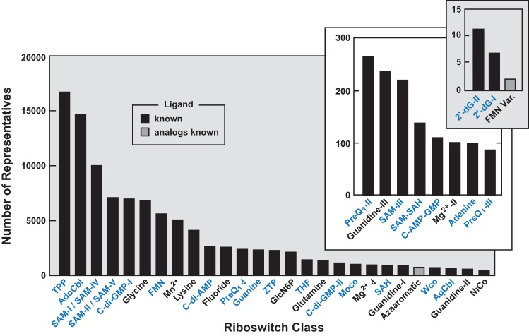
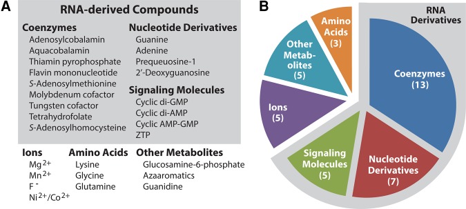
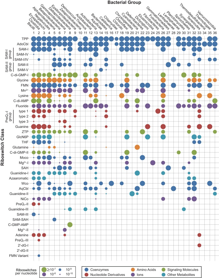
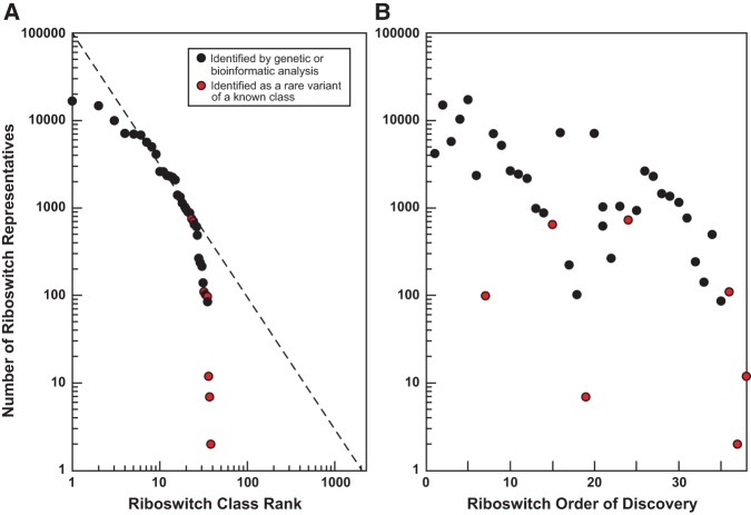
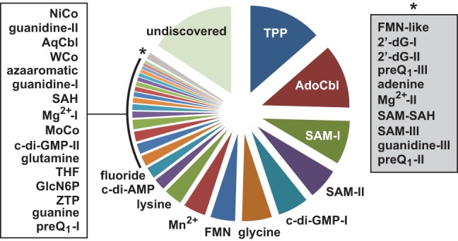
Riboswitches are commonly used by bacteria to detect a variety of metabolites and ions to regulate gene expression. To date, nearly 40 different classes of riboswitches have been discovered, experimentally validated, and modeled at atomic resolution in complex with their cognate ligands. The research findings produced since the first riboswitch validation reports in 2002 reveal that these noncoding RNA domains exploit many different structural features to create binding pockets that are extremely selective for their target ligands. Some riboswitch classes are very common and are present in bacteria from nearly all lineages, whereas others are exceedingly rare and appear in only a few species whose DNA has been sequenced. Presented herein are the consensus sequences, structural models, and phylogenetic distributions for all validated riboswitch classes. Based on our findings, we predict that there are potentially many thousands of distinct bacterial riboswitch classes remaining to be discovered, but that the rarity of individual undiscovered classes will make it increasingly difficult to find additional examples of this RNA-based sensory and gene control mechanism.

Keywords: RNA World; aptamer; coenzyme; ligand; noncoding RNA.


Figures

FIGURE 1.
FIGURE 1.
Rank order of riboswitch classes based on their abundance in genomic databases. Black and gray bars represent validated riboswitches with known and unknown natural ligands, respectively. Blue text identifies riboswitch classes whose ligands are derived from RNA nucleotides or their precursors. Each riboswitch class is named according to its ligand, wherein multiple structural classes for the same ligand are identified by Roman numerals. Inset plots include data for rarer riboswitch classes. (TPP) Thiamin pyrophosphate, (AdoCbl) adenosylcobalamin or coenzyme B12, (SAM) S-adenosylmethionine, (C-di-GMP) cyclic-di-GMP, (FMN) flavin mononucleotide, (Mn2+) divalent manganese, (C-di-AMP) cyclic-di-AMP, (PreQ1) prequeuosine1, (ZTP) 5-aminoimidazole-4-carboxamide ribonucleoside-5′-triphosphate, (GlcN6P) glucosamine-6-phosphate, (THF) tetrahydrofolate, (Moco) molybdenum cofactor, (Mg2+) divalent magnesium, (SAH) S-adenosylhomocysteine, (Wco) tungsten cofactor, (AqCbl) aquacobalamin, (NiCo) divalent nickel and divalent cobalt, (c-AMP-GMP) cyclic AMP-GMP, (2′-dG) 2′-deoxyguanosine, (FMN-Var.) FMN riboswitch variant.
FIGURE 2.
FIGURE 2.
Distinguishing characteristics of TPP riboswitches. (Left) Chemical structure of thiamin pyrophosphate, the natural ligand for TPP riboswitches. (Center) Consensus sequence and secondary structure model for TPP riboswitch aptamers. Note that the model is based on all types, including those that carry mutations in the P4–P5 region and that cannot substantially discriminate against thiamin, thiamin monophosphate, and TPP. Red, black, and gray letters represent nucleotides conserved in 97%, 90%, and 75% of the representatives, respectively, wherein R is a purine and Y is a pyrimidine. Similarly, circles represent any nucleotide, wherein its presence is reflected by the same color-coding, with open circles representing a nucleotide that is present in at least 50% of the representatives. Green, blue, and red shading of base pairs reflects strong evidence for covariation, compatible mutations, or no covarying mutations, respectively. P1 through P5 represent base-paired substructures. Number highlighted in black is the total collection of representatives for this riboswitch class in all three domains of life. (Right) Ribbon diagram representing an atomic-resolution structure of a representative TPP riboswitch aptamer bound to its ligand (space-filling model). (Bottom) The distribution of riboswitches among 36 divisions (phyla or orders) of bacteria. Circle size represents the relative number of riboswitch representatives per nucleotide of sequenced DNA as annotated elsewhere (Fig. 5). See Supplemental File 1 for a complete collection of similar data and imagery for all riboswitch classes.
FIGURE 3.
FIGURE 3.
The ligands for known riboswitch classes. (A) List of the known riboswitch ligands grouped by ligand type. (B) Pie chart presenting the number of distinct riboswitch classes for the various ligand groups as presented in A. Note that the majority of riboswitches sense ligands derived from RNA monomers or their precursors.
FIGURE 4.
FIGURE 4.
Fluoride riboswitches use their polyanionic RNA backbone to form a structure that selectively binds a fluoride anion. (A) Characteristics of fluoride riboswitches. Annotations are as described for Figure 2. (B) The atomic-resolution model for the ligand-binding site of a fluoride riboswitch. Fluoride (red) resides at the center of a Mg2+ triangle (blue), which is created by five negatively charged phosphates of the RNA aptamer backbone.
FIGURE 5.
FIGURE 5.
Grid depicting the presence and abundance of experimentally validated riboswitch classes. Abbreviations are as described for Figure 1 and data point sizes are as described for Figure 2. There are 38 general riboswitch classes, which are those that either bind a distinct ligand or use a distinct architecture to bind a ligand that is already known to be bound by another class. For example, guanine and adenine riboswitches are distinct or “general” classes because they bind distinct ligands, even though they exploit near-identical aptamer architectures. The type 1, 2, and 3 RNAs of the preQ1-I riboswitch class use only modestly different aptamer sequences and structures to bind the ligand preQ1, and thus constitute members of a single general class. Likewise, the RNAs called SAM-I (or S box), -IV, and -I/-IV are all close structural types of the general SAM-I class, and the RNAs originally called SAM-II and -V are types of the general SAM-II class. Notes: All known representatives of the 2′-dG-II riboswitch class are derived from metagenomic data, and there is insufficient DNA sequence information in these sequencing reads to confidently assign the hits to bacterial divisions. Caution should be used in interpreting rare instances of a riboswitch class in certain bacterial lineages, because some bioinformatics hits could be false positives.
FIGURE 6.
FIGURE 6.
Evidence that riboswitch classes conform to a power law distribution. (A) A log–log plot depicting the validated riboswitch classes ranked (x-axis, X) in order of their abundance (y-axis, Y). The dashed line represents an ideal power law distribution from the equation Y = mXb where m and b were set to 100,000 and −1.6, respectively, to yield a line that best tracks the data. (B) Plot of the abundance of each riboswitch class on a log scale versus the order of discovery. Red points represent classes discovered as rare variants of more common classes.
FIGURE 7.
FIGURE 7.
Distribution of IGRs carrying riboswitches. Abbreviations are as described for Figure 1, wherein the boxed riboswitch names are listed from top to bottom as the least common to most common, respectively. The size of the plot segment labeled “undiscovered” was established by summing the numbers of riboswitch classes remaining to be discovered as presented in Figure 6A and as predicted by the power law distribution wherein m = 100,000 and b = −1.6.

Similar articles

Cited by

References

    1. Ames TD, Breaker RR. 2010. Bacterial riboswitch discovery and analysis. In The chemical biology of nucleic acids (ed. Mayer G). Wiley, Chichester, UK.
    1. Ames TD, Breaker RR. 2011. Bacterial aptamers that selectively bind glutamine. RNA Biol 8: 82–89. - PMC - PubMed
    1. André G, Even S, Putzer H, Burguière P, Croux C, Danchin A, Martin-Verstraete I, Soutourina O. 2008. S-box and T-box riboswitches and antisense RNA control a sulfur metabolic operon of Clostridium acetobutylicum. Nucleic Acids Res 36: 5955–5969. - PMC - PubMed
    1. Baker JL, Sudarsan N, Weinberg Z, Roth A, Stockbridge RB, Breaker RR. 2012. Widespread genetic switches and toxicity resistance proteins for fluoride. Science 335: 233–235. - PMC - PubMed
    1. Barrick JE, Breaker RR. 2007. The distributions, mechanisms, and structures of metabolite-binding riboswitches. Genome Biol 8: R239. - PMC - PubMed

Publication types