. 2017 Apr 10;7(1):779.
doi: 10.1038/s41598-017-00900-7.

Sphingosine kinase 1-interacting protein is a novel regulator of glucose-stimulated insulin secretion

Affiliations

Sphingosine kinase 1-interacting protein is a novel regulator of glucose-stimulated insulin secretion

Yu Wang et al. Sci Rep. .

Abstract

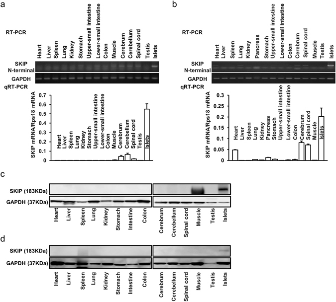
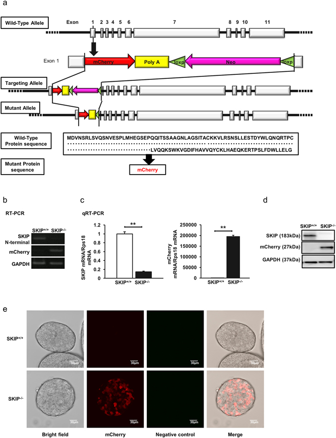
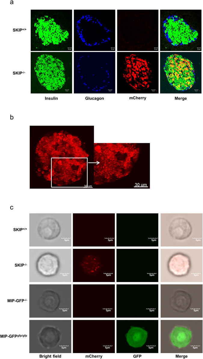
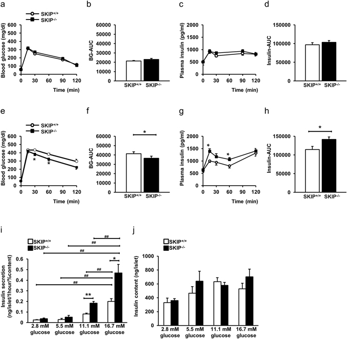
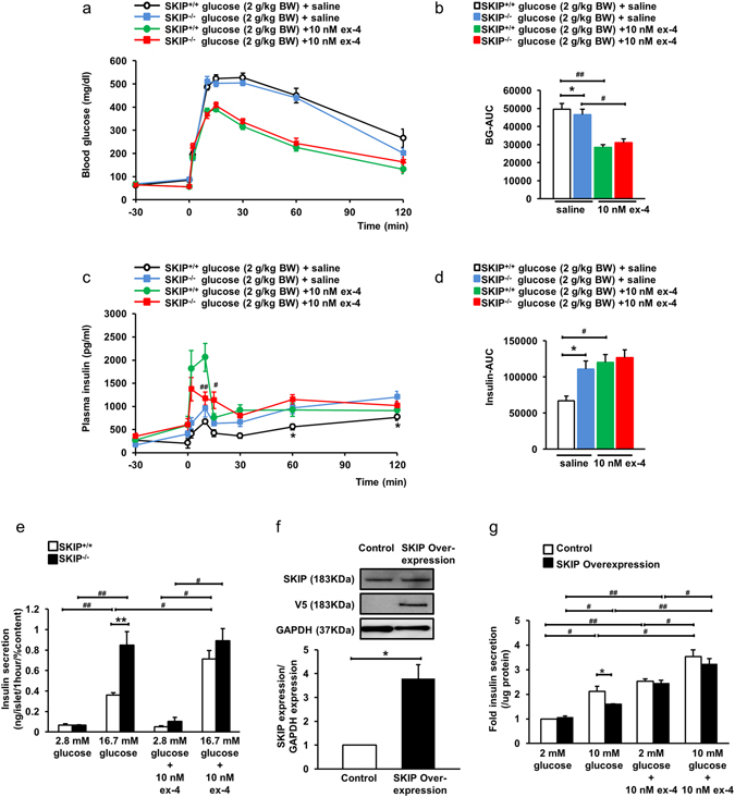
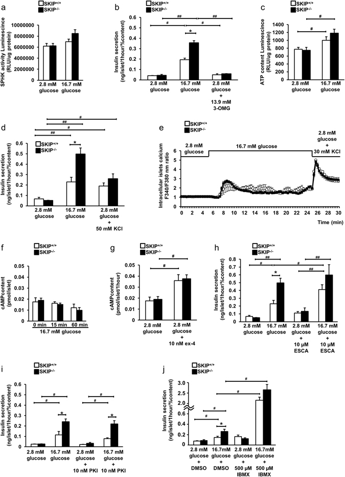
Glucose-stimulated insulin secretion (GSIS) is essential in keeping blood glucose levels within normal range. GSIS is impaired in type 2 diabetes, and its recovery is crucial in treatment of the disease. We find here that sphingosine kinase 1-interacting protein (SKIP, also called Sphkap) is highly expressed in pancreatic β-cells but not in α-cells. Intraperitoneal glucose tolerance test showed that plasma glucose levels were decreased and insulin levels were increased in SKIP-/- mice compared to SKIP+/+ mice, but exendin-4-enhanced insulin secretion was masked. GSIS was amplified more in SKIP-/- but exendin-4-enhanced insulin secretion was masked compared to that in SKIP+/+ islets. The ATP and cAMP content were similarly increased in SKIP+/+ and SKIP-/- islets; depolarization-evoked, PKA and cAMP-mediated insulin secretion were not affected. Inhibition of PDE activity equally augmented GSIS in SKIP+/+ and SKIP-/- islets. These results indicate that SKIP modulates GSIS by a pathway distinct from that of cAMP-, PDE- and sphingosine kinase-dependent pathways.


Conflict of interest statement

The authors declare that they have no competing interests.

Figures

Figure 1
Figure 1
SKIP expression in various tissues. (ad) mRNA expression of SKIP in several tissues from C57BL/6 mice (a) or Wistar rats (b) detected by RT-PCR and qRT-PCR; protein expression of SKIP in several tissues from C57BL/6 mice (c) or Wistar rats (d) detected by western blot with anti-mouse SKIP (c) and anti-rat SKIP antibody (d), respectively. The membrane was reprobed with anti-GAPDH antibody as control. All gels were run under the same experimental conditions. Uncropped images of blots/gels are shown in Supplemental Figures 2–4. (ad) 12-week-old animals were used for experiments, n = 4.
Figure 2
Figure 2
Generation of SKIP-mCherry knock-in (KI) mice. (a) Construct of SKIP-mCherry KI mice. mCherry-poly(A)-loxp-Neo-loxp was inserted into exon 1 of wild-type SKIP gene, and later, loxp-Neo was deleted to generate the mutant allele. In the mutant protein sequence, SKIP expression was deleted by mCherry expression. mRNA expression of SKIP and mCherry in isolated islets from homo SKIP-mCherry KI (SKIP−/−) mice and wild type (SKIP+/+) mice detected by RT-PCR (b) and qRT-PCR (c); data are expressed as average ± standard error of the mean (SEM). **p < 0.005 SKIP−/− vs SKIP+/+, significance was determined by student’s t-test (c). (d) Protein expression of SKIP in isolated islets from SKIP−/− mice and SKIP+/+ mice detected by western blot with anti-mouse SKIP antibody. The membrane was reprobed with anti-mCherry antibody, and reprobed with anti-GAPDH antibody as control. All gels were run under the same experimental conditions. Uncropped images of blots/gels are shown in Supplementary Figure 5. (e) Expression of mCherry in isolated islets from SKIP−/− mice and SKIP+/+ mice. Living islets were observed by incubator two-photon excitation microscopy. Two-photon excitation was effected at 1040 nm, the fluorescence of mCherry and negative controls were measured at 575–650 nm and at 460–495 nm, respectively. (be) n = 4–6 mice per group and 3 samples per group, 12-week-old mice were used for experiments.
Figure 3
Figure 3
Localization of SKIP in the islets. (a) Immunohistochemical images in the pancreas from SKIP−/−mice and SKIP+/+ mice; green, anti-insulin; blue: anti-glucagon; and red, anti-mCherry. (b) 3D imaging by incubator two-photon microscopy in isolated islets from SKIP−/− mice. (c) Incubator two-photon excitation microscopy images of living β-cells from SKIP−/− (SKIP-mCherry KI) mice and MIP-GFP mice; left column, bright field; second column, mCherry; third column, GFP; right column, merged images. 12-week-old mice were used for the experiments, n = 3.
Figure 4
Figure 4
SKIP-regulated glucose-stimulated insulin secretion (GSIS). (ad) Blood glucose levels (a), BG-AUC (b), plasma insulin levels (c) and insulin-AUC (d) during 1 g/kg body weight IpGTT. (eh) Blood glucose levels (e), BG-AUC (f), plasma insulin levels (g) and insulin-AUC (h) during 2 g/kg body weight IpGTT. (i) GSIS in islets from SKIP+/+ mice and SKIP−/− mice. Insulin secretion was measured at 2.8 mM, 5.5 mM, 11.1 mM, and 16.7 mM glucose. (j) Insulin content in the islets from SKIP+/+ mice and SKIP−/− mice. (aj) 12-week-old mice were used for the experiments. (ah) n = 7–8 mice per group, *p < 0.05 vs SKIP+/+, significance was determined by student’s t-test. (i,j) n = 7–8 mice per group and 5–6 samples per group, with 10 islets per sample, *p < 0.05, **p < 0.005 vs SKIP+/+, #p < 0.05, ##p < 0.005 for different glucose conditions in the same genotype mice of insulin secretion, significance was determined by one way ANOVA with Tukey test. All data are expressed as average ± SEM.
Figure 5
Figure 5
Exendin-4-enhanced Insulin secretion in SKIP+/+ mice and SKIP−/− mice. (ad) Blood glucose levels (a), BG-AUC (b), plasma insulin levels (c) and insulin-AUC (d) during 2 g/kg body weight IpGTT 30 min after administration of 10 nM ex-4. (e) Ex-4-induced insulin secretion in the islets from SKIP+/+ mice and SKIP−/−mice. Insulin secretion from isolated islets measured at 2.8 mM and 16.7 mM glucose with or without 10 nM ex-4. (f) Protein expression of SKIP in control and SKIP-overexpressed INS-1D cells after 48 h transfection detected by western blot with anti-rat SKIP antibody. The membrane was reprobed with anti-V5 antibody, and reprobed with anti-GAPDH antibody as control. All gels were run under the same experimental conditions. Uncropped images of blots/gels are shown in Supplemental Figure 6 (n = 3). (g) Insulin secretion in SKIP-overexpressed INS-1D cells measured at 2 mM and 10 mM glucose with or without 10 nM ex-4. (ae) 12-week-old mice were used for the experiments. (a,c) #p < 0.05 SKIP−/− ex-4 vs SKIP+/+ ex-4. (b,d) *p < 0.05, **p < 0.005 SKIP−/− saline vs SKIP+/+ saline, #p < 0.05, ##p < 0.005 for different stimulated conditions in the same genotype mice. (e) n = 7–8 mice per group and 5–6 samples per group, with 10 islets per sample, *p < 0.05, **p < 0.005 vs SKIP+/+, #p < 0.05, ##p < 0.005 for different stimulated conditions in the same genotype mice of insulin secretion. (g) Data from 4 experiments are shown, *p < 0.05 vs control, #p < 0.05, ##p < 0.005 for different stimulated conditions in the same cells. All data are expressed as average ± SEM. Significance was determined by one way ANOVA with Tukey test (ae,g) and student’s t-test (f).
Figure 6
Figure 6
The mechanism of SKIP-involved insulin secretion. (a) Sphingosine kinase activity from isolated islets measured at 2.8 mM and 16.7 mM glucose. (b) Insulin secretion measured at 2.8 mM, 16.7 mM glucose and 2.8 mM with 13.9 mM non-metabolic glucose (3-OMG). (c) ATP content from isolated islets measured at 2.8 mM and 16.7 mM glucose. (d) Potassium-evoked insulin secretion in isolated islets from SKIP−/− mice. Insulin secretion measured at 2.8 mM, 16.7 mM glucose and 2.8 mM with 50 mM KCl. (e) Calcium influx measured in islets from SKIP+/+ and SKIP−/− mice. (f) cAMP content in isolated islets from SKIP+/+ and SKIP−/− mice stimulated at 16.7 mM glucose for 0 min, 15 min, 60 min. (g) cAMP content in isolated islets from SKIP+/+ and SKIP−/− mice at 2.8 mM glucose with or without 10 nM ex-4. (h) Epac2 selective activator (ESCA)-stimulated insulin secretion in islets from SKIP+/+ and SKIP−/− mice. Insulin secretion measured at 2.8 mM or 16.7 mM glucose with or without 10 μM ESCA in the isolated islets. (i) PKA-dependent insulin secretion in islets from SKIP+/+ and SKIP−/− mice. Insulin secretion from isolated islets measured at 2.8 mM or 16.7 mM glucose with or without 10 nM PKI. (j) GSIS treated with 3-isobutyl-1-metylxantine (IBMX) in islets from SKIP+/+ and SKIP−/− mice. Insulin secretion was measured at 2.8 mM or 16.7 mM glucose with or without 500 μM IBMX in the isolated islets. (aj) 12-week-old mice were used for the experiments. *p < 0.05, **p < 0.005 vs SKIP+/+, #p < 0.05, ##p < 0.005 for different stimulated conditions in the same genotype mice of insulin secretion. (ad, hj) n = 7–8 mice per group and 5–6 samples per group, 10 islets per sample. (e) n = 3 mice per group and 3 samples per group, 12–20 islets per sample. (f,g) n = 6–7 mice per group and 5–6 samples, 25 islets per group. Data are expressed as average ± SEM. Significance was determined by one way ANOVA with Tukey test (ad, fj) and student’s t-test (e).

Similar articles

Cited by

References

    1. Rorsman P, Braun M. Regulation of insulin secretion in human pancreatic islets. Annu Rev Physiol. 2013;75:155–79. doi: 10.1146/annurev-physiol-030212-183754. - DOI - PubMed
    1. Henquin JC. Triggering and amplifying pathways of regulation of insulin secretion by glucose. Diabetes. 2000;49:1751–1760. doi: 10.2337/diabetes.49.11.1751. - DOI - PubMed
    1. Henquin JC. Regulation of insulin secretion: A matter of phase control and amplitude modulation. Diabetologia. 2009;52:739–751. doi: 10.1007/s00125-009-1314-y. - DOI - PubMed
    1. Inagaki N, et al. Reconstitution of IKATP: an inward rectifier subunit plus the sulfonylurea receptor. Science. 1995;270:1166–1170. doi: 10.1126/science.270.5239.1166. - DOI - PubMed
    1. Bryan J, Crane A, Vila-Carriles WH, Babenko AP, Aguilar-Bryan L. Insulin secretagogues, sulfonylurea receptors and K(ATP) channels. Curr Pharm Des. 2005;11:2699–2716. doi: 10.2174/1381612054546879. - DOI - PubMed

Publication types

MeSH terms