. 2017 Mar 27:8:111.
doi: 10.3389/fneur.2017.00111. eCollection 2017.

New-Onset Refractory Status Epilepticus with Claustrum Damage: Definition of the Clinical and Neuroimaging Features

Affiliations

New-Onset Refractory Status Epilepticus with Claustrum Damage: Definition of the Clinical and Neuroimaging Features

Stefano Meletti et al. Front Neurol. .

Abstract

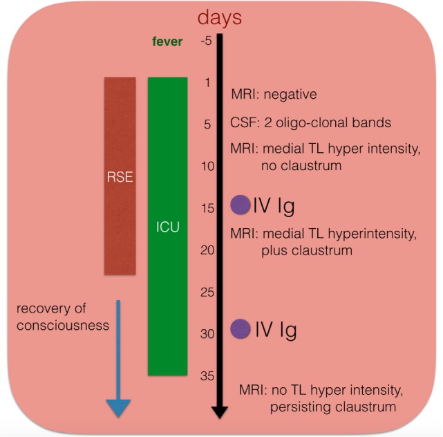
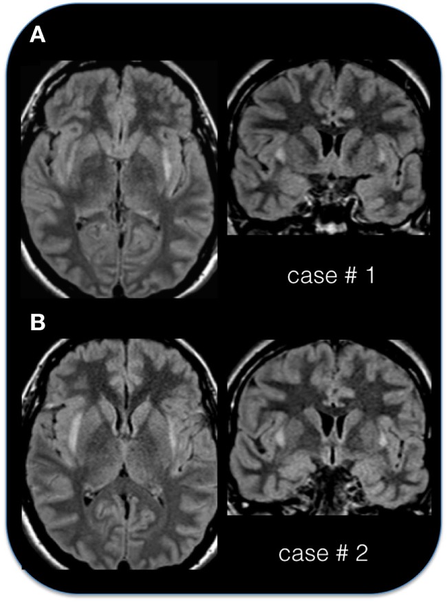
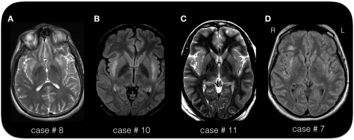
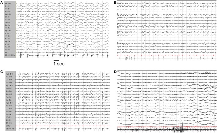
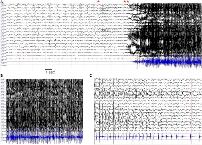
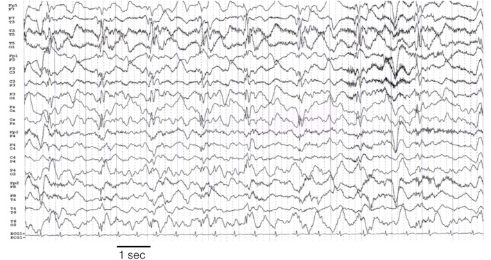
New-onset refractory status epilepticus (NORSE) is a rare but challenging condition occurring in a previously healthy patient, often with no identifiable cause. We describe the electro-clinical features and outcomes in a group of patients with NORSE who all demonstrated a typical magnetic resonance imaging (MRI) sign characterized by bilateral lesions of the claustrum. The group includes 31 patients (12 personal and 19 previously published cases; 17 females; mean age of 25 years). Fever preceded status epilepticus (SE) in 28 patients, by a mean of 6 days. SE was refractory/super-refractory in 74% of the patients, requiring third-line agents and a median of 15 days staying in an intensive care unit. Focal motor and tonic-clonic seizures were observed in 90%, complex partial seizures in 14%, and myoclonic seizures in 14% of the cases. All patients showed T2/FLAIR hyperintense foci in bilateral claustrum, appearing on average 10 days after SE onset. Other limbic (hippocampus, insular) alterations were present in 53% of patients. Within the personal cases, extensive search for known autoantibodies was inconclusive, though 7 of 11 patients had cerebrospinal fluid lymphocytic pleocytosis and 3 cases had oligoclonal bands. Two subjects died during the acute phase, one in the chronic phase (probable sudden unexplained death in epilepsy), and one developed a persistent vegetative state. Among survivors, 80% developed drug-resistant epilepsy. Febrile illness-related SE associated with bilateral claustrum hyperintensity on MRI represents a condition with defined clinical features and a presumed but unidentified autoimmune etiology. A better characterization of de novo SE is mandatory for the search of specific etiologies.

Keywords: claustrum; epilepsy; fever; new-onset refractory status epilepticus; refractory status epilepticus; status epilepticus.


Figures

Figure 1
Figure 1
Schematic temporal evolution, procedures, and immuno-modulating treatment in one patient (case # 11). IV Ig, intravenous immunoglobulins; CSF, cerebrospinal fluid; ICU, intensive care unit; RSE, refractory status epilepticus.
Figure 2
Figure 2
The “claustrum sign” in two patients. (A) Axial and coronal FLAIR sequence in case # 1, 29-year-old female, acquired 10 days after status epilepticus (SE) onset. (B) Axial and coronal FLAIR sequence in case # 2, 24-year-old female, acquired 4 days after SE onset. Note the hyperintense appearance of the capsula esterna/claustrum region in the two cases.
Figure 3
Figure 3
The “claustrum sign” in four patients. (A) Axial T2 sequence in case # 8, 25-year-old male, acquired 15 days after status epilepticus (SE) onset. (B) Axial FLAIR sequence in case # 10, 19-year-old female, acquired 6 days after SE onset. (C) Axial T2 image in case # 11, 33-year-old female, acquired 21 days after SE onset. (D) Axial FLAIR image in case # 7, 38-year-old female, acquired 25 days after SE onset. Note the hyperintense appearance of the capsula esterna/claustrum region in the four cases. Despite the different MRI scanner and image parameters, the hyperintensity of the claustrum is easily recognizable in each case. The exact location and extension of the signal alteration varies across the four cases. In all patients is present a bilateral involvement. Note the signal alteration extending to the insular/peri-insular cortex and subcortical white matter in panel (D).
Figure 4
Figure 4
EEG evolution in case # 1. (A) EEG during the initial hours of status epilepticus (SE). The patient was awake but confused with sub-continuous myoclonic jerks involving predominantly left limbs. The last channel records the surface EMG activity from the left anterior tibialis muscle. The EEG traces show a general slowing of the background activity, more evident on the right hemisphere, where periodic sharp complex are evident, often time-locked with myoclonic jerks. (B,C) EEG during the evolution in refractory SE, in the from of a myoclonic SE. The patient is under general anesthesia with barbiturates plus antiepileptic drugs poly-therapy. (D) Represents the EEG evolution during recovery. EEG recording parameters: high-pass filter: 0.3 Hz; low-pass filter 50 Hz. EMG parameters: high-pass filters 50 Hz; low-pass filter 250 Hz. Sampling frequency 1,024 Hz.
Figure 5
Figure 5
Representative seizure in case # 7. (A) Periodic theta burst involving the right fronto-temporal leads evolving into a right ictal rhythmic discharge (*) and then in a diffuse EEG “flattening” (**). (B) Diffuse EMG artifacts during the tonic–clonic phase of the seizures. (C) Terminal part of the seizures with predominant involvement of the left fronto-temporal region. In each panel, the EMG trace is recorded from the orbicularis oris muscle. EEG recording parameters: high-pass filter: 0.3 Hz; low-pass filter 50 Hz. EMG parameters: high-pass filters 50 Hz; low-pass filter 250 Hz. Sampling frequency 1,024 Hz.
Figure 6
Figure 6
Left hemisphere periodic discharges in case # 10. EEG recording parameters: high-pass filter: 0.3 Hz; low-pass filter 30 Hz. Sampling frequency 128 Hz.

References

    1. Hesdorffer DC, Logroscino G, Cascino G, Annegers JF, Hauser WA. Incidence of status epilepticus in Rochester, Minnesota, 1965–1984. Neurology (1998) 50:735–41.10.1212/WNL.50.3.735 - DOI - PubMed
    1. Mayer SA, Claassen J, Lokin J, Mendelsohn F, Dennis LJ, Fitzsimmons BF. Refractory status epilepticus: frequency, risk factors, and impact on outcome. Arch Neurol (2002) 59:205–10.10.1001/archneur.59.2.205 - DOI - PubMed
    1. Holtkamp M, Othman J, Buchheim K, Meierkord H. Predictors and prognosis of refractory status epilepticus treated in a neurological intensive care unit. J Neurol Neurosurg Psychiatry (2005) 76:534–9.10.1136/jnnp.2004.041947 - DOI - PMC - PubMed
    1. Van Lierde I, Van Paesschen W, Dupont P, Maes A, Sciot R. De novo cryptogenic refractory multifocal febrile status epilepticus in the young adult: a review of six cases. Acta Neurol Belg (2003) 103:88–94. - PubMed
    1. Wilder-Smith EP, Lim EC, Teoh HL, Sharma VK, Tan JJ, Chan BP, et al. The NORSE (new-onset refractory status epilepticus) syndrome: defining a disease entity. Ann Acad Med Singapore (2005) 34:417–20. - PubMed

LinkOut - more resources